Hematology Exam 1: Poikilocytes, Distributions, Inclusions, Parasites, Crystals
1/22
There's no tags or description
Looks like no tags are added yet.
Name | Mastery | Learn | Test | Matching | Spaced |
|---|
No study sessions yet.
23 Terms
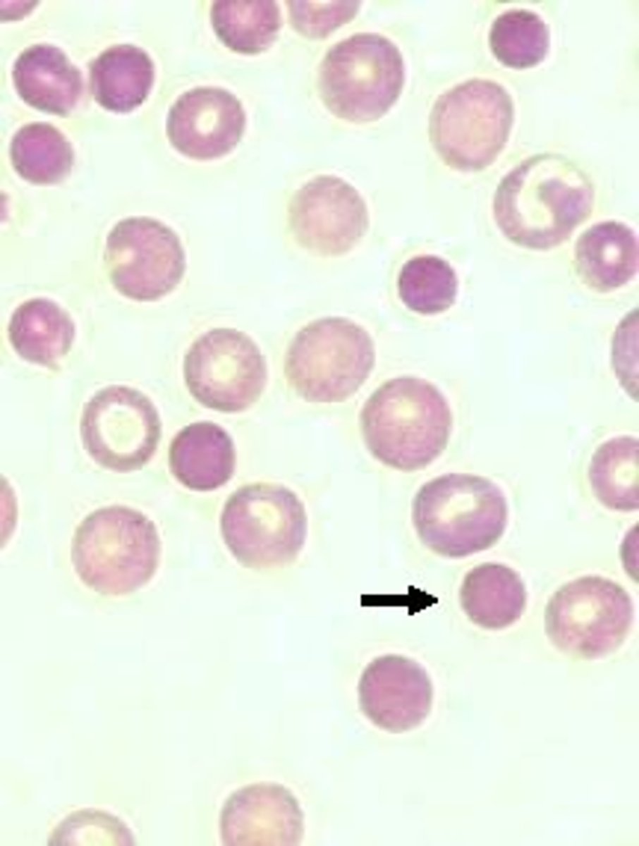
Spherocytes
- Ball with no pallor
- Decreased SA/Volume ratio (loss of biconcavity)
- Increase in MCHC
- Etiologies: Hereditary spherocytosis, autoimmune hemolytic anemia, burns \
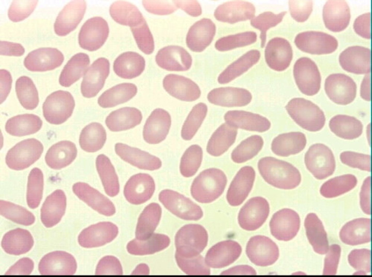
Ovalocytes
- oval/cigar shaped
- contains central pallor
- decrease in membrane cholesterol and/or spectrin
- cells cannot reform into binconcave erythrocytes
-Etiology: hereditary ovalocytosiselliptocytosis
Target Cells
- Bull's eye shape w dark central pallor
- Sombrero
- increase cholesterol/phospholipids
- decreased Hgb
- Increased SA/Volume ratio
-Etiologies: Hemoglobinopathies, Thalassemia (3-4+ cells), Liver disease (1-2+ cells)
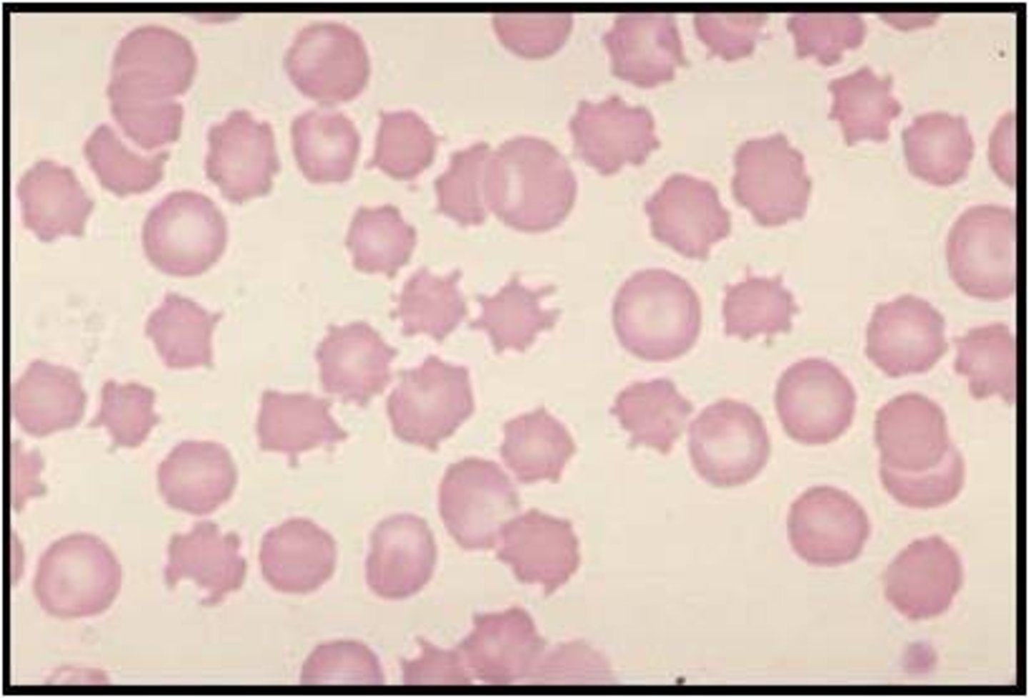
Burr cells
-short/wavy projections
-multitude of diseases
- Uremia (3-4+ cells)
- Look similar to crenated RBCs, however, crenated cells are due to lab (H2O or heparin)
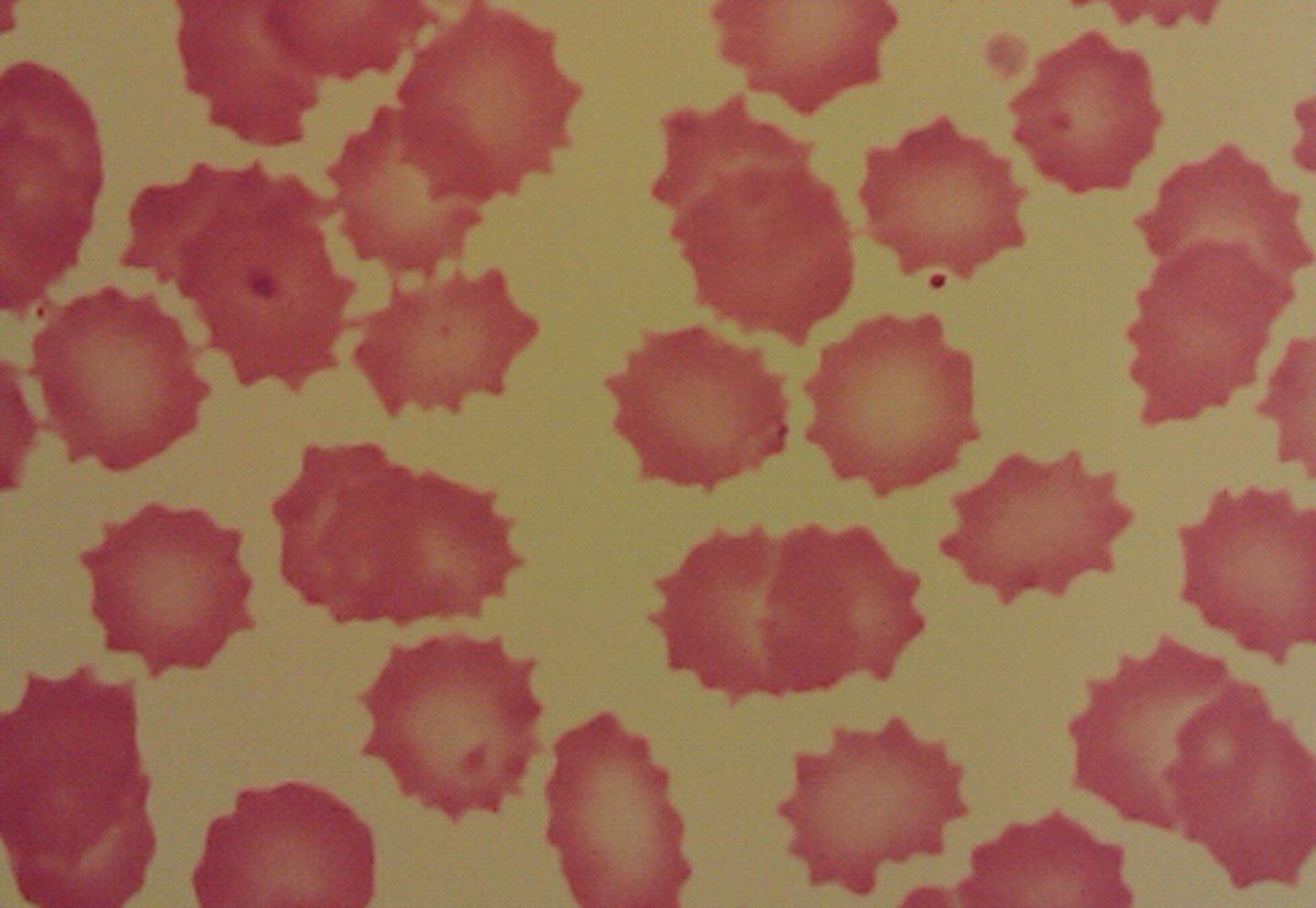
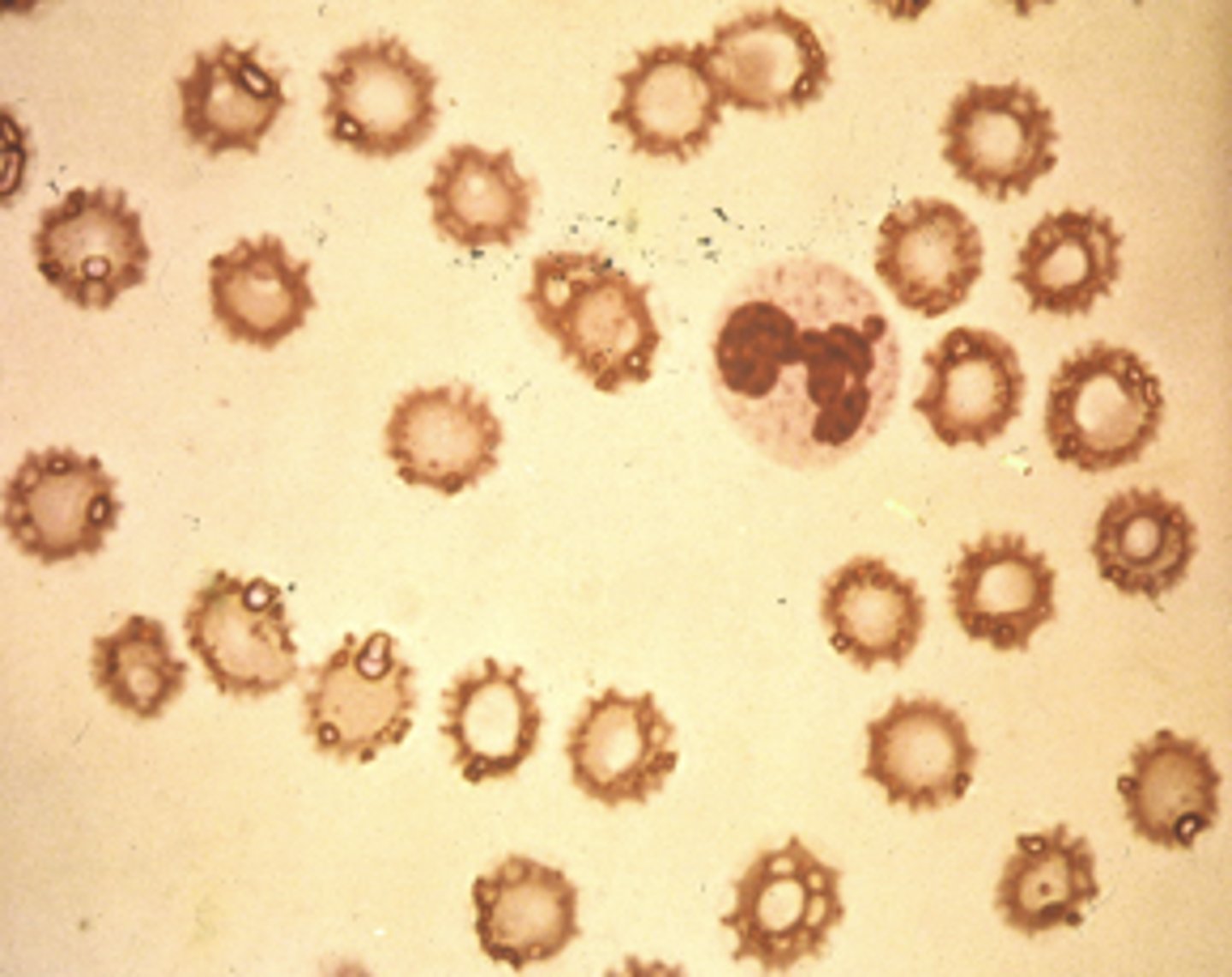
Acanthocytes
- Spherical RBCs with uneven spiny projections
- Increased cholesterol
- Decreased lecithin
- Etiologies: Abetalipoproteinemia (3-4+), Liver disease (2-3+), Retinitis pigmentosa (1-2+)
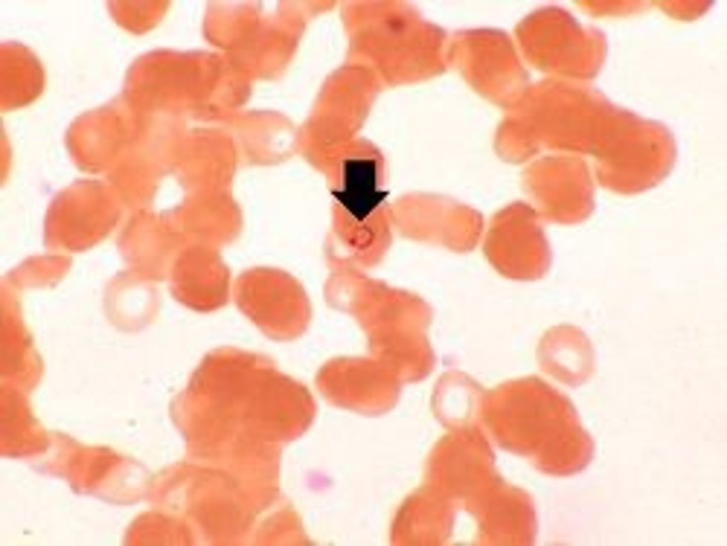
Schistocytes (1-4+)
-RBC fragments
- Both small and large fragments make up schistocytes
- Shape is due to loss of cell membrane
- Etiologies: Hemolytic, uremia, DIC, Heart valve problem, burns (2-3+)
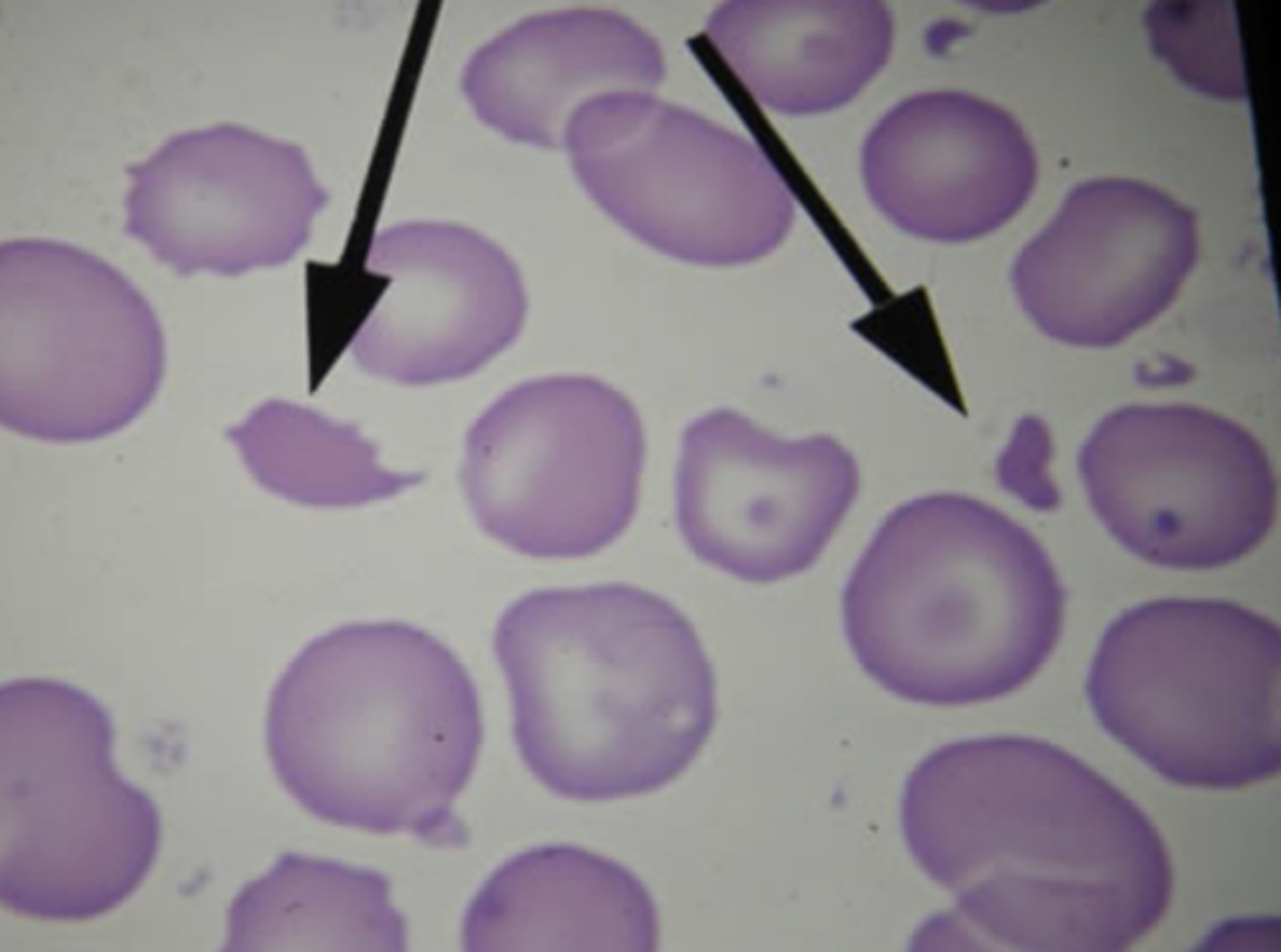
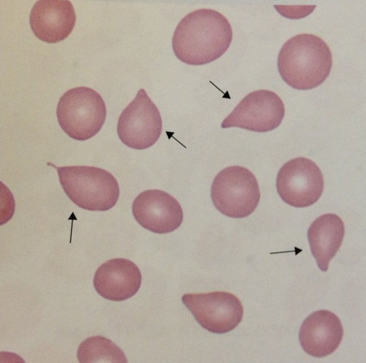
Tear Drop Cells
- Tear drop shaped
- RBCs pushed through bone marrow fibrosis
- Etiology: Primary myelofibrosis (2-3+)
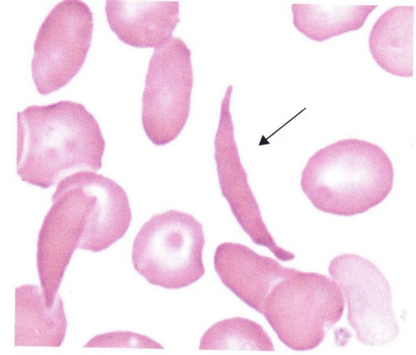
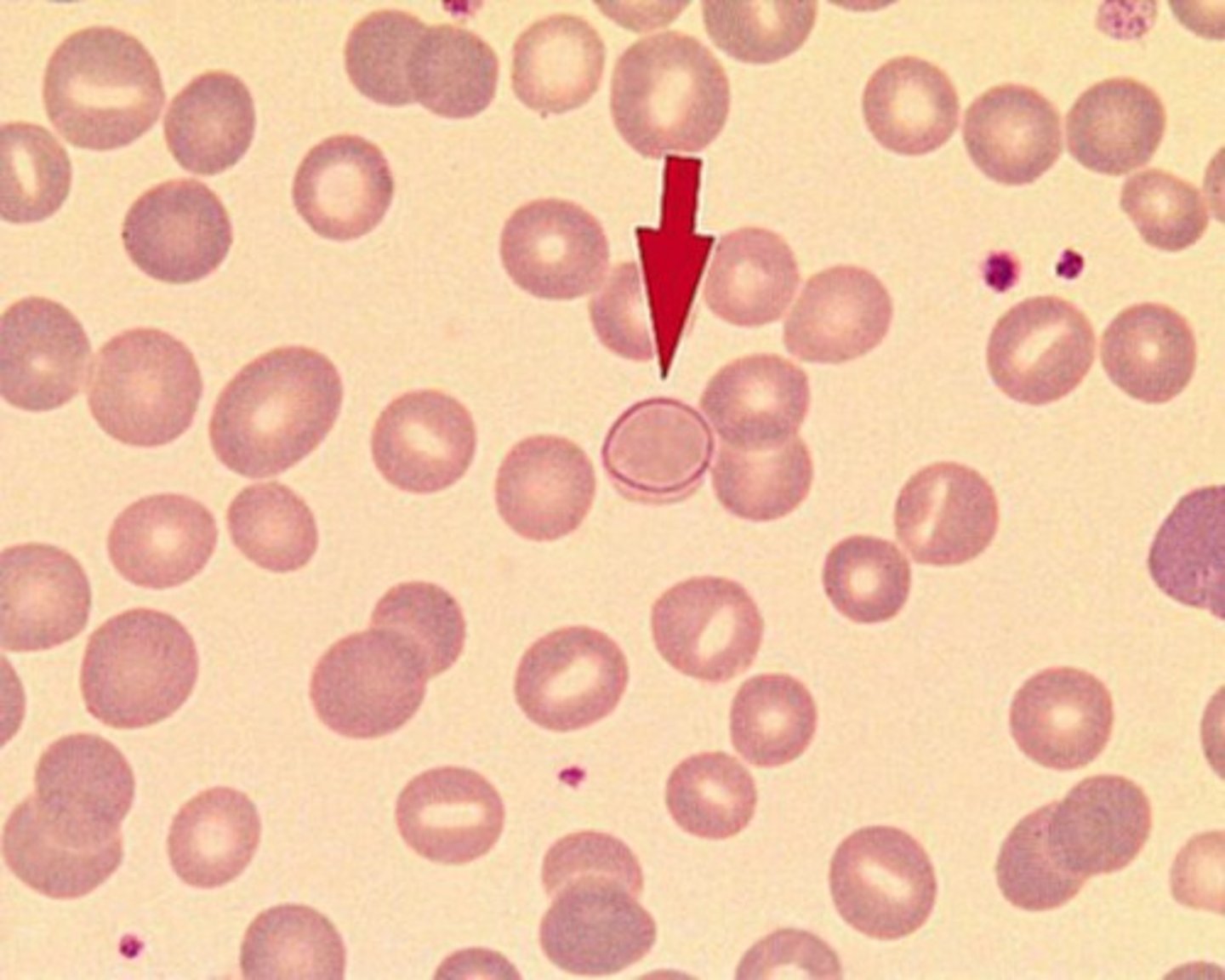
Sickle Cells
- Shaped like sickle/crescent moon
- Low O2 > HbS polymerizing > sickle cells
- Diagnostic for: HbS
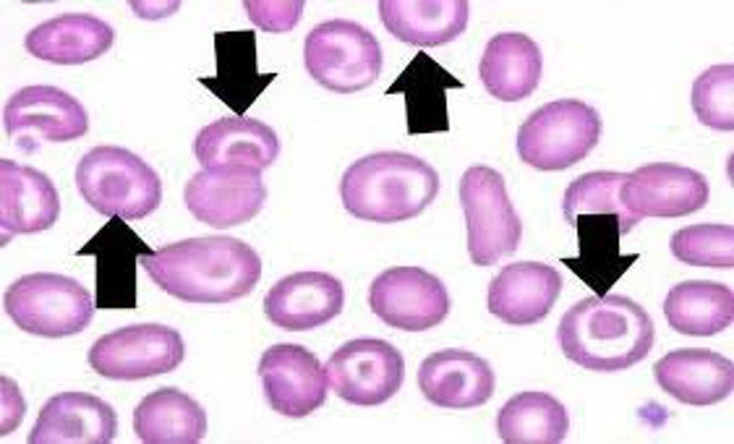
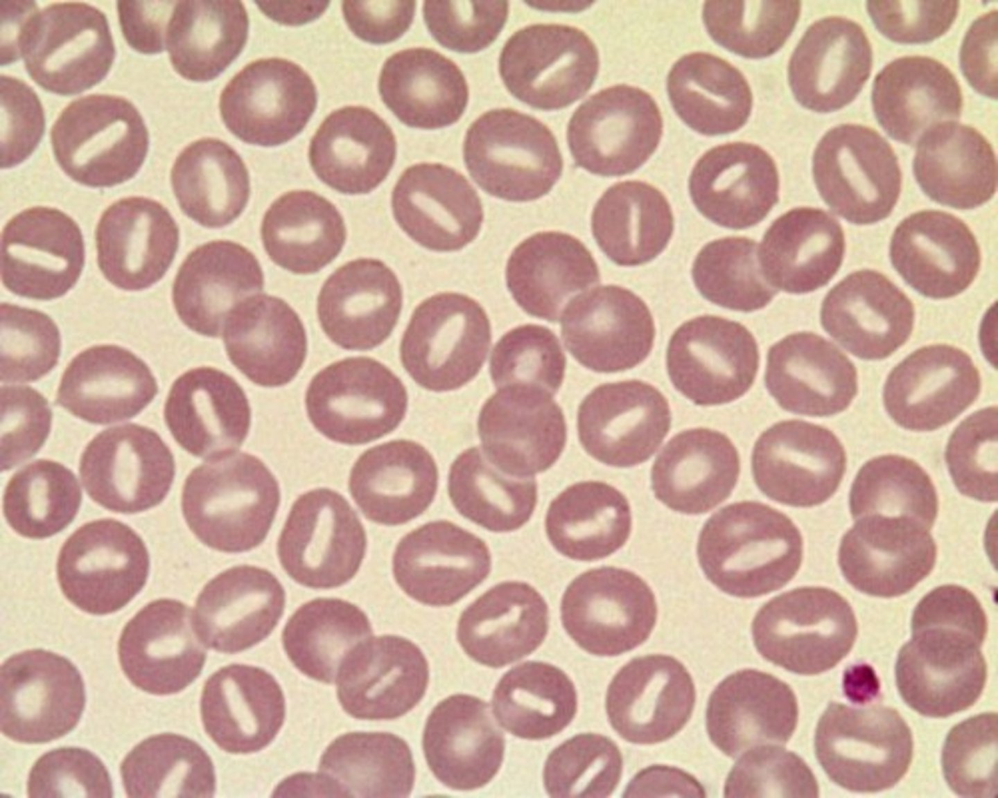
Stomatocytes
- Characterized by slit in central pallor ( concavity in 1 direction)
- Shaped like a bowl
- Defective stomatin & Na+/K+ pumps -> swelling of cell
- Etiologies:
Hereditary Stomatocytosis (3-4+)
Liver Disease (1-2+)
Alcoholism
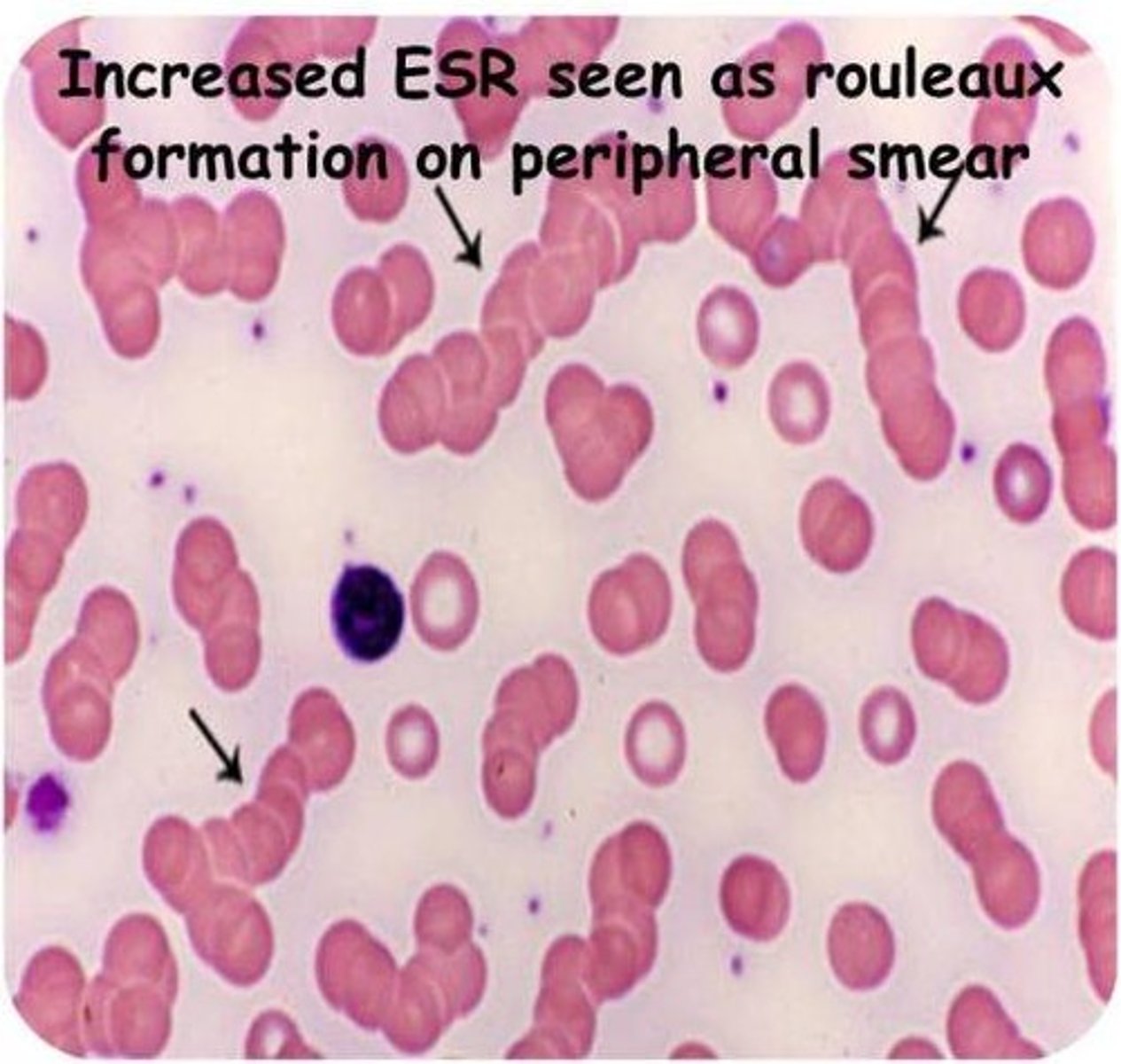
Rouleaux (1-4+)
- RBCs are stacked like rolls of coins (think grapes)
- increase plasma proteins > sticking at RBCs > neutrlizing of sialic acid > stacking of RBCs
- Etiologies:
Multiple myeloma
Waldenstrom's
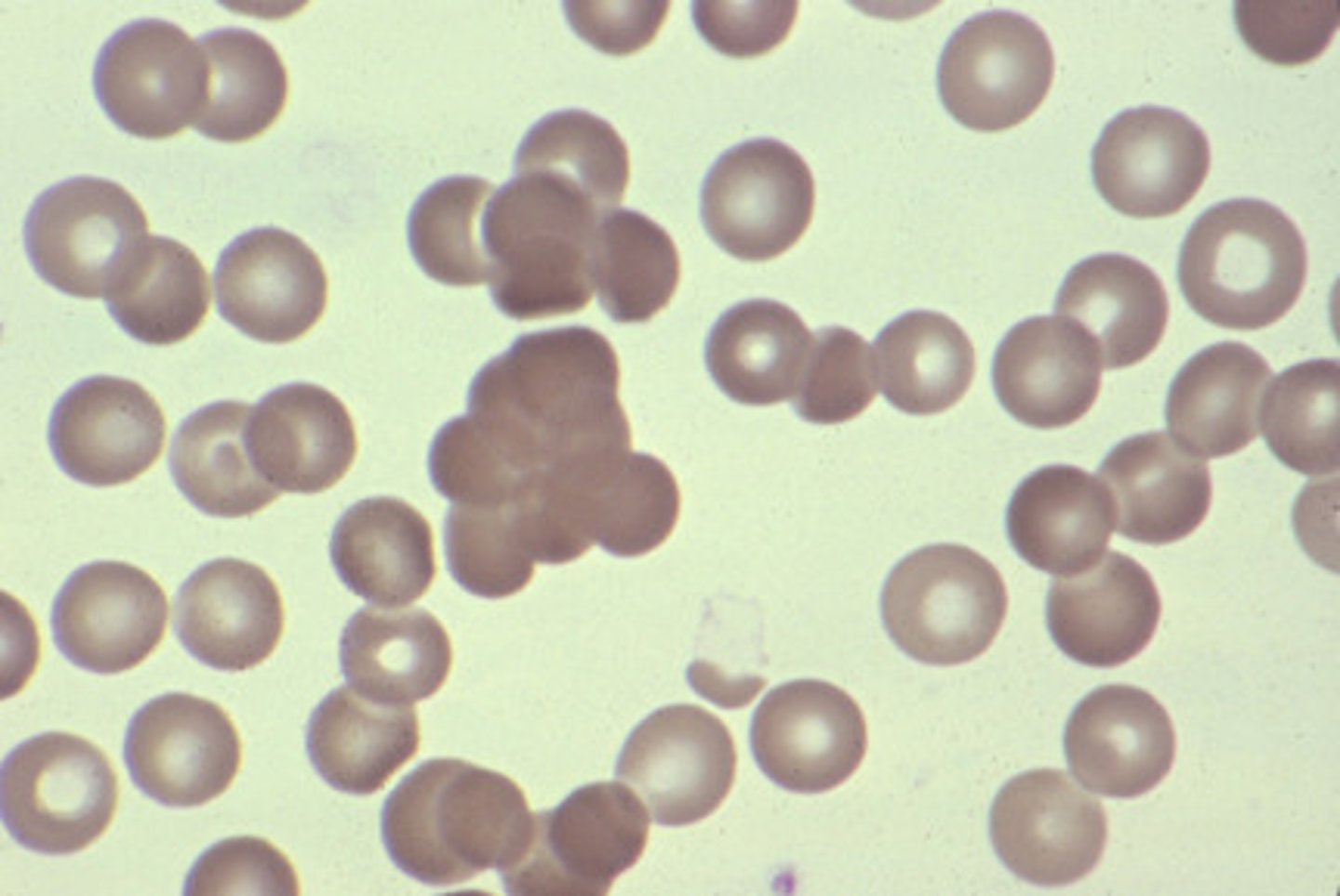
Agglutination
- RBCs clump due to antibodies
- Etiologies: Cold agglutinin syndrome
Either: Warm autoimmune hemolytic anemia OR cold autoimmune hemolytic anemia
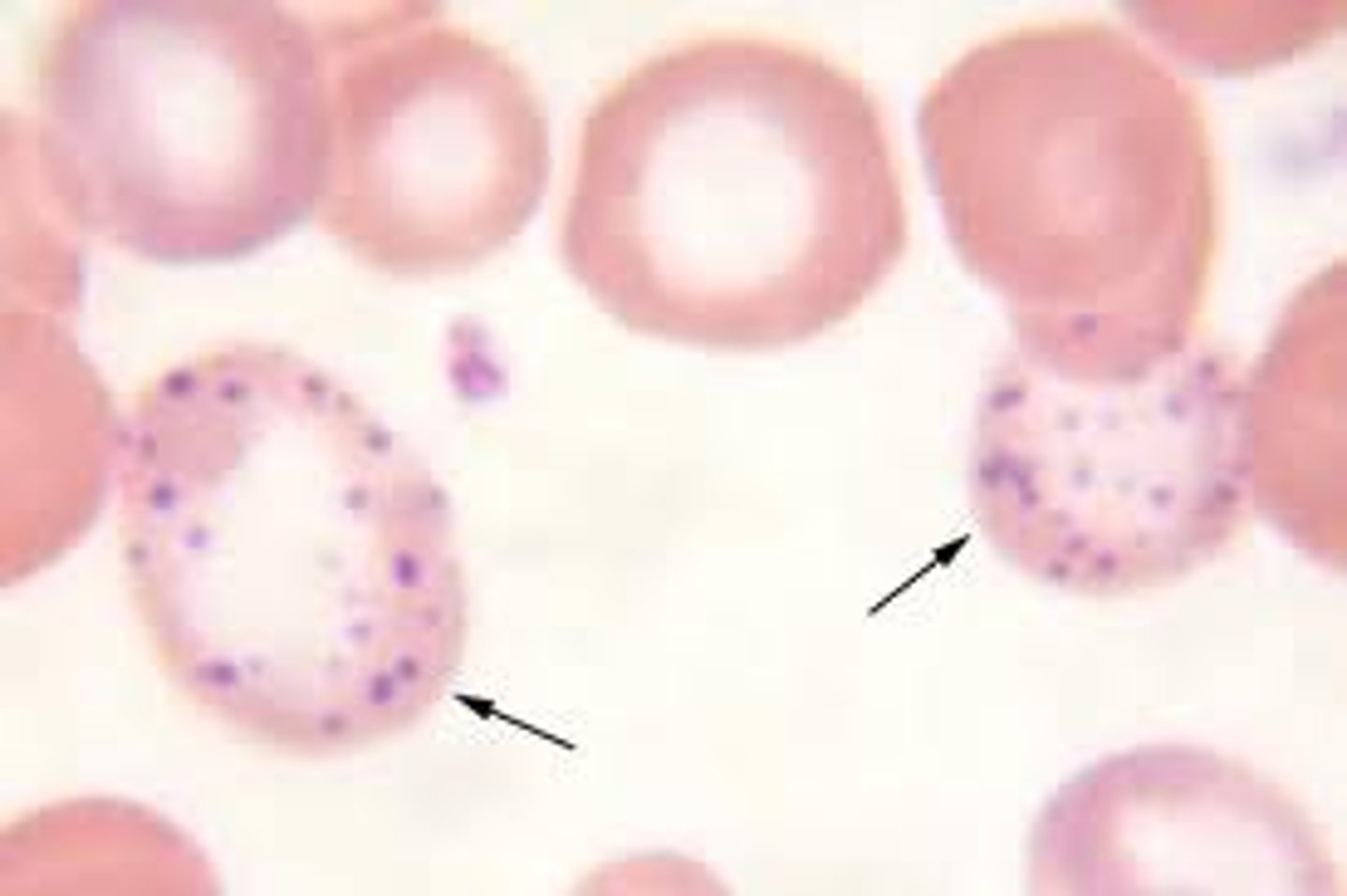
Basophilic Stippling
- Multiple, course bluish granules
- Hgb synthesis defect > RNA, ribosomes, mitochondria (within blue granules)
- Etiologies:
Lead poisoning (2-3+)
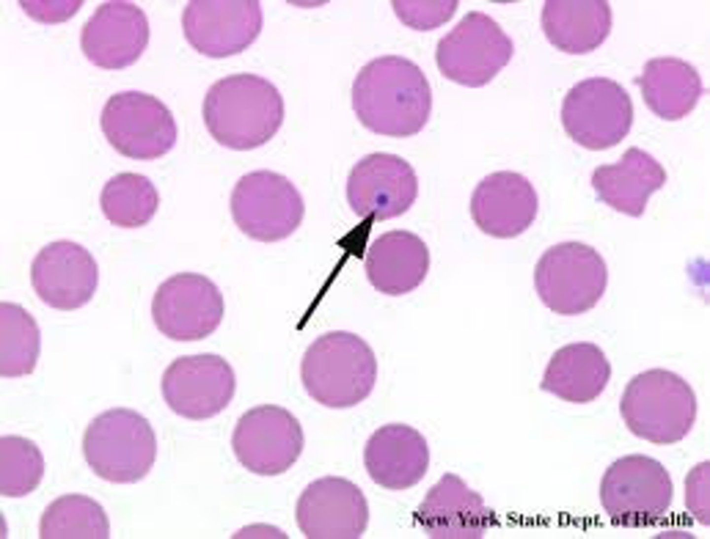
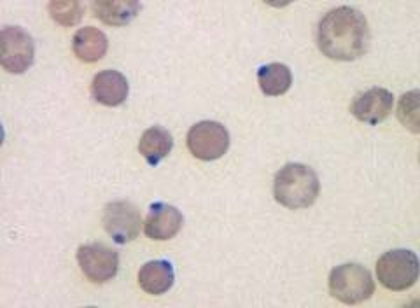
Howell-Jolly Bodies
- 'belly button'
- Larger, round blue/purple spheres
- Residual DNA isnt expelled after RBC maturation
- Etiologies
Hemolytics anemia (2-3+)
Splenectomy (2-3+)
Megaloblastic anemia
Thalassemia
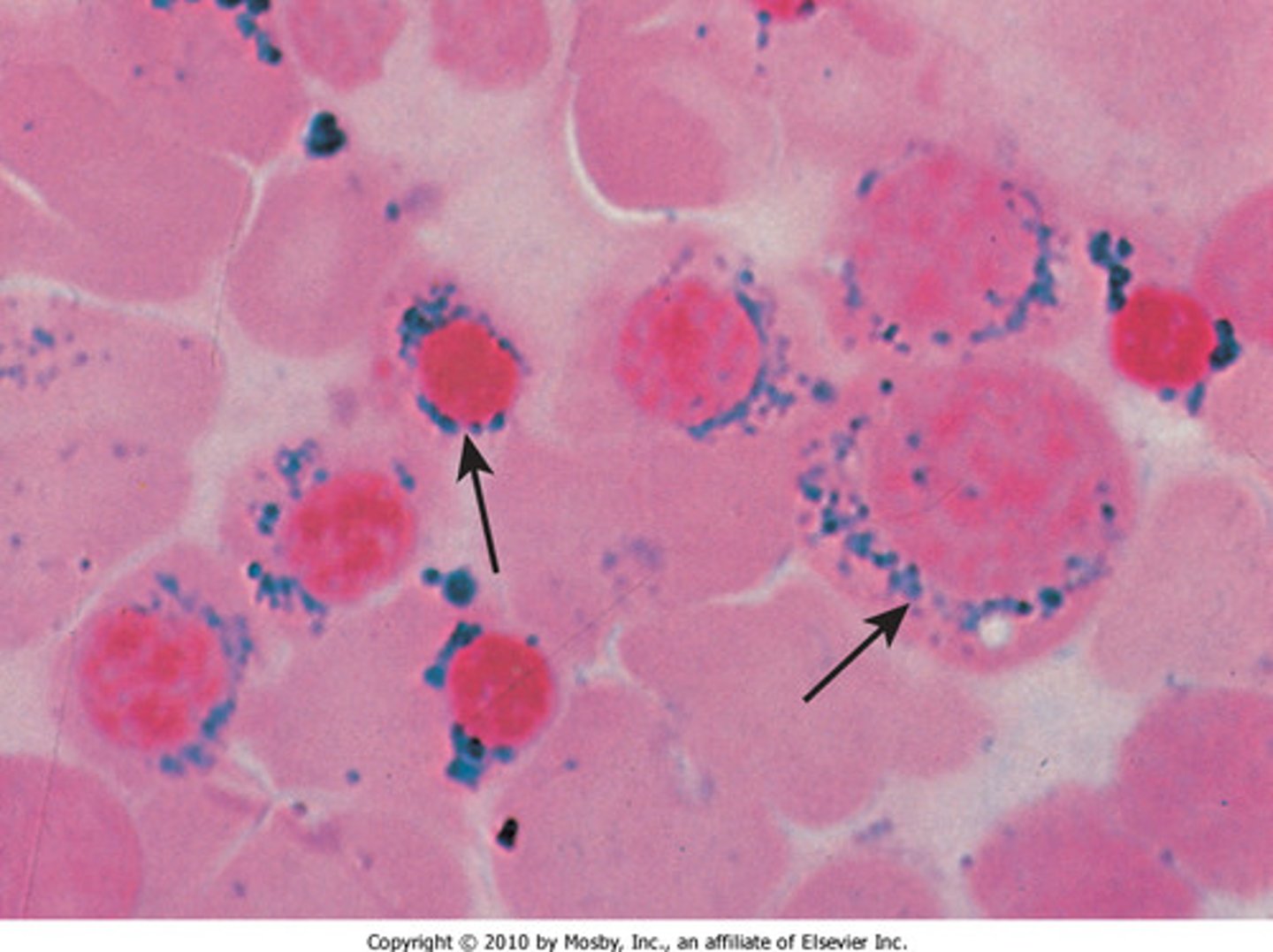
Siderotic granules
- Single/multiple greenish-blue iron granules
-Prussian blue stained iron granules
- Etiology: Sideroblastic anemia
- From impaired Hgb synthesis
Siderocyte
- RBC with NO nucleus w prussian blue stained iron granules
Sideroblast
- RBC with a nucleus w prussian blue stained iron granules
Heinz Bodies
- Round/oval bodies in VITAL STAINS
-NOT SEEN IN WRIGHT'S STAIN
-Etiologies: (3-4+)
G6PD deficiency
unstable Hgb
Thalassemias
Cabot Rings
- Fine red/violet ring structure
- Arg - rich histones w iron (rare occurence)
- Etiologies: megaloblsstic anemias, metal poisoning
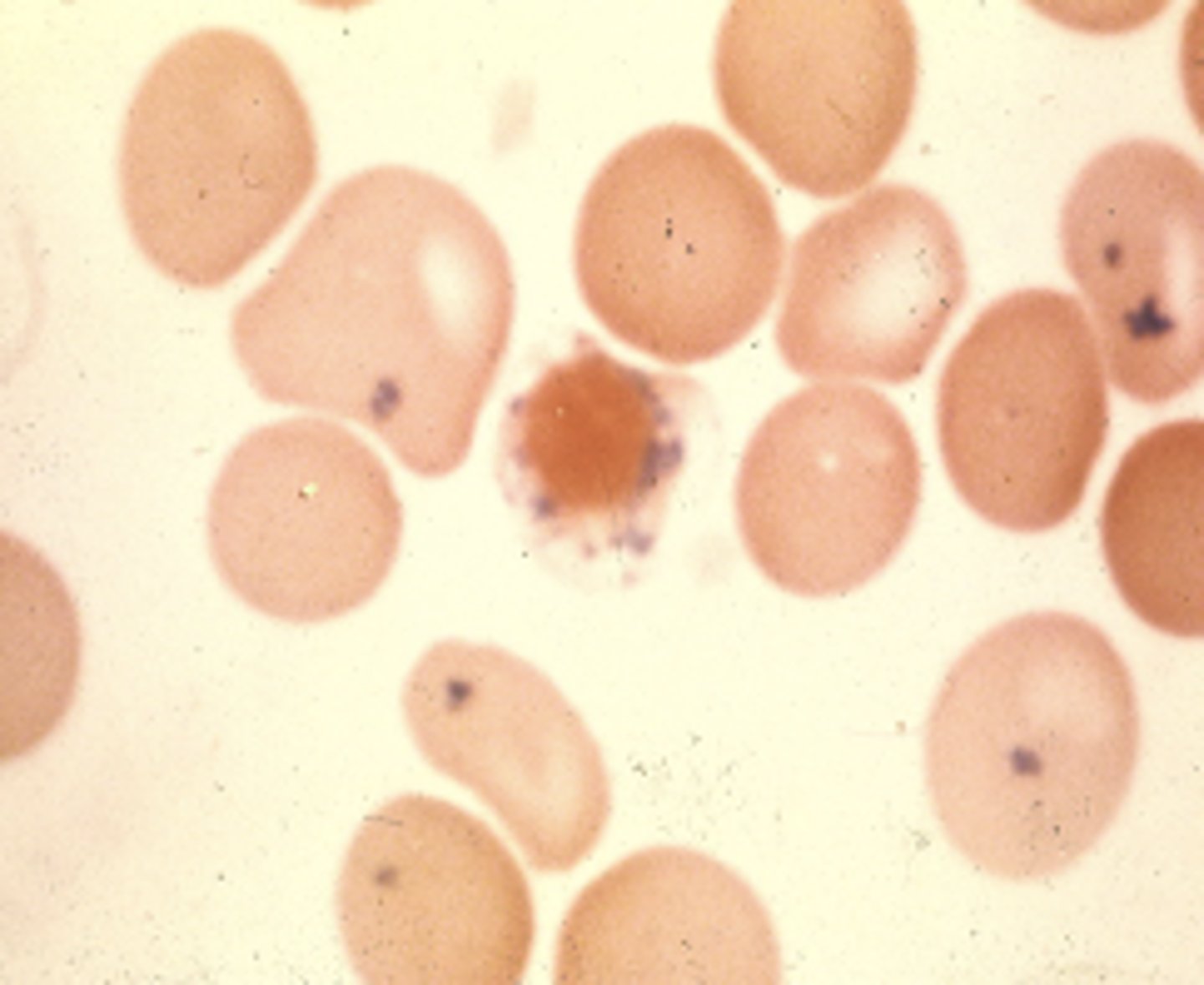
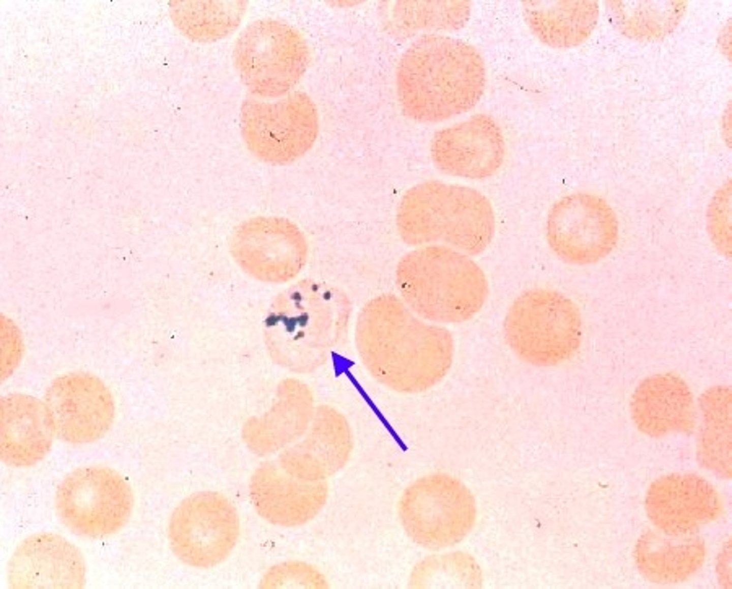
Malaria
-Malaria transmitted by mosquitoes
- Trophozoite: IDs for malaria
reddish/purple signet ring
-Schizont: reddish/purple clumps
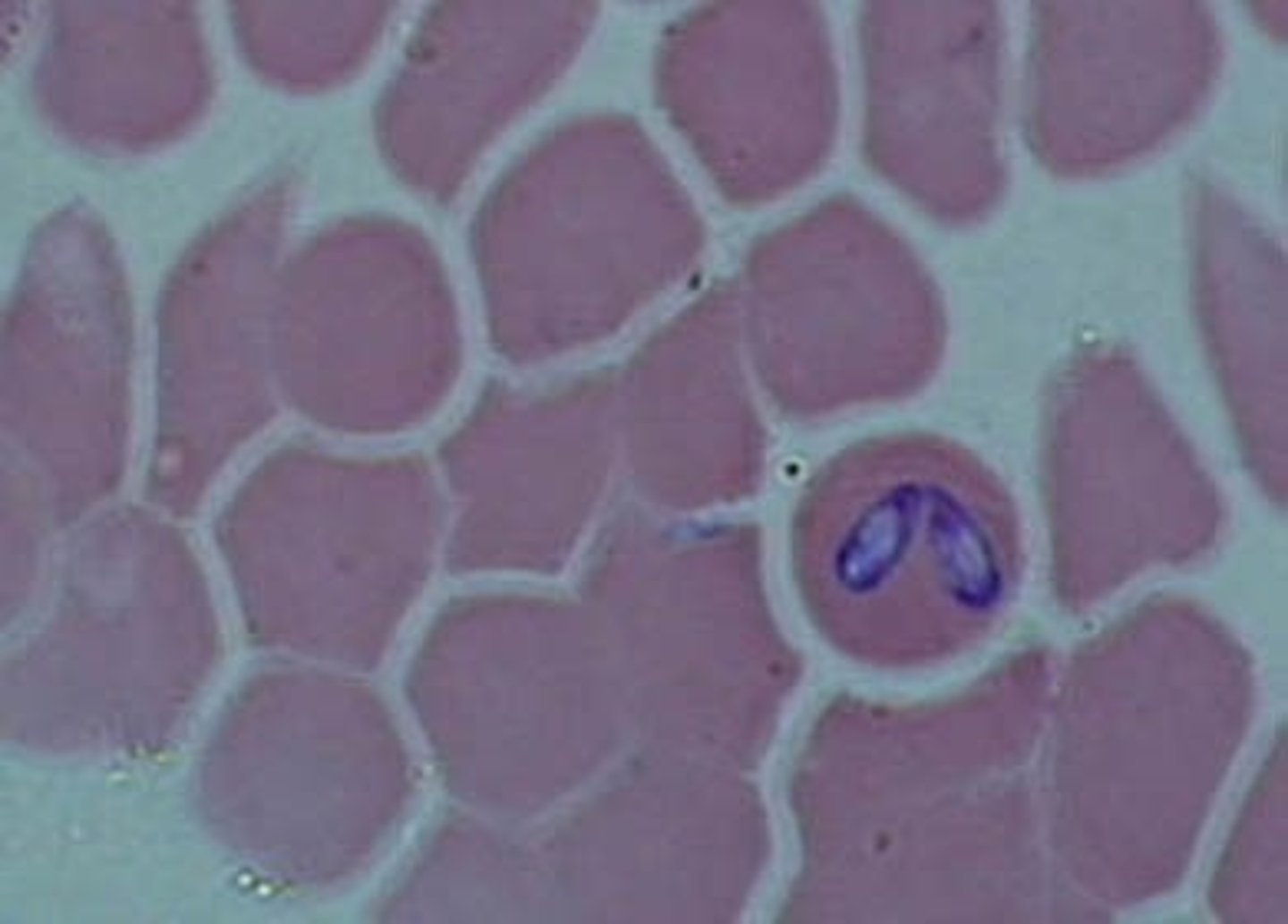
Gametocyte: shaped like a banana
Babesia
- Transmitted by ticks
- Forms trophozoite, similar to malaria
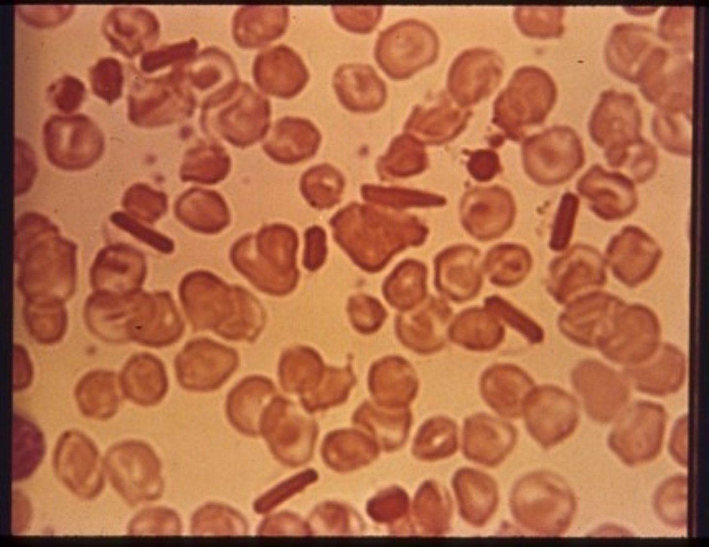
Hemoglobin C crystals
-Intracellular red rod-shaped crystals
- Beta chain mutation -> OxyHbC -> crystals
*Diagnostic for HbC
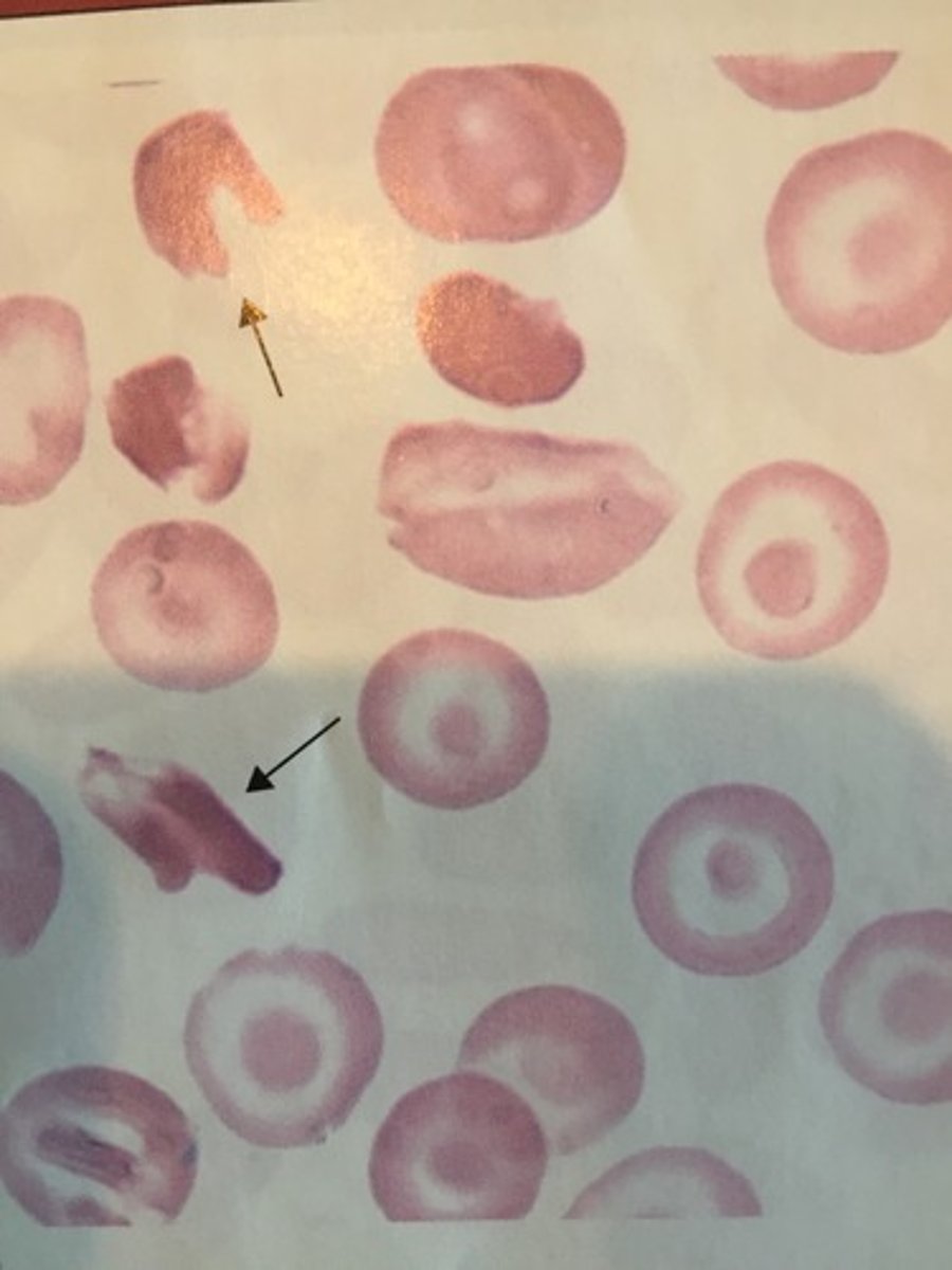
Hemoglobin SC Crystals
- Crystals are irregular shaped
- Partially HbS and partially HbC
* Diagnostic for HbSC
- Child of person with traits for HbS and HbC
Water Damage
- Due to lab error
- Stain must be changed
-Appears moth-eaten