Dental Radiography
 Call Kai
 Learn
Learn Practice Test
 Spaced Repetition
 Match
Match Flashcards
 Knowt Play
1/51

Earn XP
Description and Tags
Lecture 4
Name | Mastery | Learn | Test | Matching | Spaced | Call with Kai | Chat |
|---|
No analytics yet
Send a link to your students to track their progress
52 Terms
List indications for dental radiography.
diagnosis of periodontal disease
diagnosis of endodontic disease
“missing” teeth
extractions( prior to, sometimes during, and after)
resorptive tooth lesions
screening radiographs during routine dental cleaning
periorbital swelling, gingival swellings and oral neoplasia
developmental abnormalities of teeth
trauma
malocclusions
R/O dental cause of “nasal” disease
What are advantages to using a conventional radiograph unit for dental radiographs?
may be available in practices without dental radiograph unit
useful in imaging mandibular/maxillary fractures, trauma, masses, temporomandibular joint (TMJ)
What are disadvantages to using a conventional radiograph unit for dental radiographs?
inconvenient (usually not in location where dental procedures are performed)
can’t take intra-operative films without moving patient from dental treatment table
superimposition problems
difficult to isolate individual teeth
How do we set exposure on dental radiograph units?
kVp and mA are often fixed and only time is selected
most units have anatomic interface where operator sets the size and tooth, and the exposure is adjusted automatically
Dental X-Ray units are required to be registered and inspected by who?
the State Department of Health
In Virginia, dental X-Ray operators must stand how far from the tube when making the exposure?
at least 9 feet
What angle should you stand from the primary beam when shooting dental radiographs?
90-135 degrees
What can be used to help stabilize the sensor/film during dental radiographs to prevent exposure of staff by holding it?
positioning aides such as gauze sponge or play-doh
What should always be worn by personnel in areas where radiographs are taken to measure the extent of exposure?
dosimetry badge
What are the three options for obtaining an image during dental radiographs?
conventional intraoral film
sensor-based radiography (DR) - “digital radiography”
photostimulable phosphor plates (CR) - “computed radiography”
What are the advantages of using conventional intraoral film for obtaining an image?
least expensive ($500)
requires no automation
several sizes available
What are the disadvantages of using conventional intraoral film for obtaining an image?
takes more time during procedure
requires developing chemicals
must store and catalog films as part of the patient record
processing errors can be a problem
What are the advantages of using a hard wired sensor “DR” for obtaining an image?
immediate images available (5 seconds)
digital images - easy to store
more consistent quality images than film
less radiation required
What are the disadvantages of using a hard wired sensor “DR” for obtaining an image?
sensors can be expensive and somewhat fragile ($2000+)
only #1 and #2 size available
What are the advantages of using a Phosphor screen “CR” for obtaining an image?
digital images - easy to store
#4 plate allows images of larger teeth and multiple teeth
#1 plate works better in cats
quality images
What are the disadvantages of using a Phosphor screen “CR” for obtaining an image?
slower than hard-wired sensor (30 seconds to 1 minute)
Radiographic studies with conventional x-ray units typically use a ______ when possible to prevent distortion of the structure being imaged.
parallel technique
The parallel technique can be used in dental radiography only when imaging the _____ teeth.
distal mandibular (distal to 307/407)
When using parallel technique for dental radiographs, the film (sensor) can be placed ____ to the tooth and ____ to the primary beam.
parallel
perpendicular
The _____ is required to prevent distortion of the image and duplicate the size of the tooth on the film or sensor.
bisecting angle technique
Why is it not possible for the film to be aligned parallel with the long axis of the tooth and its roots in most cases in the oral cavity?
anatomical interference
palate causes interference with all maxillary teeth
mandibular symphysis and angle of roots, relative to the oral surfaces in the rostral mandibular teeth
Describe how to obtain the bisecting angle when positioning for dental radiograph.
orient the beam half way between the two extremes
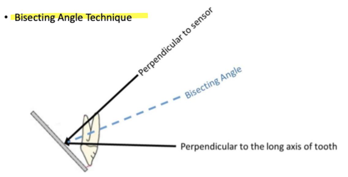
What view should be used for radiographs of the maxillary incisors in the dog?
70 degree rostral-caudal in sternal recumbency
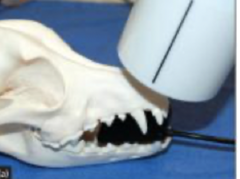
What view should be used for radiographs of the maxillary canines in the dog?
70 degree rostral-caudal oblique in sternal recumbency
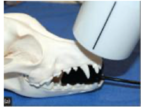
What view should be used for radiographs of the maxillary premolars in the dog?
45-55 degree lateral in sternal recumbency
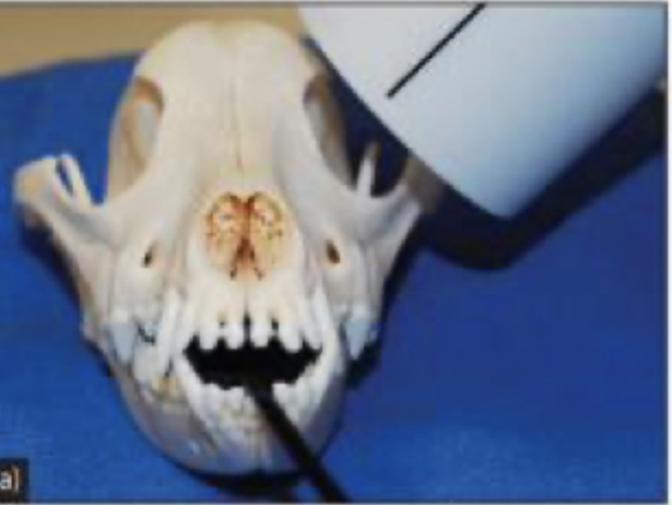
When the xray tube is positioned laterally, the mesio-buccal and mesio-palatal root of the 4th maxillary premolars are superimposed. How can you separate the mesiobuccal and mesiopalatal roots of the 4th maxillary premolar?
shift the tube head to a rostral to caudal oblique in sternal recumbency
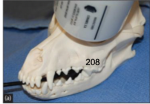
What view should be used for radiographs of the maxillary molars in the dog?
45-55 degree caudal rostral oblique in sternal recumbency
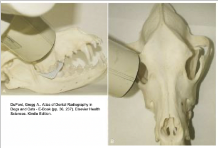
What view should be used for radiographs of the mandibular incisors in the dog?
70 degree rostral caudal in dorsal recumbency
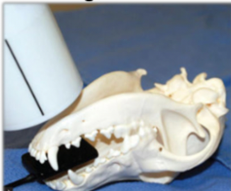
What view should be used for radiographs of the mandibular canines in the dog?
70 degree rostral caudal oblique in dorsal recumbency
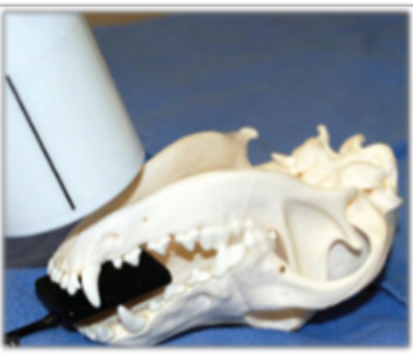
What view should be used for radiographs of the mandibular premolars in the dog?
45 degree lateral in dorsal recumbency
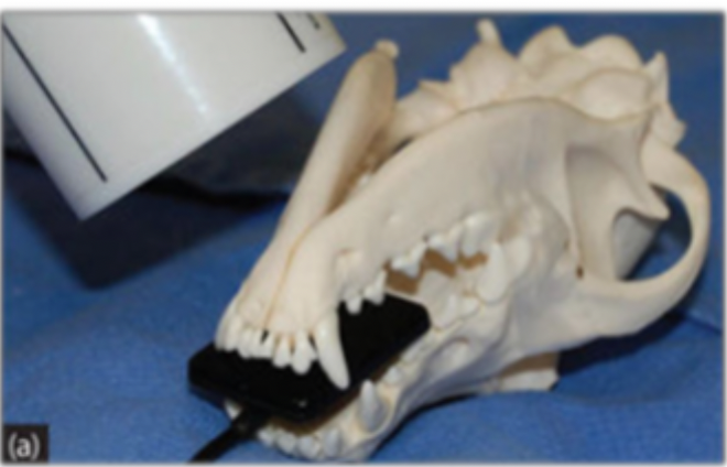
What view should be used for radiographs of the mandibular molars in the dog?
parallel technique, 90 degree lateral in dorsal recumbency
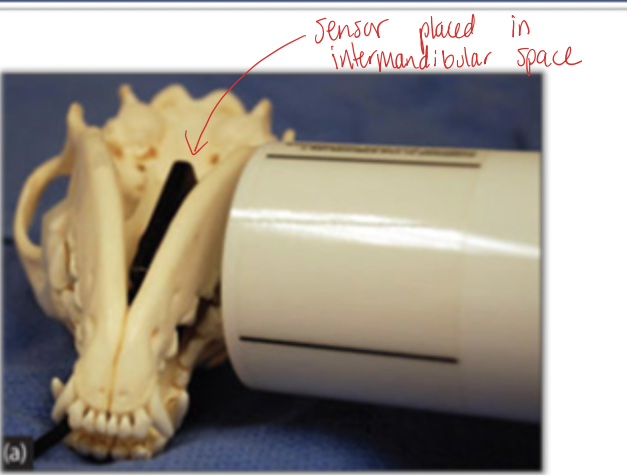
What view should be used for radiographs of the maxillary incisors in the cat?
70 degree rostral-caudal in sternal recumbency
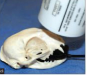
What view should be used for radiographs of the maxillary canines in the cat?
70 degree rostral-caudal oblique in sternal recumbency
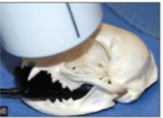
Which technique results in the superimposition of the mesial roots of the 4th maxillary premolar and zygomatic arch interference?
standard bisecting angle technique
What view should be used for radiographs of the mandibular incisors and canines in the cat?
70 degree rostral caudal in dorsal recumbency
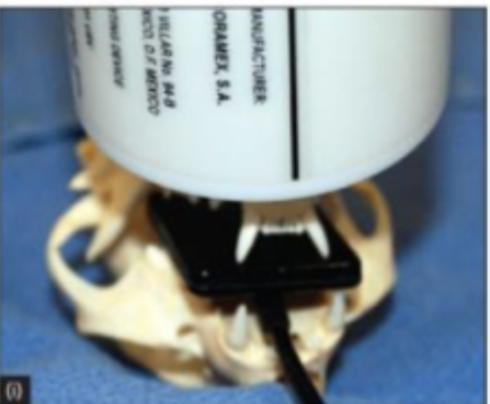
What view should be used for radiographs of the mandibular premolars in the cat?
45 degree lateral in dorsal recumbency
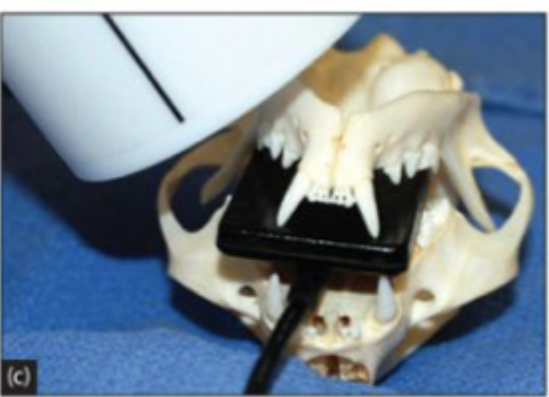
What view should be used for radiographs of the mandibular molar in the cat?
parallel technique, 90 degree lateral in dorsal recumbency
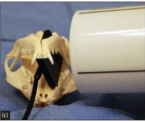
Which technique results in the superimposition of the mesial roots of the 4th maxillary premolar but avoids zygomatic arch interference?
extraoral technique
When the x-ray beam is too perpendicular to the sensor or film, it results in a _____ image.
foreshortened
How can you correct foreshortening?
drop angle to a more lateral position
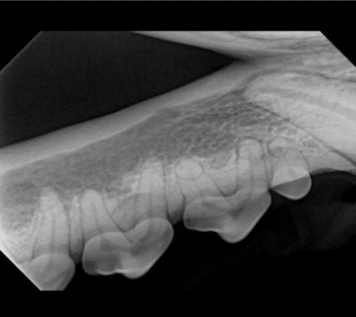
What error is shown in this image?
foreshortening
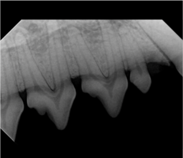
What error is shown in this image?
elongation
When the x-ray beam is too perpendicular to the tooth, it results in an ____ image.
elongated
How can you correct elongation?
increase the angle to a more vertical position
What error is caused by the tube not being aligned with sensor or sensor not aligned with the tooth?
area of interest not on image
How can you correct the area of interest not being on image?
change tube positioning or sensor position or both
If the sensor size is small when taking x-rays, what may be required?
multiple images of large teeth
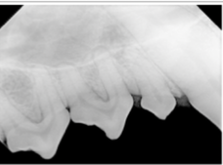
What error is shown in this image?
underexposure
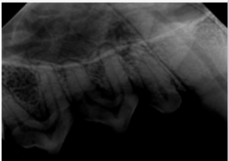
What error is shown in this image?
overexposure
How can you correct underexposure?
increase time
How can you correct overexposure?
decrease time
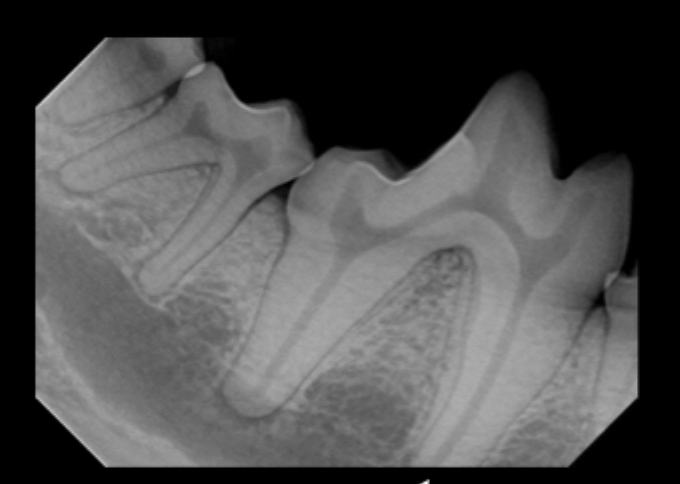
What error is shown in this image?
area of interest not on image (missing mesial root apex)