Lab 1: Anatomy
 Call Kai
 Learn
Learn Practice Test
 Spaced Repetition
 Match
Match Flashcards
 Knowt Play
1/53

Earn XP
Description and Tags
Vocabulary practice flashcards covering microscopy, primary tissue types, histology, and the integumentary system based on Seeley's Anatomy & Physiology Chapters 4 and 5.
Name | Mastery | Learn | Test | Matching | Spaced | Call with Kai | Chat |
|---|
No analytics yet
Send a link to your students to track their progress
54 Terms
Histology
The study of the microscopic structure of cells, tissues, and organisms in relation to their function.
Four Major Tissue Categories
Epithelial, Muscular, Nervous, and Connective tissue.
Epithelial Tissue
A highly cellular tissue that covers or lines parts of the body, creates boundaries between different environments, and forms glands.

What is this and what is its three typical parts
Its a spinal cord smear (neuron). Its three typical parts are the axon, dendrite, and cell body (soma)
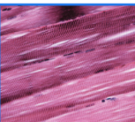
What tissue
skeletal

what tissue
cardiac

what tissue
smooth
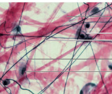
what tissue
Aerolar
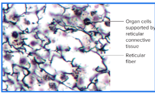
What tissue
Reticular
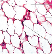
what tissue
Adipose
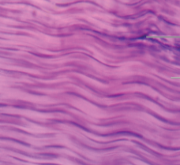
What tissue
Dense regular (dessert sand)
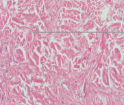
What tissue
Dense irregular

What tissue (cartilage)
Fibrocartilage
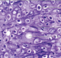
What tissue (cartilage)
Elastic
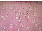
What tissue (cartilage)
Hyaline

What type of tissue
blood

What type of tissue
Bone marrow
Characteristics of Epithelial Tissue
Key traits including regeneration, polarity (apical and basal surface), cellularity and specialized contacts, support by connective tissue (reticular lamina), avascularity, and innervation.
Epithelial Tissue Functions
Primary functions including filtration, absorption, secretion, protection, excretion, and sensory reception.
Stratum Basale
The deepest layer of the epidermis where cells divide by mitosis and contain the youngest cells.
Stratum Spinosum
The epidermal stratum located superficial to the stratum basale where keratin fibers and lamellar bodies accumulate.
Stratum Granulosum
The epidermal stratum where keratohyalin granules accumulate, a hard protein envelope forms beneath the plasma membrane, lamellar bodies release lipids, and cells die.
Stratum Lucidum
The epidermal stratum in which the cells are dead and contain dispersed keratohyalin.
Stratum Corneum
The most superficial epidermal layer containing the oldest dead cells, which have a hard protein envelope, contain keratin, and are surrounded by lipids.
Eccrine Sweat Glands
The most common type of sweat glands, sensitive to temperature, that produce normal body perspiration.
Apocrine Sweat Glands
Sweat glands that secrete water and higher concentrations of organic acids than eccrine glands.
Integumentary System
An organ system comprised of skin, hair, glands, and nails.
Main Tissue Layers of the Skin
The two primary tissue layers of the skin: the Epidermis and the Dermis.
Mesenchyme
The common embryonic tissue from which all connective tissues originate.
Three Groups of Connective Tissue
Connective tissue proper (loose and dense), fluid connective tissue (blood and lymph), and supporting connective tissue (bone and cartilage).
Connective Tissue Functions
Functions primary to structural support and joining structural components to one another.
Primary Muscular Tissue Types
The three muscle types: Skeletal muscle, Cardiac muscle, and Smooth muscle.
Skeletal Muscle Characteristics
Muscular tissue exhibiting prominent striations and skeletal muscle fibers with nuclei located near the periphery of the cell.
Cardiac Muscle Characteristics
Muscular tissue exhibiting striations, cardiac muscle fibers, cell nuclei, and intercalated discs.
Smooth Muscle Locations
Muscular tissue found in internal organ walls, specifically the wall of the stomach, wall of the large intestine, and wall of the small intestine.
Action Potentials
Electrical signals conducted by neurons and glial cells in nervous tissue.
Nervous Tissue Components
Tissue comprised of neurons and glial cells that conduct electrical signals throughout the body.
Axon Hillock
The region of a neuron connecting the neuron cell body to the axon.
Types of Hair
Terminal, Vellus, and Intermediate.
Hair Bulb
The base of the hair root containing the matrix growth zone, hair papilla, and melanocytes.
Hair Shaft
The region of the hair extending above the skin surface.
Hair Root
The portion of the hair situated below the skin surface.
Arrector Pili
A smooth muscle attached to the hair follicle.
Dermis Layers
The two distinct layers of the dermis: the Papillary layer and the Reticular layer.
Primary Epithelial Tissue Types
Simple Squamous, Simple Cuboidal, Simple Columnar, Pseudostratified Columnar, Stratified Squamous, and Transitional epithelium.
Connective Tissue Proper Types
Loose (areolar), adipose, reticular, dense regular, dense irregular, and elastic connective tissue.
Supporting Connective Tissue Types
Hyaline cartilage, fibrocartilage, elastic cartilage, ground bone, and cancellous bone.
Adipose Tissue Characteristics
A connective tissue type characterized by a plasma membrane, a nucleus pushed to the periphery, and an empty space where lipids are stored.
Intercalated Discs
Special junctions between cells found uniquely in cardiac muscle tissue.
Osteon
The structural unit of compact bone composed of central canals, concentric lamellae, and lacunae.
Bone Cells: Osteoblasts, Osteocytes, and Osteoclasts.
The cell types involved in bone tissue:
Components of Blood
Fluid connective tissue consisting of red blood cells, white blood cells, platelets, and plasma.
Goblet Cell
A specialized cell containing mucus, found in pseudostratified ciliated columnar epithelium.
Working Distance
The distance between the microscope objective lens and the microscope slide coverslip.