RADI 442 EXAM 3
 Call Kai
 Learn
Learn Practice Test
 Spaced Repetition
 Match
Match Flashcards
 Knowt Play
1/106

There's no tags or description
Looks like no tags are added yet.
Name | Mastery | Learn | Test | Matching | Spaced | Call with Kai | Chat |
|---|
No analytics yet
Send a link to your students to track their progress
107 Terms
What are the accepted rules of conduct like integrity, compassion, honesty, and respectfulness?
Moral principles
What are some descriptive findings vocab?
echogenic foci in the gallbladder; positive for shadowing; mobile; measure if necessary; twinkle sign with color
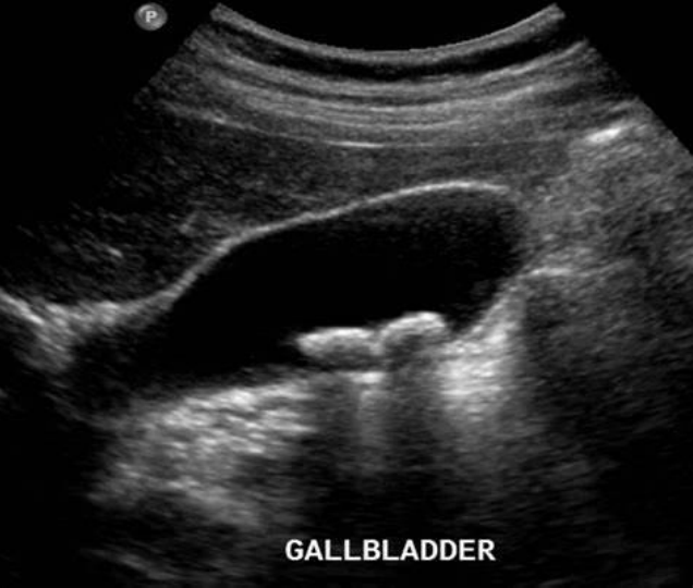
finding in the gallbladder
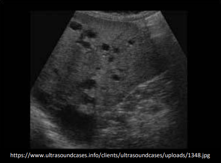
example of abnormal finding
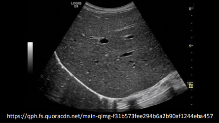
example of normal
What is it called when an entire organ affected by an infiltrative disease or some process that is affecting the cells in the organ?
Diffuse disease
What is it called when a mass or masses are easily seen and measured?
focal or localized
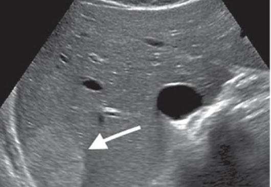
focal (lesion can be seen)
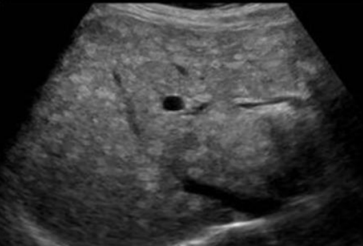
diffuse
How do you describe abnormalities?
How many; origin; adjacent organs/structures involved; composition (sonographic descriptors); size (always 3 measurements in 3 planes LWD); always shown in two planes; vascularity
Understand the echotexture of an image. Is it…
homo/heterogeneous, mixed, isoechoic, hypo/hyperechoic, echogenic, cystic, solid, complex
Understand the borders of the finding. Are they…
regular/smooth, irregular, well defined, not well defined, thickness
Artifact that is fluid, cyst/cystic
posterior enhancement
Artifact that is calcified, solid
shadowing
artifact that is showing air
dirty shadowing
artifact that shows reverberation (EX: gallbladder pathologies)
comet tail artifact
What is a cyst criteria?
round/oval, smooth, well-defined borders, COMPLETELY anechoic, thin walls, increased transmission — true cyst. if does not meet these requirements it is a cystic/complex lesion
What is complex criteria?
has both cystic and solid characteristics (abscess); most lesions happen here
What is solid criteria?
irregular borders, internal echoes, decreased transmission
What is vascularity?
does it have blood flow
What could a not well defined border mean?
bowel gas
Do we want to see thick borders?
no
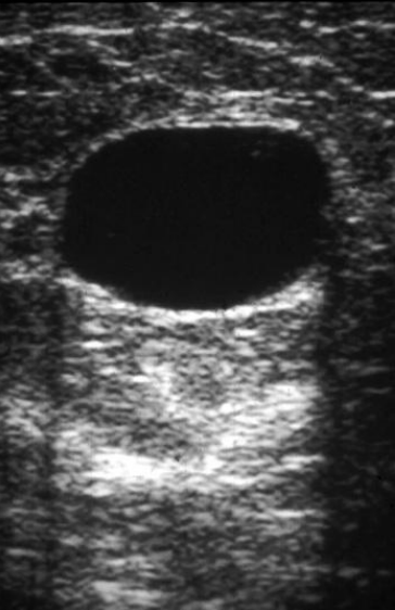
cyst
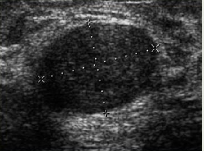
cystic lesion
Questions to consider asking yourself when trying to describe an abnormal finding:
how many? location — what organ, where is located within the organ? are any other organs/structures involved? describe — complex, mixed echogenic, anechoic, well-defined, etc…
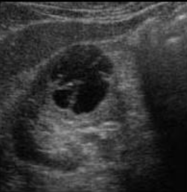
How would you describe this finding?
solitary, complex but mostly cystic, well-circumscribed cortical lesion within the right kidney with internal septation
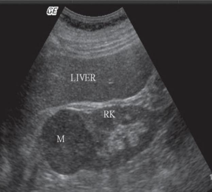
how would you describe this finding?
solitary, well-circumscribed hypoechoic lesion within the upper pole of right kidney
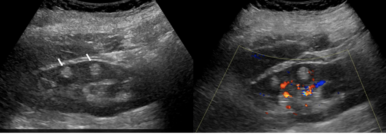
How would you describe this finding?
multiple hypovascular (avascular) echogenic cortical lesions within the right kidney
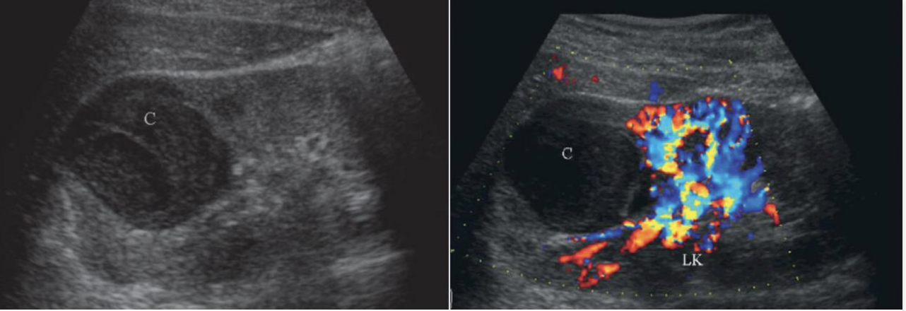
How would you describe this finding?
solitary, well-circumscribed avascular, hypoechoic lesion within the upper pole of left kidney
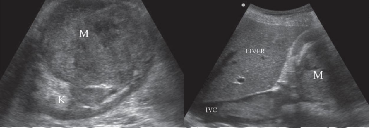
How would you describe this finding?
heterogenous, solid mass in the upper pole of right kidney with irregular borders. echogenic structure within the central lumen of the inferior vena cava at the level of the right renal vein which may reflect non-occlusive thrombus versus abutting or invading mass
The color doppler can be used for what?
to look for vascularity internally
What are findings that require immediate notification and do not let the patient leave
DVT, severe carotid findings, ectopic, fetal demise, appendicitis, air in the portal system
Step 1 of Sonographic Examination
assessment of relevant documentation
step 2 of sonographic examination
examining reports and images
step 3 of sonographic examination
preparing the exam room and protocol review
step 4 of sonographic examination
introducing yourself
step 5 of sonographic examination
gathering clinical history
Anything the patient or family member tells you regarding their history (EX: “I’m having epigastric pain and back pain”)
subjective information
information the sonographer can clearly see, hear, feel, or read. it is factual and concrete (EX: lab values in patient chart confirm that patient amylase and lipase levels are elevated)
objective information
step 6 of sonographic examination
patient education
step 7 of sonographic examination
conducting the sonogram
step 8 of sonographic examination
completing the sonography report
step 9 of sonographic examination
sonographer interaction with interpreting physician
step 10 of sonographic examination
discharging the patient (ensure they don’t have any additional exams, tests, or imaging studies before letting them go)
What is the placement of the liver?
extends from RT hypochondrium across the epigastric area into LT hypochondrium
What is the fibrous connective tissue with nerves and blood vessels that covers the liver?
glisson’s capsule
What is the measurement that is considered hepatomegaly in the liver?
more than 15-17 cm
Riedel’s lobe
right lobe of liver extends below inferior pole of right kidney
What is helpful in visualization of pancreas?
elongated left tip of liver
Where the main portal vein and hepatic artery enter the liver and common bile duct exits the liver
porta hepatis
What are the 3 fossa?
gallbladder, porta hepatis, and IVC
The pancreas is what to the liver?
inferior to the LT lobe
The duodenum is what to the liver?
adjacent and medial to RT lobe
The gallbladder is what to the liver?
posterior
What is the smallest lobe of the liver?
caudate
What is the largest lobe of the liver?
right
Hepatic veins, portal veins, ligaments, and fissures
what divides the lobes of the liver
What forms upper border and attaches to diaphragm, separates RT from LT lobes?
falciform ligament
What does the right lobe border?
anterolateral abdominal wall
Where does the right lobe lay?
right lateral undersurface of diaphragm
The right lobe is divided into anterior and posterior segments by what?
right portal vein and right hepatic vein
What separates the right lobe from the left lobe?
middle hepatic vein and main portal vein
What divides right from left lobes?
main lobar fissure
Where does the left lobe lay?
undersurface of diaphragm
The left lobe is what to the pancreas?
anterior (anatomy dependent)
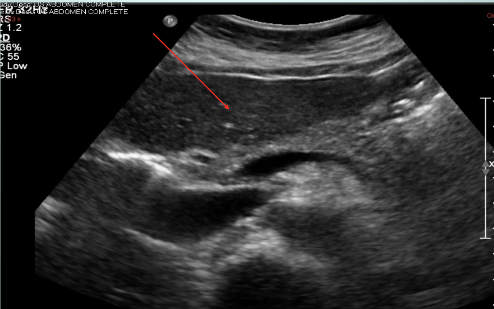
transverse left lobe
The left lobe is divided into medial and lateral segments by what?
ligament of teres, left hepatic vein, and left portal vein
What separates the left lobe from caudate?
ligament venosum
What are all the anatomy that separates RT lobe from LT lobe?
falciform ligament, main lobar fissure, middle hepatic vein, and main portal vein
Where does the caudate lobe lay?
posterior liver
What is the anterior border of the caudate that separates it from the left lobe?
ligament venosum
What forms the posterior border of the caudate lobe?
inferior vena cava
The caudate lobe can be enlarged with pathology such as…
cirrhosis
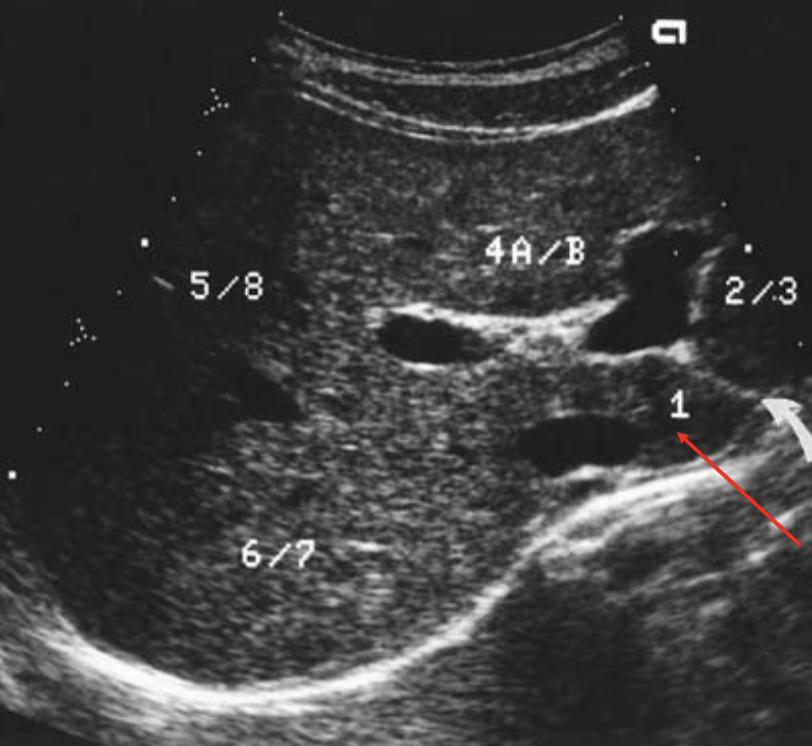
what’s the arrow pointing at
caudate lobe
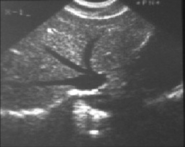
what is the image showing?
hepatic veins
What meets the falciform to connect liver to diaphragm (usually do not see)?
coronary ligament
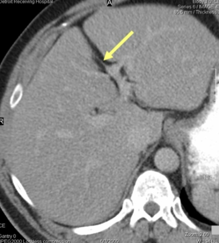
what is the arrow pointing to?
falciform ligament
What has anterior and posterior branch
right portal vein
what enters the liver anterior to IVC and posterior to hepatic artery
main portal vein
what has lateral and medial branch
left portal vein
How to designate MPV and RPV
mpv is outside of liver, rpv inside liver
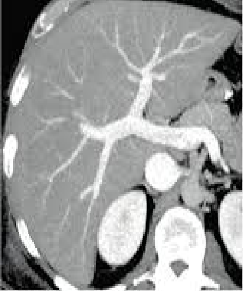
rpv/lpv confluence
oxygen-rich blood flows into the liver through what?
hepatic artery
nutrient-rich blood coming from the bowel flows into the liver through the what?
portal vein
bile flows out of the liver through what?
bile duct
blood flows out of the liver through 3 hepatic veins into a big vein called the what?
inferior vena cava
What is the large duct at the porta hepatis that drains bile?
common bile duct
all scan should begin with what?
a survey
sagittal liver left lobe protocol
lateral margin
aorta
caudate lobe
ivc
ivc with color
transverse liver left lobe protocol
lateral margin/dome
left hepatic vein
caudate lobe
left portal vein
sagittal liver right protocol
ivc
gallbladder
2 mids
dome
kidney
measurement
transverse liver right protocol
hepatic veins
portal rt/lt confluence
kidney
dome
mpv
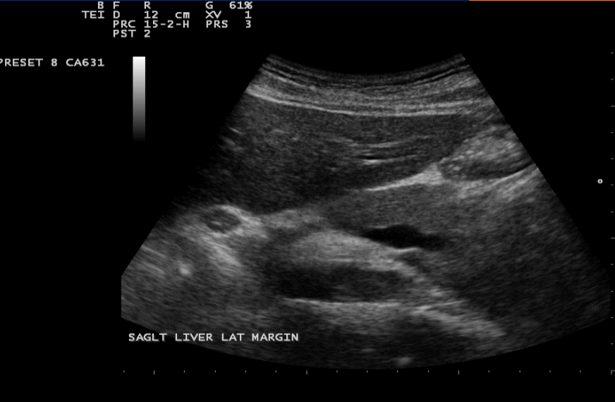
sagittal liver left lateral margin
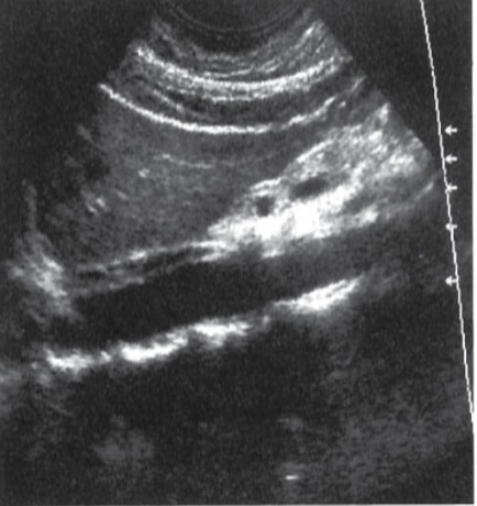
sagittal liver left with aorta
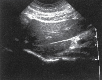
sagittal liver left with caudate
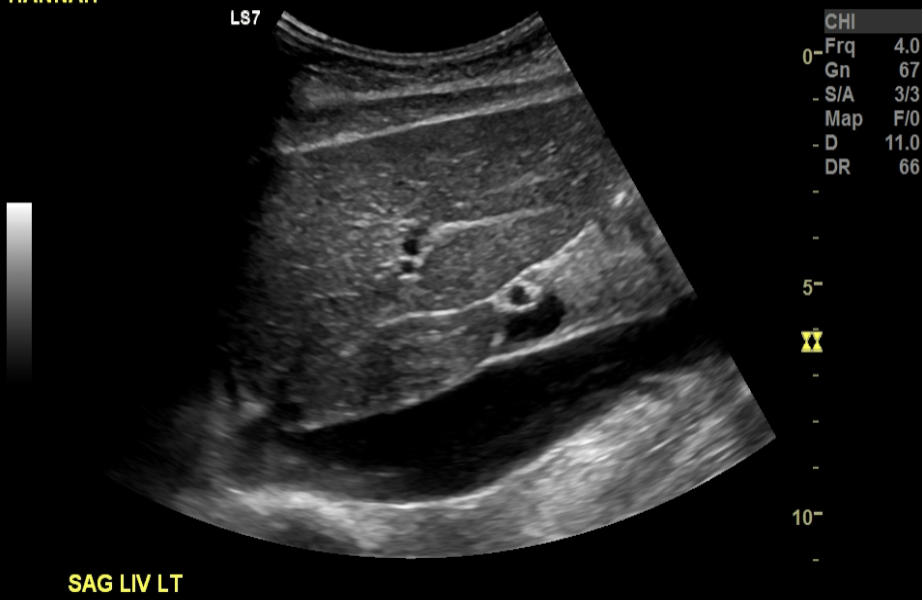
sagittal liver left with ivc
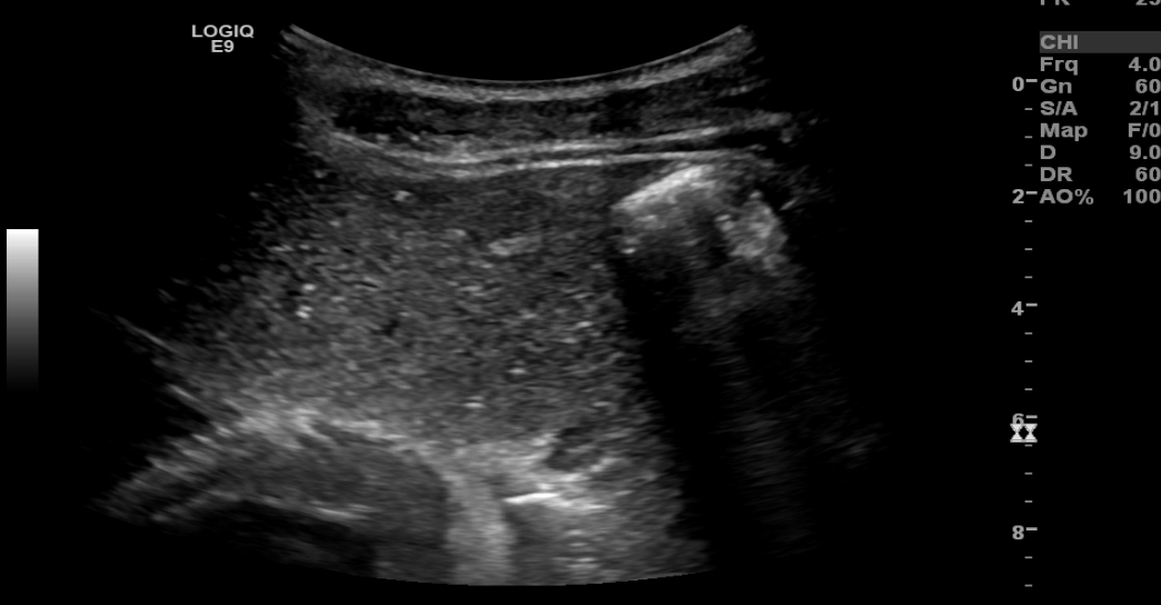
transverse liver left dome
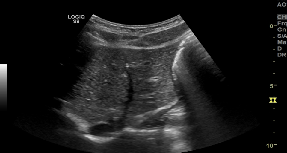
transverse liver left with hepatic vein
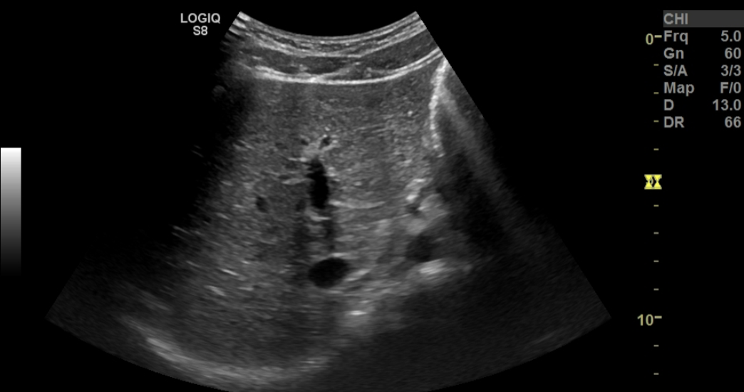
transverse liver left with left portal vein
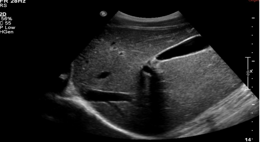
sagittal liver right with gallbladder