Orbital Contents
1/69
There's no tags or description
Looks like no tags are added yet.
Name | Mastery | Learn | Test | Matching | Spaced |
|---|
No study sessions yet.
70 Terms
what is located in the superolateral aspects of the orbit and has 9-13 openings allowing the tears to be secreted into the conjunctival sac?
lacrimal gland
what does the lacrimal gland do?
it keeps the orbit moist
where are tears secreted from the lacrimal gland?
conjunctival sac
what does blinking do?
it sweeps the fluid across eye medially keeping cornea and conjunctiva moist and lubricated
how is fluid drained from the medial corner of the eye?
through a small opening (punctum) on the medial aspect of the eye
once fluid gets drained into the puncta what happens?
it opens into the nasolacrimal duct that opens into the nasal cavity under the inferior concha on the lateral wall known as the inferior meatus
(this is why we blow our nose when we cry from all the Menty B's :)
each eyelid contains a semilunar shaped plate of dense connective tissue called what?
a tarsal plate (eyeliner location)
embedded in each tarsal plate are a number of coiled BLANKS that do what?
secrete a fatty substance (waxy) that functions in preventing the eyelids from sticking together when closed
how are the tarsal plates anchored to the orbital margin?
by medial and lateral palpebral ligaments
the levator palpebrae superioris is skeletal or smooth? what does this mean?
its a voluntary skeletal muscle that raises the upper eyelid that can tire out quickly
in the raised position of the levator palpebrae superiororis there is a smaller muscle called what? what type of muscle is this?
a smaller muscle called the superior tarsal muscle that is involuntary smooth muscle
what is responsible for the tone of the eyelid?
the superior tarsal muscle
(this should rest just above the iris (color) of the eye, if weak can allow fatty orbit to come down into eyeball)
the superior tarsal is innervated by what?
sympathetic fibers traveling with the frontal nerve
a lesion of the superior cervical ganglion (Horners syndrome) can cause what?
the tone of the upper lid to relax and cause it to fall just above the pupil
innervation of the ciliary gland located just above the eyelid when inflamed becomes what?
a stye
what are the symptoms of Horners?
UNILATERAL
- miosis
- ptosis
- hyperemia
- anhidrosus
what is miosis? why does it happen in Horners?
constricted pupil due to deinnervation of dilator pupillae
(loss of sympathetics so cannot dilate)
what is ptosis? why does it happen in Horners?
droopy eyelid from deinnervation of smooth muscle of the eyelid (superior tarsal)
what is hyperemia? why do we have it in Horners?
flushed skin from denervation of vasomotor fibers, so you have dilated blood vessels
loss of sympathetics that would normally allow you to constrict those vessels
what is anhidrosus? why do we have it in Horners?
lack of sweating, this is from denervation of sudomotor fibers
what is the major way to get Horners syndrome?
lesion to superior cervical ganglia
no postganglionic innervation from sympathetic that would normally travel V1 nasociliary to dilator pupillae via long and short ciliary nerves
we still have post-ganglionic parasympathetics constricting the eyes via sphincter pupillae UNLESS something affects nasociliary nerve in V1.
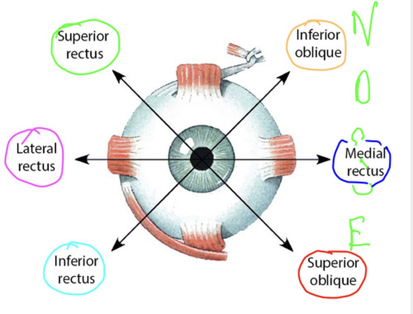
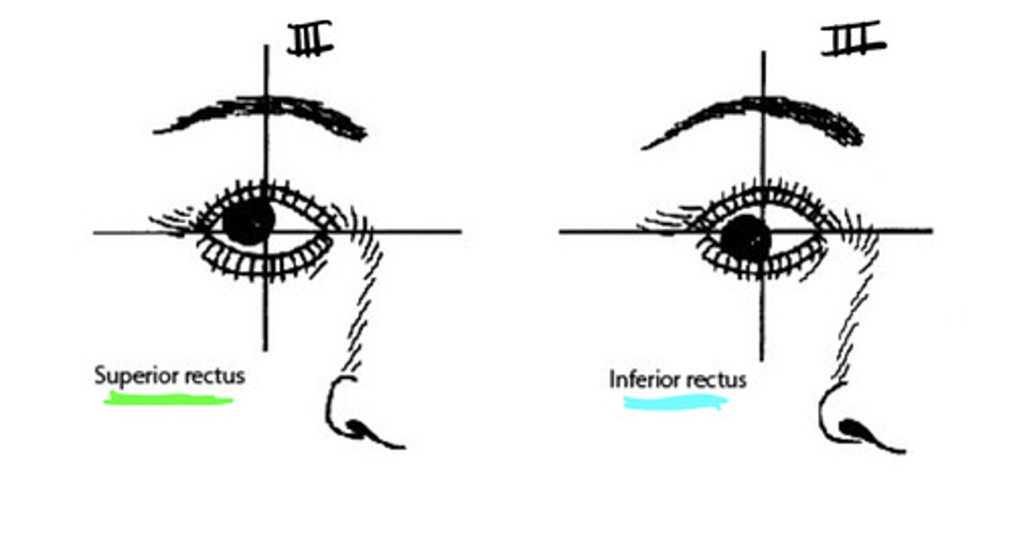
what are our 4 straight (rectus) extraocular muscles?
- superior rectus
- inferior rectus
- medial rectus
- lateral rectus
what are our 2 oblique extra-ocular muscles?
- superior oblique
- inferior oblique
what is the innervation to the extra-ocular muscles? what is the exception of this innervation of extra-ocular muscles?
- lateral rectus (Abducens)
- Superior Oblique (trochlear)
all others are oculomotor!!
what moves the eye toward the nose (medially)
medial rectus

what moves the eye away from the nose (laterally)
lateral rectus
what moves the eye up and lateral?
superior rectus

what moves the eye down and lateral?
inferior rectus

what rotates the eye so top of the eye moves toward the nose (intortion) pupil points down and medial?
superior oblique

what rotates the eye so the top of the eye moves away from the nose (extortion) so pupil points up and medial?
inferior oblique

how would we need to test each muscle individually?
you ask then patient to isolate each muscle by having them move their eye into the line of gaze position at 22 degrees lateral from the visual axis
if you just test a muscle group functionality you could do what test? where is this tested?
you could do the quick H test that tests in the visual axis rather than the line of gaze position
the actions of the medial and lateral recti are what?
- medial: adducts the eyeball making you cross eyed
- lateral: abducts the eyeball away from the nose
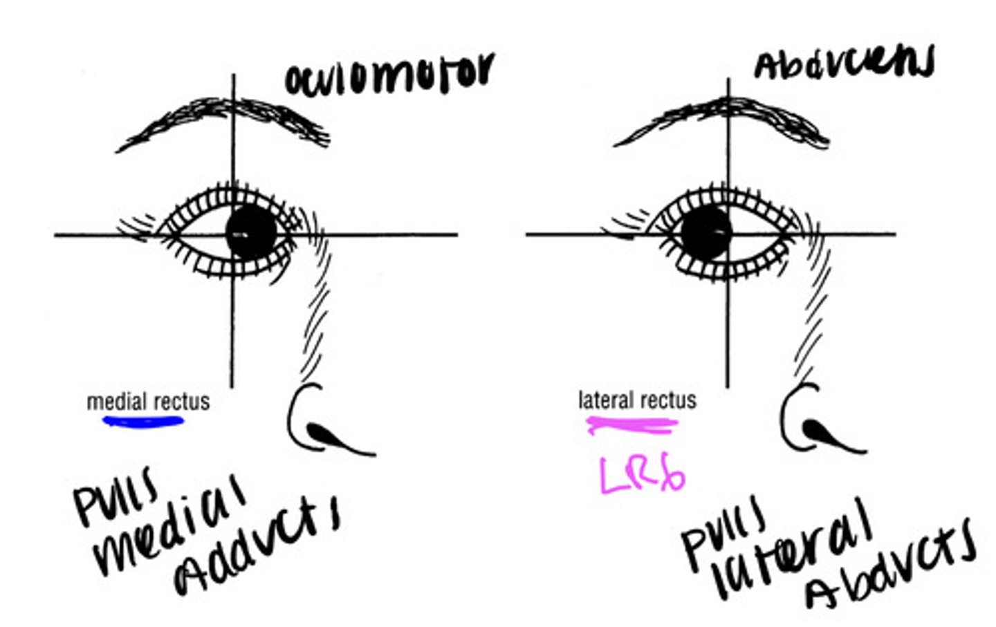
if you lesioned the abducens nerve (CN VI) what would this result in?
you would have a paralyzed lateral rectus and an inability to abduct the pupil.
this patient would present with an adducted pupil due to the unopposed pull of the medial rectus of the affected eye
so one eye huntin an one eye fishin
the lateral direction of the superior rectus and inferior rectus is due to what?
the fact that the superior and inferior rectus muscles are attached posteriorly and medial to the position of the eyeball in the orbit
the oblique muscles are attached to the posterior half of the globe of each eyeball, so wen the obliques act they want to do what?
rotate the eyeball in the orbit
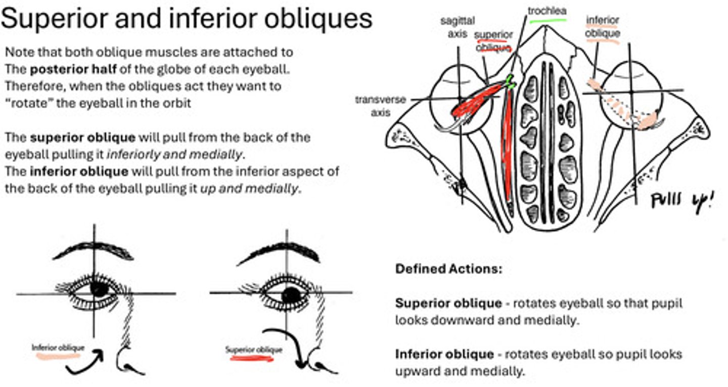
the primary action of the superior and inferior oblique muscles (without the visual axis aligned with the line of pull) is to do what?
rotate the eyeball around its visual axis
the superior oblique will produce what rotation?
intorsion (medial inward rotation to the top of the eyeball)
the inferior oblique will produce what rotation?
extorsion (lateral outward rotation of the bottom of the eyeball)
the secondary action of superior oblique is what?
depression
the secondary action of inferior oblique is what?
elevation
a lesion of the trochlear nerve (IV) would result in what in the eye?
in the inferior oblique being unopposed so intorsion is not possible and the eyeball is extorted resulting in diplopia (or double vision)
how would a patient with a trochlear nerve lesion present?
they would exhibit a head tilt with a paralysis of the superior oblique in an attempt to maintain the vertical axis of the eye to minimize double vision
so you are basically having to manually rotate the eye to prevent a double vision
if your right trochlear nerve is damaged you would tilt your head to the left to manually rotate your eye (right?)
the eyeball is composed of what three layers?
- an external fibrous layer: sclera/cornea
- a middle/vascular layer: choroid, ciliary body, iris
- internal/retinal layer: very thin delicate membrane

the posterior 5/6 of the external fibrous layer is what?
the opaque sclera
the anterior 1/6 of the external fibrous layer is what?
the transparent cornea
what is the portion of the middle layer of the eye that is adherent to the retina terminating at the ciliary body?
the choroid
what is the portion of the middle layer of the eye that is connecting the choroid to the iris?
ciliary body
what is the contractile diaphragm anterior to the lens?
iris
what is a very thin delicate membrane containing bipolar neurons responsible for relaying visual stimuli to the brain?
the internal/retinal layer
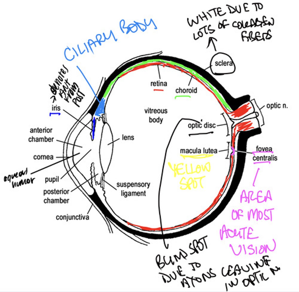
what is a clear watery fluid in the anterior and posterior chambers of the eyeball?
aqueous humor
what produces aqueous humor continually?
the ciliary processes
how does the aqueous humor get from the posterior chamber of the eye into the anterior chamber of the eye?
via the pupil
how is the aqueous humor drained once it reaches the pupil?
its drained away through spaces at the iridocorneal angle that drains into a circular venous canal
blockage of the drainage into the circular venous canal causes increased pressure in the compartment also known as what?
glaucoma
what does the aqueous humor provide nutrients for?
the avascular cornea and lens
what is a transparent biconvex structure held within a transparent capsule?
the lens
the lens is surrounded by what?
ciliary processes
what attaches to the ciliary processes of the eye?
highly elastic and radially arranged suspensory ligaments of the lens
curvature of the lens is altered by what?
contraction or relaxation of this ligament
loss of transparency in the lens is called what?
cataracts
what fills the eyeball posterior to the lens and is a jelly-like matrix with a fine meshwork of collagen fibers?
vitreous body
what fills the vitreous chamber of the eyeball?
gel
what contracts circular reducing the diameter of the pupil by pulling the iris, ciliary body, and processes medially (centrally)?
the ciliary muscle
the pupil constricts as circular muscles of the iris contract via sympathetic or parasympathetics?
parasympathetics
the pupil dilates as radial muscles of the iris contract via sympathetics or parasympathetics?
sympathetics
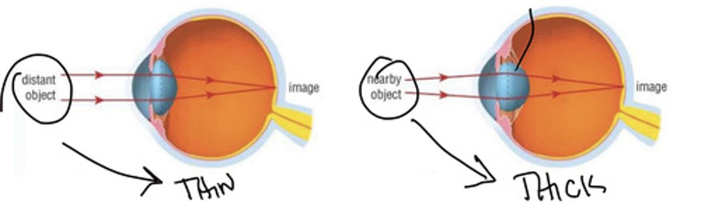
what is the changing of the shape of the lens by ciliary muscles to allow a sharp focused image to form on the retina?
accommodation
changing of the shape of the lens, the focal length changes and allows us to do what?
focus on nearby and distant objects
to view nearby objects what muscles contract? what happens to the suspensory ligaments? what happens to the lens?
the ciliary muscles contract and the suspensory ligaments become slack, the lens becomes more rounded
the point at which a nearby object becomes blurred occurs when what happens?
the lens reaches its maximum curvature