IB Psychology - Learning and Cognition 2.0
 Call Kai
 Learn
Learn Practice Test
 Spaced Repetition
 Match
Match Flashcards
 Knowt Play
1/115

There's no tags or description
Looks like no tags are added yet.
Name | Mastery | Learn | Test | Matching | Spaced | Call with Kai | Chat |
|---|
No analytics yet
Send a link to your students to track their progress
116 Terms
Biological approach on human behvaiour
most basic assumption is that complex human behaviours may be reduced to biological origins → if a researcher only focuses on a single biological factor to explain a complex human behaviour, this is a reductionist approach
Using animals to understand the human brain
Animal research can provide insight into human behaviour → our brains, nervous system + endocrine systems work very much like other mammals
Human brain diagram
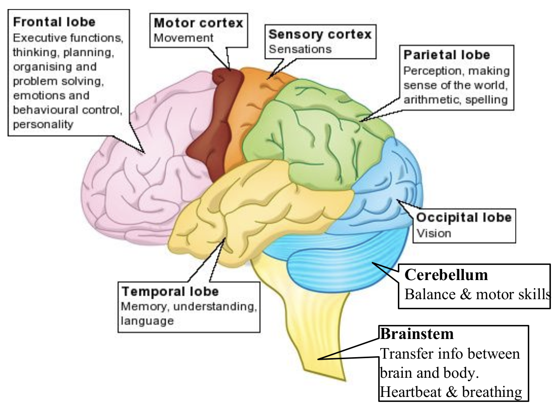
Frontal lobe
responsible for production/formation of corrext speech
Broca’s aphasia (frontal lobe)
Person can understand language
Prevents person from producing speech
Words slurred + not properly formed
patients also struggled with written words in the same way as speaking
Way of remembering
BROca → BROnt → FRONT → BROken speech
Temporal lobe
Language comprehension is essential as it enables the brain to interpret spoken and written language → allows individuals to understand words, sentences + their meanings
Wernicke’s aphasia (temporal lobe)
Loss of ability to understand language
Person speaks clearly but words don’t make sense ‘word salad’
Localization of function
the theory that specific parts of the brain are responsible for specific functions + reflects the idea that behaviour, emotion and/or thoughts originate in specific regions of the brain → damage to that region causes a drastic loss of that function
Types of localization
strict
relative
distributed
Lateralization
strict localization
A function is linked to a very specific + fixed area of the brain → e.g. hippocampus = transfer of semantic information from STM → LTM → supported by HM case study
relative localization
Certain brain areas specialise in particular functions, but other areas may also contribute → there is a primary area for the function, but it works with other areas to function properly → e.g. Broca’s + Wernicke’s areas work together in language processing, with neighbouring areas also assisting
distributed localization
Functions are not isolated to one brain area → several brain regions work together in a network → e.g. memory involves the hippocampus + prefrontal cortex + other related structures → no single area is responsible for the behaviour
Lateralization of localization
Certain processes are more dominant in 1 hemisphere than the other → e.g. language is primarily lateralised to the left hemisphere
Hippocampus - which part of memory it’s involved in
for encoding + consolidating autobiographical memories → helps bind different elements of an experience → e.g. sights, sounds, emotions into a cohesive memory
Prefrontal cortex - which part of memory it’s involved in
helps integrate autobiographical memories with emotions + self-relevance → damage here can lead to distorted memories/difficulty recalling life events coherently
Amygdala - which part of memory it’s involved in
enhances emotionally charged memories → makes them more vivid + long-lasting
This distribution of memory across multiple brain regions ensures long-term retention, even if 1 area is damaged
Study to use for localization of function
HM case study
HM Case Study Aim
To support the view of localization of function, in this case study that regions of the brain (hippocampus) are responsible for behaviours (memory)
Scanning techniques
MRI
fMRI
MRI
scans anatomical structure, can help localize brain damage → e.g. stroke

fMRI
images metabolic function → measures brain activity by detecting changes associated with blood flow
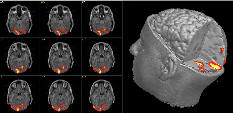
Link to HM to localization of function
The hippocampus is needed for memories to be transferred to LTM → HM case reveals the interaction of cognition (memory) + physiology (brain damage in the hippocampus) in amnesia → brain damage in relevant areas causes memory impairment → this study supports the view of localisation of specific functions as it suggests that the specific part of the brain (hippocampus) is responsible for a specific function (transferring of info from STM to LTM)
Limitations of localisation of function
Much of the research is limited → using case studies with brain damage/autopsies → impossible to assess the individual’s behaviour and/or cognitive skills before the damage → studies are retrospective + open to bias
Very few behaviours are strictly localised → research suggests that behaviour results from the interaction of different parts of the brain
Reductionist approach to human behaviour → attributing complex behaviour to a single part of the brain
Although fMRI scans reveal where activity is taking place in the brain during some behaviours, people’s ability to carry out tasks is limited → ecological validity
MRIs can be used to determine localisation by observing areas of the brain with more grey matter → however, these studies are correlational and do not determine causality → also other parts of the brain may play a role but not show a significant increase in grey matter
Consent by proxy - ethical considerations for studies about brain damage
when an individual has the legal right to make decisions on behalf of another who cannot do so → even though his mother consented to the research, is this acceptable?
Capacity to consent - ethical considerations for studies about brain damage
Assess the individual's capacity to provide informed consent, as brain damage can affect cognitive abilities + decision-making
Reconsent - ethical considerations for studies about brain damage
Regularly re-evaluate + re-confirm the participant's consent, as their cognitive abilities + understanding of the study may change over time
Anonymity - ethical considerations for studies about brain damage
Use anonymized data whenever possible to protect the participant's identity
Avoiding exploitation - ethical considerations for studies about brain damage
Be mindful of power dynamics + avoid any form of exploitation or undue influence
Reporting results - ethical considerations for studies about brain damage
Communicate the results of the study to participants + their families in an appropriate + understandable manner
Ethical reflection - ethical considerations for studies about brain damage
Regularly reflect on + discuss ethical issues with the research team to ensure that the study remains ethically sound
Damage to hippocampus
Alzheimer’s disease → symptoms are memory loss _ disorientation, have been associated with a decline in hippocampal activity → hippocampus is the earliest brain region + most severely affected → high levels of chronic stress leads to a decrease in the volume of the hippocampus
Reasons to do animal studies
shorter lifespan
breeds faster
less rigid ethics for them
similar genetic makeup
Meaney et al (1988): animal study
Rats were randomly allocated
C1: newborn rats were handled daily for 3 weeks → they were taken away from their mom for 15 mins→ researchers brushed them for an internse 15 mins to stimulate grooming
C2 (control): taken away from mum → did NOT brush (no grooming) → after 2 years, rats were put into a pool of milky water → in the pool, there was a platform, the researchers tracked the rats' route
Meaney et al (1988): results
C2 took a circuitous route to the platform
Rats were killed to examine their brain
Hippocampal cell loss + pronounced spatial memory deficits emerged with aged neglected rates but were absent in the rats groomed by the researchers → C2 had high levels of glucocorticoids – stress hormones (cortisol)

Domino causality
mother's neglect can cause memory impairment in old age → but is not a direct causality, but a domino causality → in human samples, not everyone who experiences neglect has the same health/psychological problems → a series of causal relationships and interaction with the environment that may lead to memory loss
Domino causality in Meaney et al
Neglect > lack of gene expression > inability to regulate the stress response > higher levels of stress hormones > hippocampal cell loss > memory impairment
hippocampus contains high levels of glucocorticoid receptors → more susceptible to long-term stress → “inability” of C2 rats to turn off stress receptors results in hippocampal cell death → eventually leads to rats’ inability to recall spatial info → affecting memory
How to answer P1A (4m) for localization of function
define localization of function
explicitly link to a behaviour/cognition process → memory
example → how we know the part of the brain has a specific function
Neuroplasticity
The ability of the brain to change itself in response to environmental demands through the making + breaking of synaptic connections between neurons.
Making synaptic connections
neural branching
Breaking synaptic connection
neural pruning
The process of neuroplasticity
The brain is a dynamic system that interacts with the environment. In a sense, experience physically shapes the brain. Not only can the brain determine + change behavior, but behavior + environment can also change the brain.
Brain plasticity refers to the brain’s ability to rearrange the connections between its neurons → changes in the brain's structure due to learning or experience. High levels of stimulation + numerous learning opportunities lead to an increase in the density of neural connections.
Neural branching
Every time we learn something new, the neurons connect to create a new trace in the brain → called dendritic branching because the dendrites of the neurons grow in numbers and connect with other neurons.
Synapses become stronger through repeated use (practice) → the repeated firing of neurons leads to stronger connections → known as long-term potentiation. LTP leads to dendritic branching. These branches allow neurons to receive information from other neurons + transmit information back to them. The benefit of many extensions is that it allows for more connections between neurons, making communication more efficient.
Neural pruning
When a synapse is not used/under stimulated, it may undergo synaptic/neural pruning.
It is believed that this is the way for the brain to remove synapses that are no longer needed, making the functioning of neural networks more efficient.
Maguire et al (2000) aim
Maguire carried out a study to see whether the brains of London taxi drivers would be somehow different from the average person as a result of their training + extensive use of spatial memory
Maguire et al (2000) procedure
The participants for this study were male London taxi drivers. Control group: healthy male, no taxi driving experience
Average experience of taxi drivers was 14 years.
MRI scans were compared between drivers and non-drivers. Researchers also corelated the number of years of taxi driving experience with MRI scans.
In order to take part in the study, the participants had to have completed the "Knowledge" test and have their license for at least 1.5 years.
The sample included a range of ages so that age would not be a confounding variable
It was also a single-blind study- that is, the researcher did not know whether she was looking at the scan of a taxi driver or a control group.
Maguire et al (2000) results
The results showed that the taxi drivers had larger posterior hippocampi compared to the controls due to spatial navigation skills
involved.
The controls had larger anterior hippocampi compared to the taxi drivers.
They found a positive correlation between the number of years the participants had been taxi drivers and the size of the posterior hippocampus.
Maguire et al (2000) conclusion
Maguire argued that this demonstrates the plasticity of the hippocampus in response to environmental demands as the posterior hippocampus stores a spatial representation of the environment.
The volume of the posterior hippocampus expanded because of their high reliance on navigation skills and spatial memories → leading to neural branching.
However, no new spatial information was stored so this led to neural pruning in the anterior area.
Maguire et al (2000) Evaluation
Alternative view is that they could have a special talent in spatial navigation which led to the differences in the hippocampus + therefore chosen taxi driving as a profession.
However, Maguire found a positive correlation in the size of the posterior hippocampus and the time spent as a taxi driver. Hence this clarifies the bidirectional ambiguity.
Redistribution of the grey matter in the hippocampus occurs in taxi drivers in response to gaining navigational experience.
Cortical remapping after strokes
Cortical remapping is the brain's ability to reorganize itself by forming new neural connections. When a part of the body is injured or no longer used, the brain can "remap" that area's function to other, healthy parts of the cortex.
This process helps the brain adapt to changes → e.g. after injury, learning new skills, or during recovery from conditions like stroke.
The brain can recover from a stroke to a significant extent due to its neuroplasticity → the ability to reorganize and form new neural connections. Depending on the severity + location of the stroke, undamaged areas of the brain can sometimes take over lost functions, especially with rehabilitation
What is neurotransmitter categorised under in IB questions
chemical messengers = neurotransmitters
Neurotransmitters
1 of the most important chemical messengers in the brain that affect behavior and cognition → process by which these messages are sent is called neurotransmission
key neurotransmitter is Acetylcholine (ACh)
Structure of typical neuron

Where in the neuron are the messages received
Dendrite

Where in the neuron are the messages sent from
axon terminal
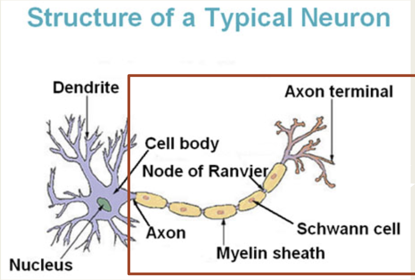
Action potential
when neurotransmitters travel down the axon

Where is the final destination for the neurotransmitters in the pre synaptic neuron
terminal button
Where are neurotransmitters stored
synaptic vesicles

What is the gap between the presynaptic and postsynaptic called
synaptic gap
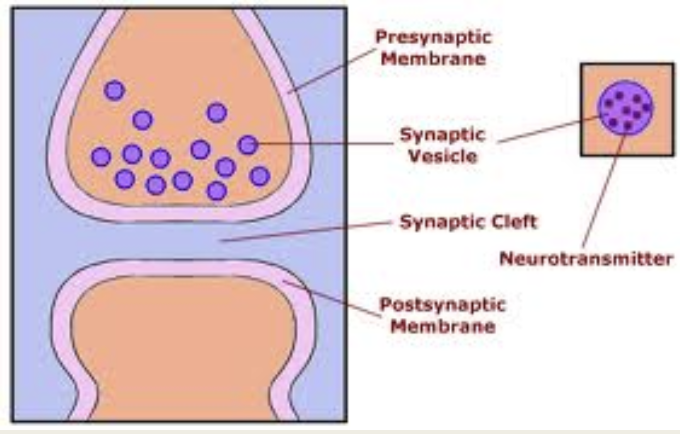
How are the neurotransmitters absorbed by the postsynaptic neuron

If the neurons do not bind, where do the neurotransmitters go
destroyed by enzymes or go back to the presynaptic neuron via the uptake pump
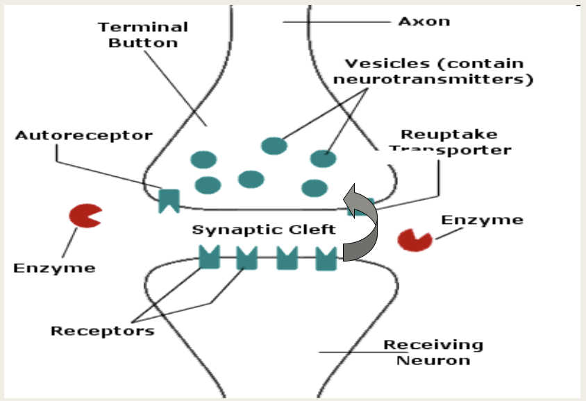
Process of neurotransmission
The electrical impulse that travels along the body of the neuron is called an action potential
When an action potential travels down the body, or axon, of the neuron, it releases neurotransmitters stored in the neuron’s terminal buttons
The neurotransmitters are then released into the gap between the neurons → called the synpatic gap. action potential terminal buttons synapse
The dendrites of the post synaptic neuron have special receptors to receive the neurotransmitter → when the right neurotransmitter is received (where the chemical 'shape' of the neurotransmitter 'fits' the receptor) the message continues up the dendrites of the receiving neuron, along the axon continuing the message
Any neurotransmitters that are not received by the dendrites are either destroyed by enzymes, or taken back into the pre-synaptic neuron via reuptake
Main neurotransmitter → ACh
Plays a role in the consolidation of memory in the hippocampus when new learning takes place + contributes to attention + arousal (when memory is aroused) → sends + receives info between motor neurons + voluntary muscles
Exogenous neurotransmitters
neurotransmitters not from your body
endogenous neurotransmitters
neurotransmtiters from your body
Characteristics of neurotransmitters
Drugs can replicate the shape of the neurotransmitter + then occupy the receptor site on the dendrites → exogenous neurotransmitters → should work as a lock and key model
neurotransmitter may be excitatory → causes the neuron to fire + binds on to the post-synaptic receptor sites
neurotransmitter may be inhibitory → prevents a neuron from firing + inhibiting binding of neurons to receptor sites
Types of neurotransmitters
excitatory
inhibitory
agonist
antagonist
excitatory
increase the likelihood of a neuron firing → e.g. ACh
inhibitory
decrease the likelihood of a neuron firing
agonist
chemical that binds to a receptor + activates the receptor to produce a biological response → ACH is an agonist for ACh receptors
antagonist
blocks the action of the agonist → scopolamine is an exogenous antagonist for ACh
Rogers and Kesner - animal model aim
to investigate the role of acetylcholine in spatial memory (encoding + retrieval)
Rogers and Kesner - animal model
The rats were randomly allocated to one of two conditions, they were either injected directly to the hippocampus with:
Scopolamine (blocks acetylcholine receptor sites)
Saline as a control (placebo)
Rogers and Kesner - animal model results
The scopolamine group (blocks acetylcholine) found that they made more errors + took longer in learning the maze → suggests acetylcholine plays a role in encoding of spatial memories
Did not affect retrieval of memories that has been created from before → hence, acetylcholine may play an important role in the consolidation of spatial memory
Measurement: animal studies - strengths
animals can serve as models for human physiology + behaviour → the basic biological structure + functioning of hippocampus + ACH systems are relatively similar
Invasive procedures → e.g. injecting scopolamine into the hippocampus cannot be ethically done in human → why animal models are valuable
Measurement: animal studies - limitations
animal behaviors differ from human → more complex than a rat
both have similar patterns of memory impairment
social, emotional + cultural factors influence memory in human cannot be tested in rats → what might be stimulating for a rat may not have the same effect on humans
since these lab rats are raised in controlled environments, it’s uncertain how valid or applicable the results are to natural behaviors
Strengths of neurotransmission
have led to successful treatments for certain behaviors (prevention + treatment) → successful drug treatments have been developed
experiments can be replicated to establish reliability → have a standard procedure
Limitations of neurotransmission
many of these studies are on animals → cannot guarantee that the neurotransmitter plays the same role in human behavior
Research is most often done indirectly (needs an inhibitory neurotransmitter, antoagonist - scopolamine to block ACh receptors)
reliance on fMRI means that the limitations of the techniques are relevant to the evaluation
Much of the research on humans is correlational → indirectional ambiguity
The argument that neurotransmitters are the cause of behavior is reductionist → although a reductionist argument may be good since such arguments could potentially lead to positive intervention strategies → explaining a complex behavior like falling in love as a "neurochemical cocktail" could be considered an oversimplification of human behavior
Method of Loci
associating the information you want to remember with specific locations in a familiar place → improves memory by combining visual imagery with spatial memory
involves placing information you want to remember along a familiar mental route → e.g. your house, your route to school → then recalling it by mentally “walking” through that route
Why does the MoL work
Humans have ability to remember spatial environments → most likely evolved as a survival mechanism → MoL taps into this by anchoring abstract/disconnected information to physical locations you already know well
Effective because it promotes elaborative encoding → not just rehearsing information → transforming it into rich, multi-sensory experience
When you take a fact and turn it into an image, place it in a location + visualize a dynamic scene, you’re engaging multiple parts of the brain: visual, spatial, emotional + linguistic → this process forms more pathways to retrieval of memory
Dresler et al
Controls (non-athletes) were then randomly assigned to the method of loci training, a working memory task, or no training at all. They were then rescanned
Dresler et al results
compared the brains of "memory athletes" to those of MoL participants → non-athletes (controls) not only achieved similar levels of memory + brains changes to look more like the memory athletes
Before + after training, the controls were given 72 words to memorize → MoL group remembered more than the initial round
Networks handling visuospatial and memory processing were more tightly linked → other 2 control groups didn’t see a significant change in their memories or brains

Spaced repetition
the brain learns best when information is reviewed at gradually increasing intervals.
When you first learn a new piece of information, you should review it shortly after to strengthen your memory → as time passes + your memory of the information starts to fade, you should review it again → by spacing out your reviews over time, you can reinforce your memory + recall of the information while reducing the time you need to spend studying
Gilbert et al
Medical students were trained to use Anki → an app that uses spaced repetition → researchers looked at the frequency of use of the app + scores on the Comprehensive Basic Science Exam
Gilbert et al results
Students who reported using Anki more for studying performed significantly better on the CBSE exams
Why does spaced repetition work
When you wait a little while before reviewing something, recalling it requires more effort → the harder your brain has to work to retrieve a memory (as long as it succeeds), the strong the memory becomes
Spaced practice allows for sleep-dependent memory consolidation → during sleep, the brain strengthens newly learned information by reorganizing + stabilizing memory tracks, making them easier to recall later → because spaced learning gives the brain multiple opportunities to sleep between study session, memories become more durable + resistant to forgetting as compared with cramming in a single session
Which perspective are memory techniques slotted under
cognitive perspective
Different perspectives in cognitive learning

Dual process theory
argues that our brain minimizes cognitive load + only uses effort when necessary
What different types of thinking systems do we have in DPT
system 1
system 2
System 1

How to describe system 1
automatic, fast, intuitive + requires little effort
System 1
uses heuristics (mental shortcuts) to make quick decisions → because humans are cognitive misers
allows for efficient processing in complex situations
However, it can lead to errors or biases when assumptions are incorrect
More likely to be used when cognitive load is high
Why do we use system 1 thinking
We are cognitive misers → want to use as little energy as we can to think
Sometimes we have too many other things going on in our mind to allocate energy for solving a problem → cognitive load is too high
System 2

How to describe system 2
slow, deliberate + conscious
System 2
rational thinking + effortful, requiring mental energy
involves careful analysis of information
Time consuming and less prone to errors
Used when tasks require focus, reasoning + critical thinking
Dual process theory
important to remember that we often use both of these systems when addressing a problem → ystem 1 will reach a quick conclusion + System 2 will go into further analysis to hopefully reach a "more correct" conclusion → because System 1 is activated before System 2 can do its work, System 1 often interferes with the effectiveness of System 2
Which research to use for DPT
stroop test