Lids 3: infection and inflammation
1/30
There's no tags or description
Looks like no tags are added yet.
Name | Mastery | Learn | Test | Matching | Spaced |
|---|
No study sessions yet.
31 Terms
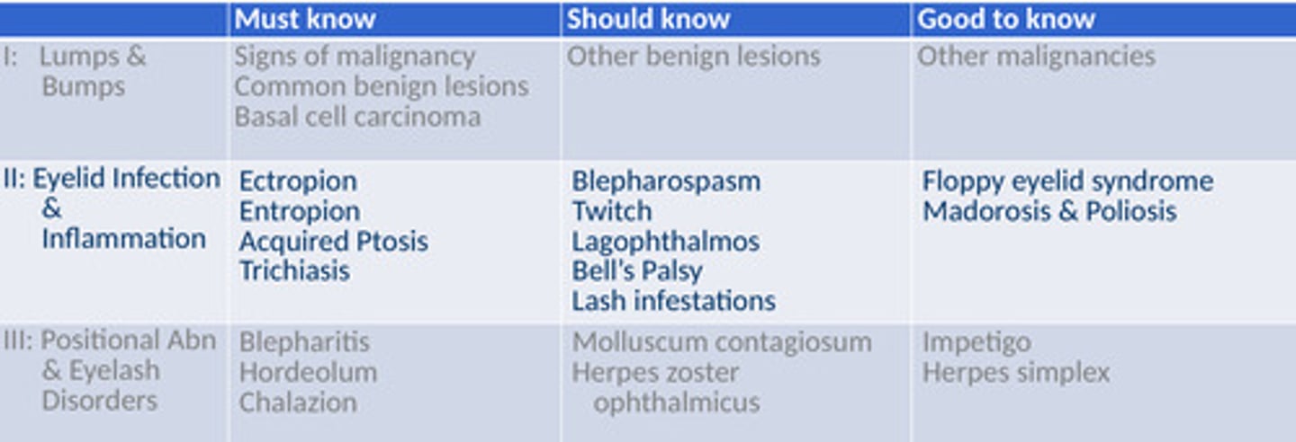
Must know/should know/ good to know
Blepharitis image
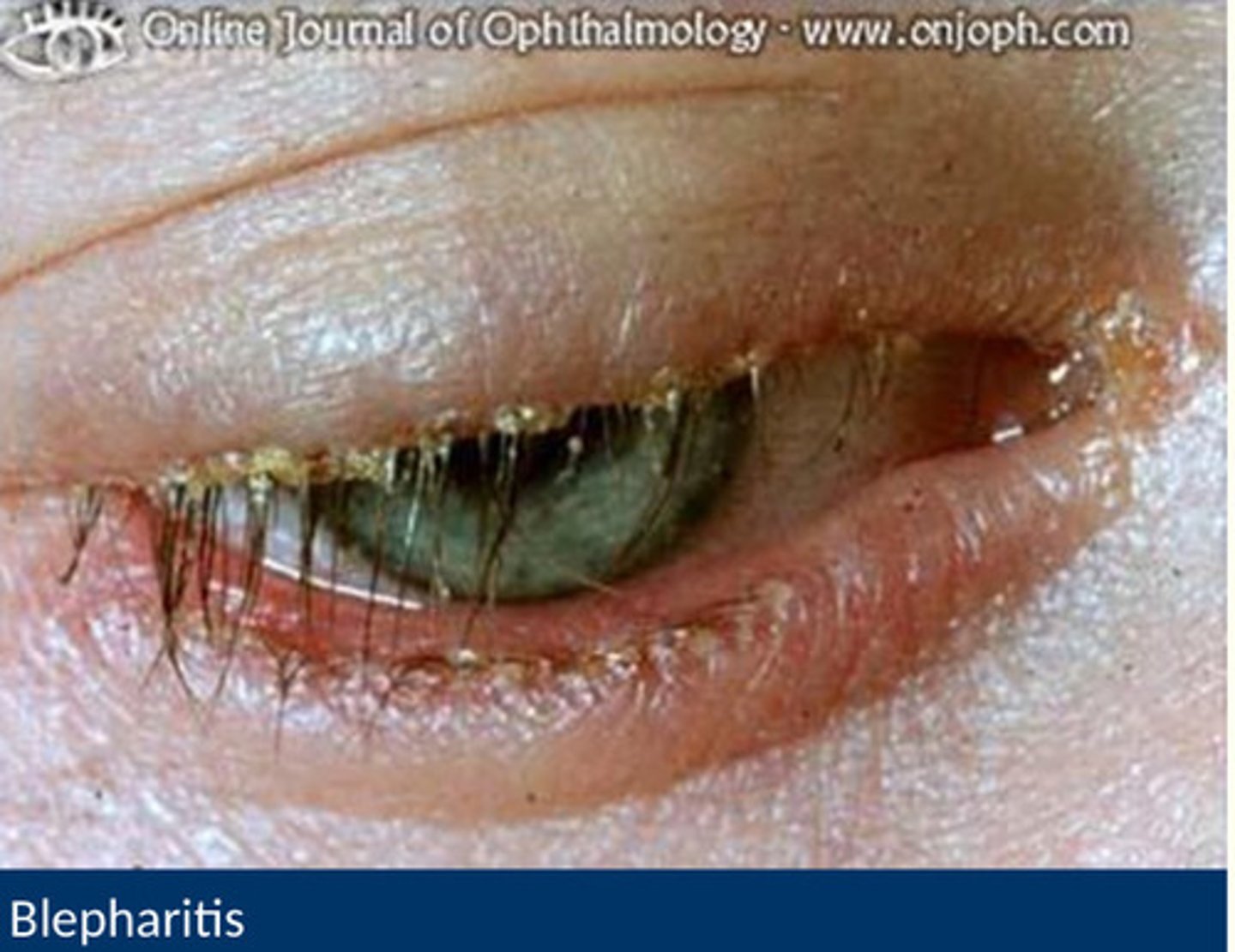
What is blepharitis?
- inflammation of the eyelid margins
- extremely common
- chromic/ relapsing LONG TERM
- typicallg bilteral
- more than 70% of people in the uk habe blepharitis at any one time
What are the predisposing/ risk factors of blepharitis?
- seborrheic dermatitis (dndruff)
- rosacea ( a chronic skin condition)
- can happen to long term contact lens wearer
- can be causes by topical eye medication (galucoma)
- demodex ( hair follicle mites)
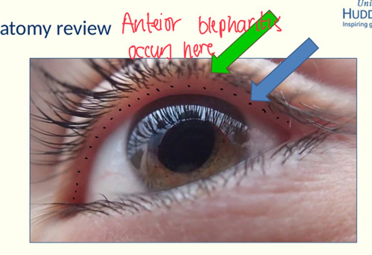
Anatomy of blepharitis
Anterior blepharitis affects the glands of zeiss and glands of moll
Posterior blepharitis affects meinomian glands
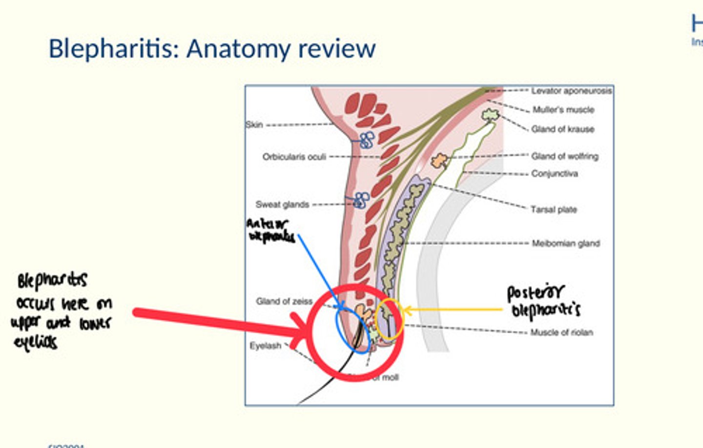
Anterior blepharitis
Posteior blepharitis ovvurs just behind the dots where the meibomian glands are, where secretions fo the meibomian glands are sectreted
Aetiology of anterior blepharitis
Why are signs of blephritis important?
They can be used to differentiate between the type of blephiritis e.g. bacterial or seborrheic anterior blephiritis
Symptoms of blepharitis
Similar symptoms for both types of blepharitis
- very variable
- chronic: lasts months or years (hard to get rid of it completely)
- symptoms dont always correspond well to signs
- ocular discomfort
- gritty
- soreness
- itching
- burning
- photophobia
- contact lens intolerance
Anterior bacterial blepharitis signs
bacterial (staphylococcal) signs
- crusting/ collarettes/ scales at the base of lashes
- telangectasia (dilated blood vessels)
- lash misdirection/ loss
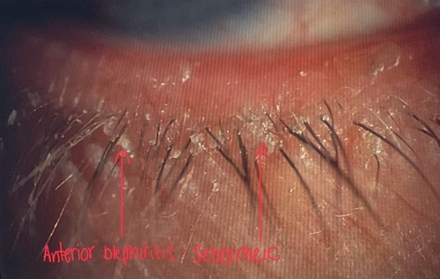
Anterior seborrheic blephiritis signs
Greasy deposits at the base if the lashes
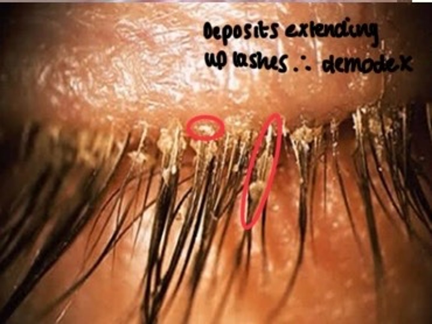
Anterior demodex blepharitis
- cylindrical deposits extending up lashes
- lash misdirection/ loss, general redness
- itching
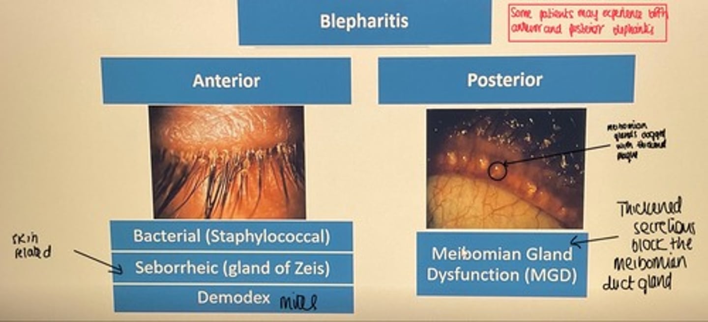
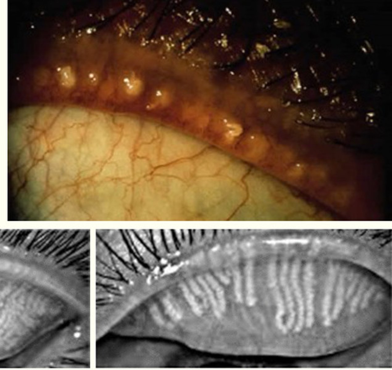
Posterior blephiritis signs
- thickened meibomian secretions
- microliths
- meibomianitis: passive retention of secretions (chalazions) - the glands are clogged so it is harder for secretions to occur
- foam in tear miniscus
- unstable tear film - evaporative tear deficiency
Meibiomisn glands secrete oils into tear film to ,ske it more stable but the secretions are not happening
Posterior blephiritis image
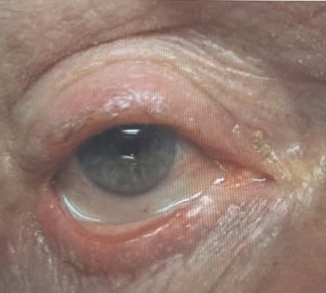
Secondary signs of all blephiritis Image
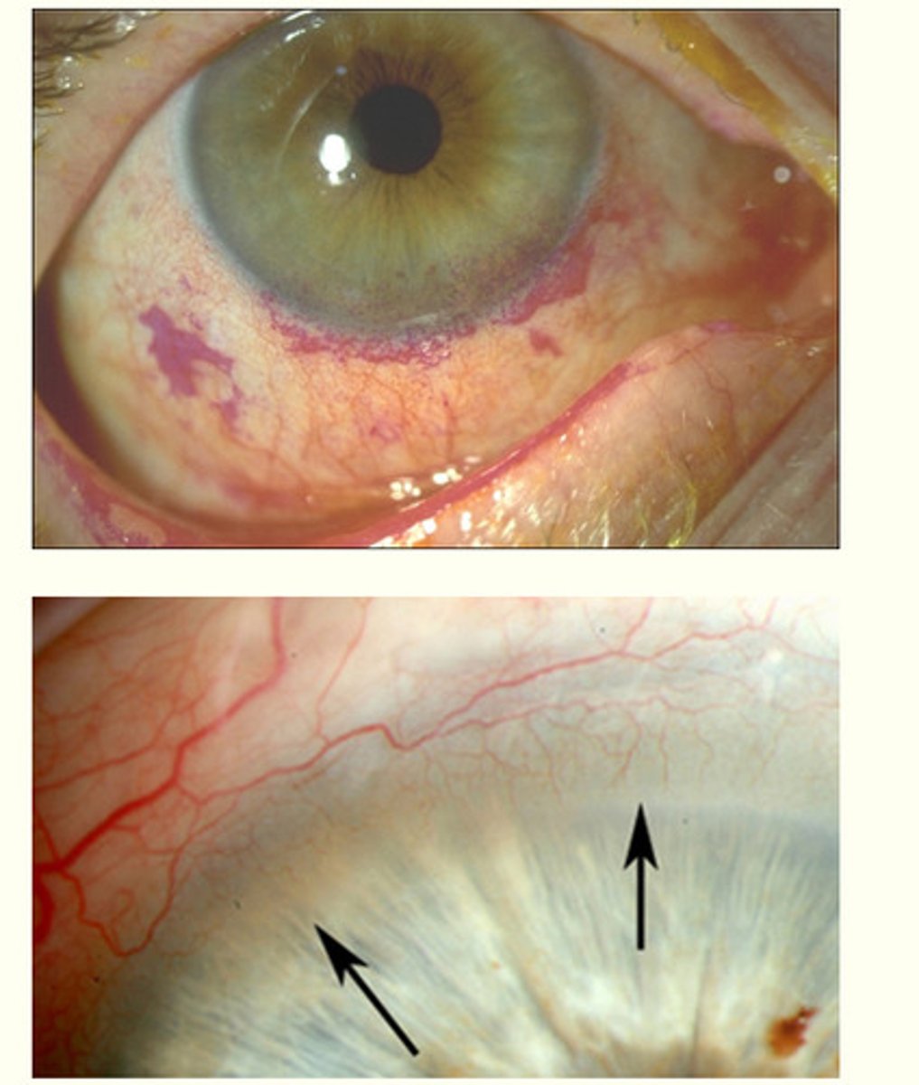
Secondary signs of all blephiritis
- chronic red eye (lid margin and conjunctival hyperaemia)
- punctate epithelial erosion ( lower 3rd of cornea)
- conjunctival staining
- marginal keratitis and scarring
- neovovascularisation (new blood celk formation) around limbus
- pannus: cornea becomes opaque
Management of blepharitis
all cases
- lid hygiene
- lid massage
- clean/ wipe
- see lid hygience video on lecture slides
Where indicated
- ocular lubricants
- drops / ointments
- vitamin supplements
- omega 3/ fish oils
Optometric management of blephiritis
Refractory cases (resistant to treatment)
- topical antibiotics (chloramphenicol ointment or doxycycline for 3 months)
- meibomian gland expression
- various lid treatments
- demodex - tea tree oil (experienced clinician needs to do this)
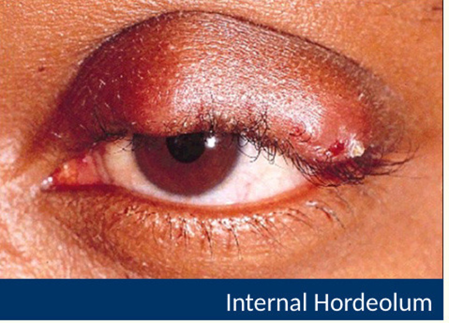
Internal hordeoleum image
- Further inside rhe lid
- affects meibomian gland tarsal plate
External hordeolum image
- stye
- infection if the gland of ziess or gland of moll (base of the eyelash)
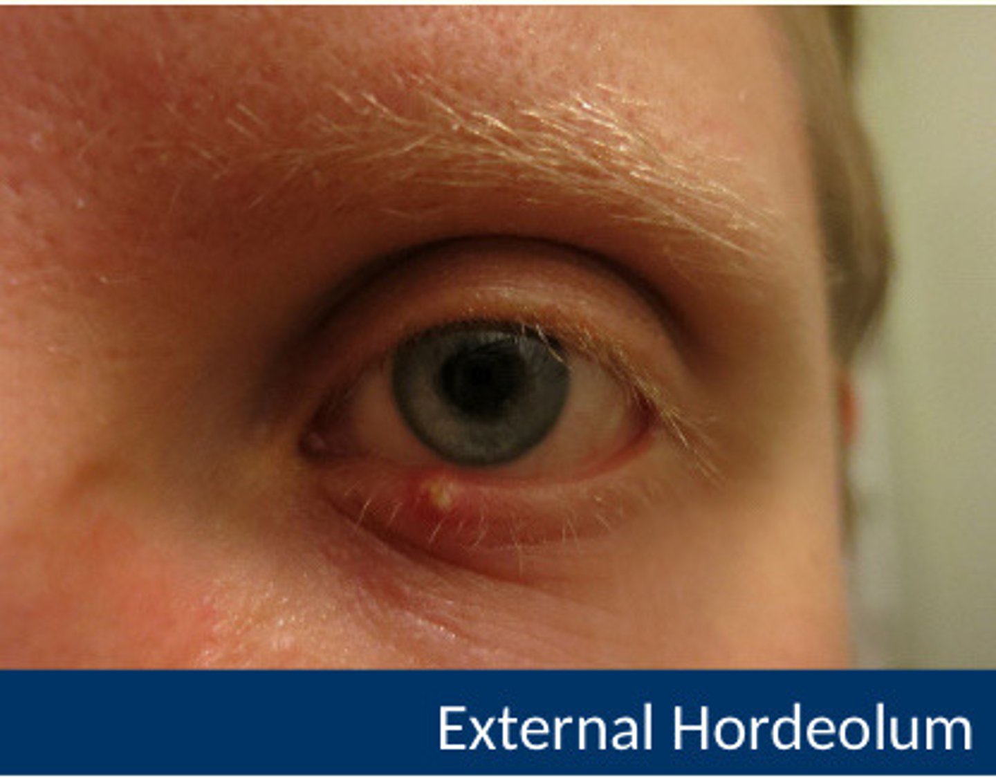
What is a hordeolum?
- accute bacterial infection (staphylococcal) of an eyelid gland
- occur in24-48 hours / red swelling
- tender eyelid lump
- may spontaneously express itself with a purulent material
- often associated with blepharitis
Hordeolum treatment
Treat as you would treat blephiritis
- warm compress
- massage
- clean
Chalazion image
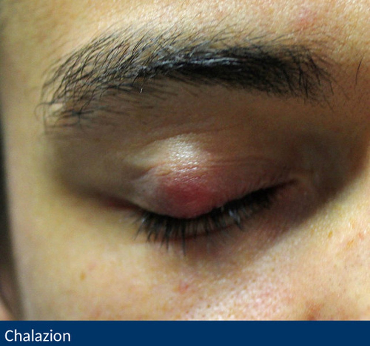
Chalazion aetiology and symptoms
- common, chronic lid lump
Aetiology (causes)
- blockage of meibomian gland duct
- inflammatory response: stagnate secretions and inflammatory cells
- spontaneous or follow hordeolum
- typically less accute amd occurs over a period of weeks
Symptoms
- usually painless lid lump
- single or multiple/ may be recurrent swelling
Chalazion signs and management
Signs
- well defined, 2-8 mm ø subcutaneous nodule in tarsal plate
- may be associated with blephirits and astigmatism
Management
- tend to resolve on their own
- lid hygiene (warm compress/ clean/ lid massage)
- resolution may take several weeks (need o refer them routinely)
- occasionally may be surgically removed
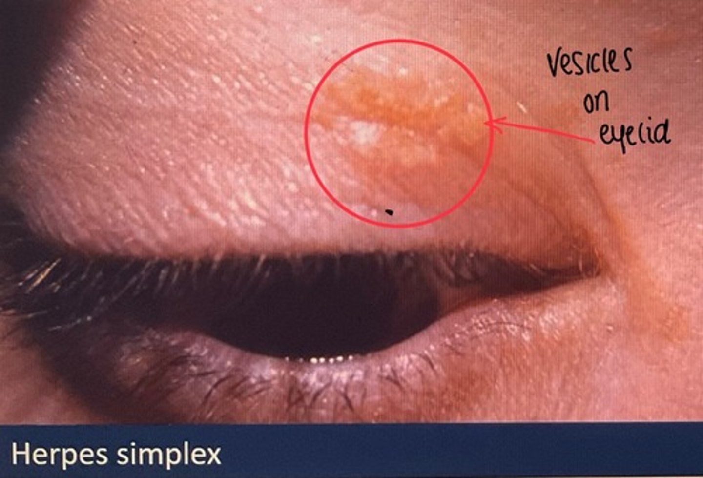
Herpes simplex virus
Swollen lids and conjunctivitis
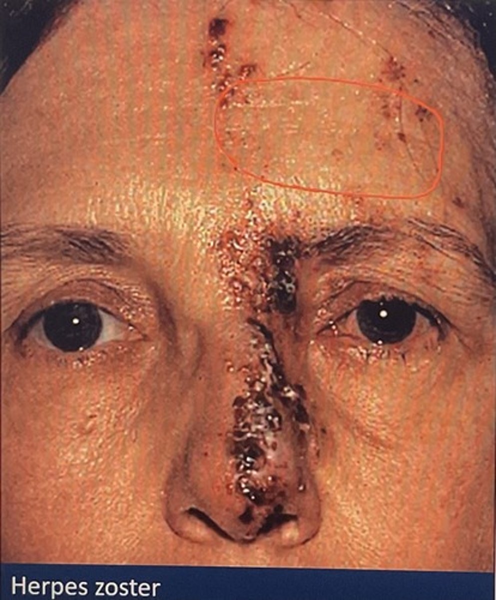
herpes zoster virus (shingles)
- originates from chicken pox virus. Varicella virus remains dorment in a nerve in the body. Expressed later in life as herpres zoster
- patients expeience pain (neuralgia- tingling pain)
- a week later a rash develops called vesicular rash
- appear on one side of the face
- a lesion on the tip of the nose (hutchinson's sign) higher risk of ocular complications
- oral acylovir (meds) within 72 hours reduces eye distorders from 50% to 20-30% and reduces pain
Herpes zoster image
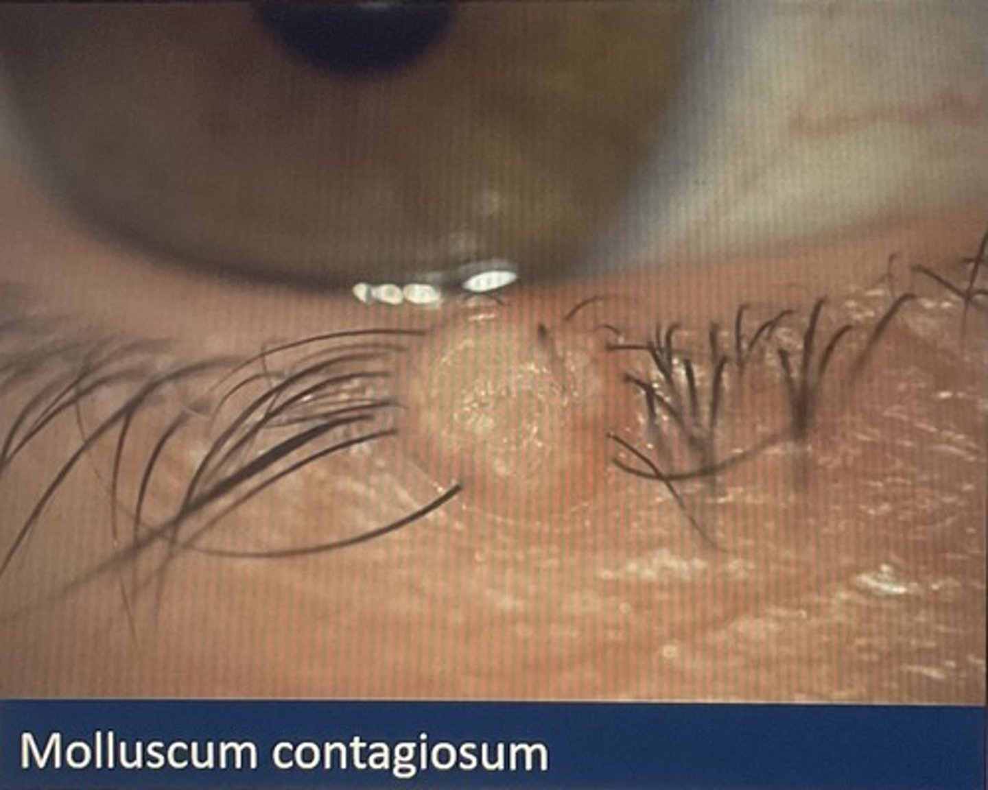
Molluscum contagiosum image
- A proxyvirus
- mildly contagious (skin to skin contact)
- umbilicated skin nodule (2-3mm ø)
- viral toxins may cause follicular conjunctivitis
- curette lesions
Fungal infections
Rare
Diffilcult to treat

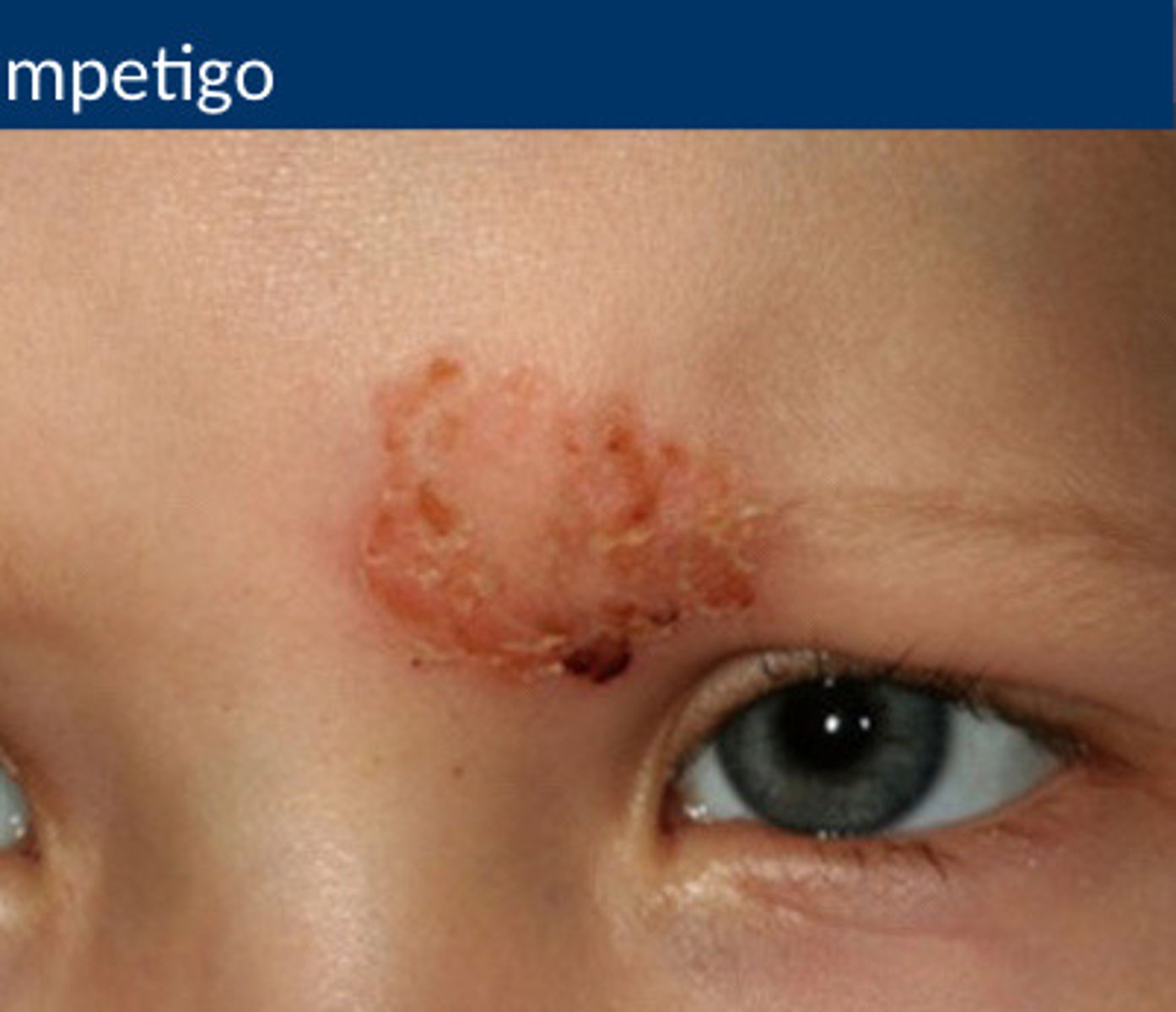
Impetigo
- staph infection
- rash
- occasionally blisters on the skin
- common in children younger than 14