Day 3: Lecture and Dry Lab 2
1/116
There's no tags or description
Looks like no tags are added yet.
Name | Mastery | Learn | Test | Matching | Spaced | Call with Kai |
|---|
No analytics yet
Send a link to your students to track their progress
117 Terms
Cranial Cruciate Ligament Rupture AKA _____ _____ in humans.
ACL tear
Cruciate ligament
Connects tibia to femur
Provides support
People rupture cranial more than caudal
There’s cranial and caudal ligaments
Rupture CrCL rupture
Partial or complete tear
CrCL etiology
Excessive force on normal joint/abnormal
CrCL diagnosis
Physical exam (Cranial Drawer Test), radiographs
Treatment for CrCL
Surgery, stop activity, weight management, pain meds
Cranial Drawer Test
Grab leg and move like fdrawer to see if there’s rupture betwene tebia and femur
TPLO
Take tibia to saw bone and rotate proximal tibia to be plateaued __> into crusched ligamnet —> plate permannet
Osteoarthritis in animals
A degenerative joint disease characterized by the breakdown of cartilage and underlying bone, often leading to pain and stiffness in affected joints.
Healthy joint has _____ cartilage.
smooth
Arthritic joint has ___ and _____ lesion.
bone, cartilage
Etiology of 1st and 2nd osteoarthritis
Idiopathic, underlying condition (other ruptures)
Diagnosis of Osteoarthritis
Physical exams, radiographs (less expensive)
Treatment of osteoarthritis
Pain meds, joint supplements, weight management, environmental modifications
Water treadmill
Helps with osteoarthritis rehab and other geriatric dogs
Synovial joint
2 parts: synovial capsule and synovial membrane (also capsule)
Bone
A dynamic connective tissue containing cells, their products, and mineral deposits
Connective
Cells (osteoclasts and osteoblasts) suspended within a tissue-specific extracellular matrix that is constantly changing
Functions of bones
Support/protection (posture)
Encase organs
Locomotion
Storage
Homostasis (keep)
Storage in bones
Stores minerals, fats, RBC, WBC, platelets; endocrine regulation of growth factor, bone formation
Hematopoiesis
Production of blood cells
External anatomy of long bones
Epiphysis
Diaphysis
Metaphysis
Physis
Internal anatomy of long bones
Outer layer = cortex
Inner layer = medula
Epiphyisis
Both end of the long bone; connecting articular cartilage
Diaphysis
Central shift of the long bne
Metaphysis
Fared areas between diaphysis and epiphysis
Physis (growth plate)
Growing cartilage; ossifies (becomes bone) and forms the epiphyseal line after growth.
Medula contains
Spongy bone: red marrow —> hematopoiesis
Medullary cavity: yellow marrow —> fat storage
Joint
A point where 2+ bones meet
Synovial joint
Where two bones meet and are connected by a fibrous capsule filled with synovial fluid, allowing for movement.
Tendon
Connects muscle to bone
Ligament
Connects bone to bone
Flexion
Brings bones closer
Extension
Brings bones farther
Muscle
Type of tissue made up of cells and muscle fibers that can contract (shorten)
Types of muscle
Skeletal
Cardiac
Smooth
Skeletal muscle
Striated (striped)
Voluntary
Multinucleated
Regenerative
Cardic muscle
Striated muscle
Only in heart
Involuntary
Intercalated discs
One nucleus
Nonregenerative
Smooth muscle
Non-striated, criss-crossed
Found in tubular organs
Involuntary
One nucleus
Regenerative
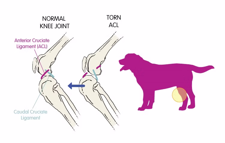
The stifle (knee) joint has two cruciate ligaments called
Cranial cruciate ligament (CrCL-”ACL”) and the caudal cruciate ligament
The 2 cruciate ligament do what
Connect the femur to the tibia
The CrCL runs where?
From the femur to the tibia in a caudal to cranial direction
Function of the CrCL
Stabilize the stifle joint and prevent cranial sliding of the tibia against the femur
Picture of CCLR (Cranial Cruciate Ligament Rupture)

What’s pink?
Scapula

What’s upper left black?
Humerus

What’s grey?
Radius

What’s upper light green?
Ulna

What’s red?
Carpals

What’s yellow?
Metacarpals

What’s lower purple?
Phalanges

What’s lower light green?
Thoracic limbs
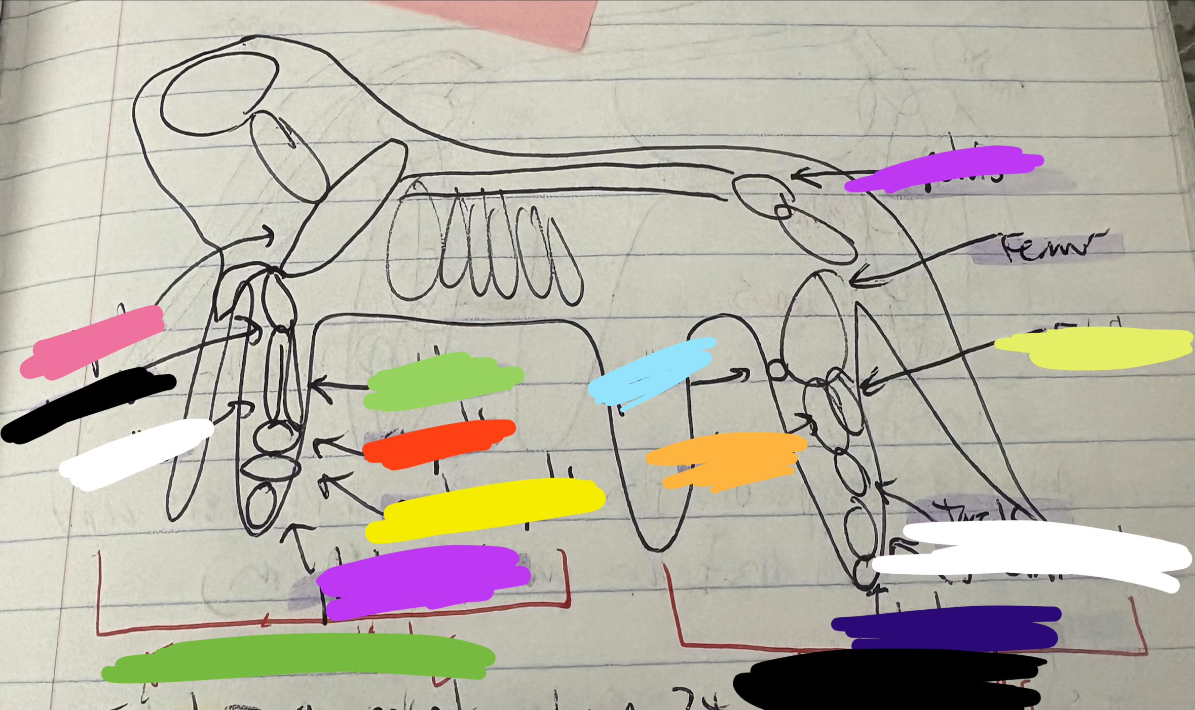
What’s light blue?
Patella

What’s orange?
Tibia

What’s upper purple?
Pelvis

What’s lime?
Fibula

What’s above lime?
Femur

What’s under the white?
Tarsals and metatarsals

What’s navy?
Phalanges

What’s lower black?
Pelvic limbs

What’s purple?
Shoulder

What’s black?
Elbow
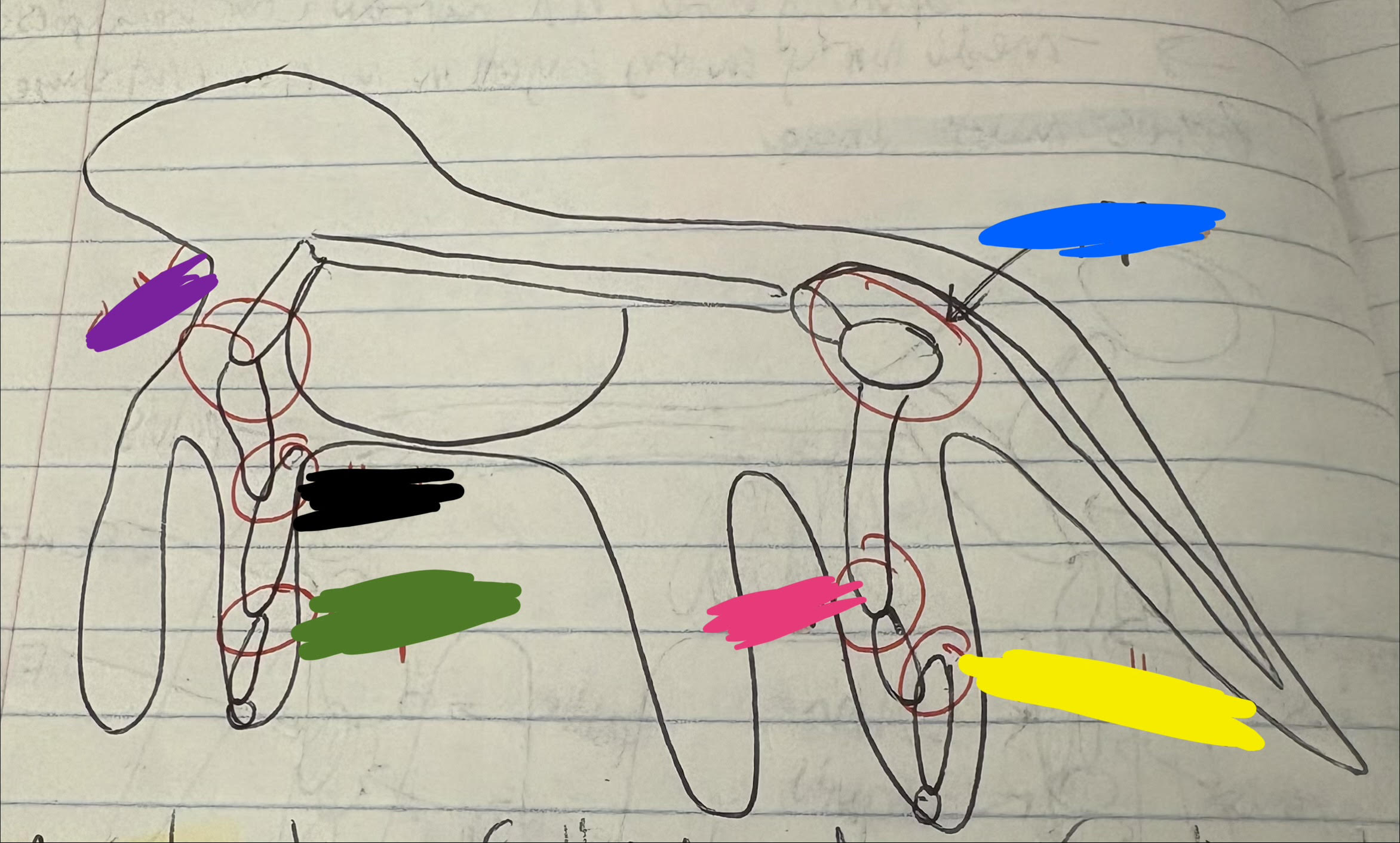
What’s green?
Carpus

What’s pink?
Stifle (knee)

What’s yellow?
Tarsus

What’s blue?
Hip
5 core imaging techs
Radiography, ultrasounds, nuclear medicine, CT scans, MRI, PET
CT is for _____ and is more ______.
internal, expensive
A ______ _____ shows the whole body in layers while an ___-____ shows the body in a few or one image.
CT scan, x-ray
Types of imaging (3)
Structural, functional, molecular
Structural imaging
Provides images depicting morphology
Functional imaging
Makes images depicting blood flow, neuronal activity, and glomerular filtration rate
Molecular imaging
Depicts activity of specific molecules (e. proteins), especially subcellular processes (e. apoptosis)
X-ray bean orientation can be _____ or ______.
perpendicular or parallel (horizontal)
Vertical aquisition
X-ray over person/small animal
Horizontal aquisition
X-ray to the side of a person/large animal
_____ amases different distributions in both vertical and horizontal aquisitions.
Gravity
Magnification
Refers to enlargement of image, but needs to be close to detector or will be fuzzy, and image doesn’t represnet true size
Distortion
When image misrepresents true shape/position of object, due to unequal magnification of different parts of the same objectThis can occur from various factors such as angle of imaging and distance from the detector, leading to inaccuracies in representation
______ ______ ______ is crucial for preventing magnification and distortion.
Standardized patient positioning (but position might be too painful)
Temporal requirements
Thoracic radiographs, or peak inhilation
Projection
How the x-ray beams from the machine when shot through the body to create image —→ described by direction from point of entrance to point of exit
DV
Dorsoventral thoracic radiographs —> 1st entered the dorsal aspect of the thorax and exited the ventral aspect
On VD or DV radiographs
"L" or "R" marker indicates the left or right side of the patient.
On lateral radiographs
"L" or "R" marker indicates whether the patient was lying left-side down or right-side down.
Radiographs of the limbs
"L" and "R" markers indicate that the left or right limb was imaged
Lateral radiographs (right or left, RLAT or LLAT) of any body part should be viewed with the cranial (or rostral, depending on where the image is being taken) aspect of the animal to the viewer's _____.
left
Ventrodorsal (VD) or dorsoventral (DV) radiographs of the head, neck or trunk should be placed with the cranial (or rostral) part of the animal pointing up. The ____ side of the animal should be on the viewer's ____, as would be the case if you were standing face-to-face with another person.
left, right
Transverse cross-sectional scans of the head, neck, or back should be placed with the ______ part of the animal pointing up, and with the left side of the animal to the viewer's ______. This is like looking at the animal while standing in front of it.
dorsal, right
Radiographs of the limb should be viewed with the proximal end of the extremity at the top, and the ______ (or dorsal) side to the ____. There is no convention regarding whether the medial or lateral side of the extremity is placed at the viewer's right or left.
cranial, left
X-rays are flipped so two x-rays on each side look the same orientation, _____ vs. _____.
left, right
3 types of info received in an image
Temporal resolution
Spatial resolution
Contrast resolution
Temporal resolution def
Precision of an observation or measurement with respect to time (e. the heart pumping and getting bigger vs. smaller)
Moving structures are harder to _______.
measure (e. the backwards wheel)
Spatial resolution def
Our ability to distinguish two points close together as separate (to see fine anatomic detail); on PACS the zoom function helps you make use of all spatial resolution originally required
Contrast resolution
Ability to distinguish between different tissues based on differences in energy intensity in image (high-diff colors vs. low-similar colors contrast)
Ideal resolution
Perfect temporal, spatial, and contrast resolution (situation specific); NEEDED for surgery to obtain precise images.
Multimodel imaging
To provide for information than individual modalities can give
Standard conventions (“hanging protocols")
To ensure consistent image interpretation and comparison across different imaging studies. These protocols dictate how images are arranged and displayed.
Most radiographs ______ displayed in their entirety at full spatial resolution since they would not fit on the monitor.
aren’t