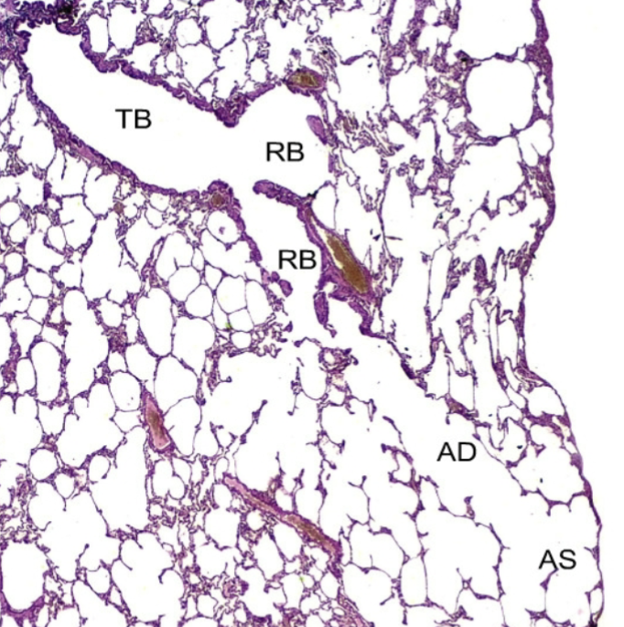
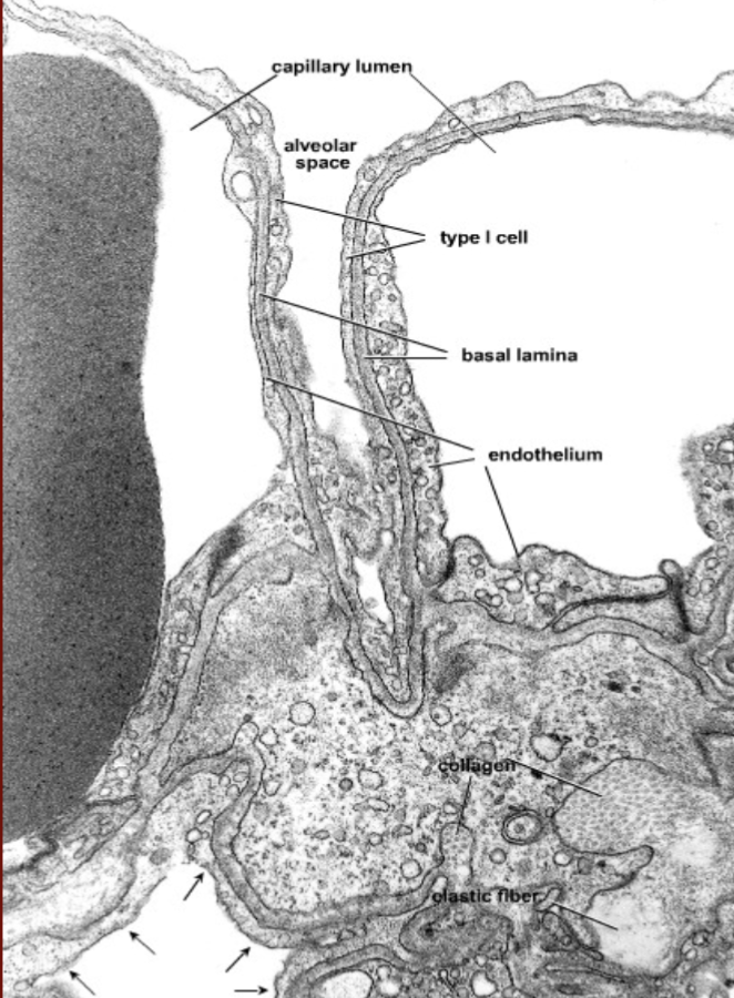
Histology Exam 2 - Images

0.0(0)
Studied by 0 people
call kaiCall Kai
Locked
learnLearn
examPractice Test
spaced repetitionSpaced Repetition
heart puzzleMatch
flashcardsFlashcards
GameKnowt Play
Card Sorting

1/92

encourage image

There's no tags or description

Looks like no tags are added yet.

Last updated 3:53 AM on 7/25/26
Name
Mastery
Learn
Test
Matching
Spaced
Call with Kai
Chat

No analytics yet

Send a link to your students to track their progress

93 Terms
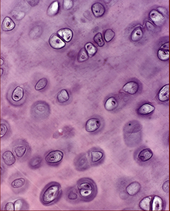
1
New cards
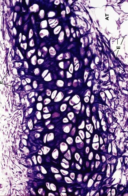
<p>What is this?</p>

What is this?

hyaline Cartilage

2
New cards
<p>What is this?</p>

What is this?

elastic cartilage

3
New cards
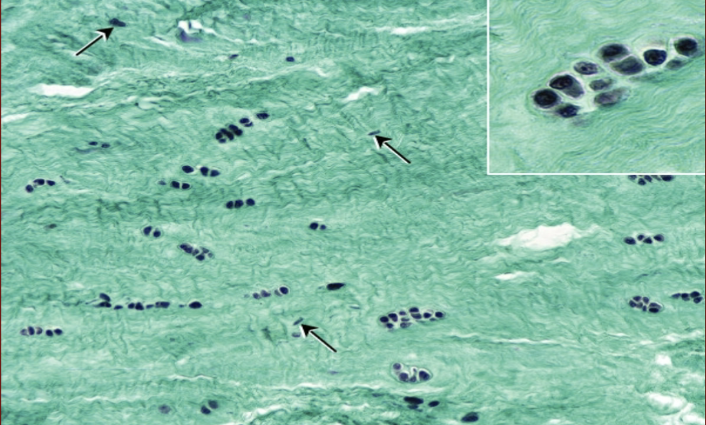
<p></p><p>What is this?</p>

What is this?

Fibrocartilage

4
New cards
<p>What is this?</p>

What is this?

bone

5
New cards
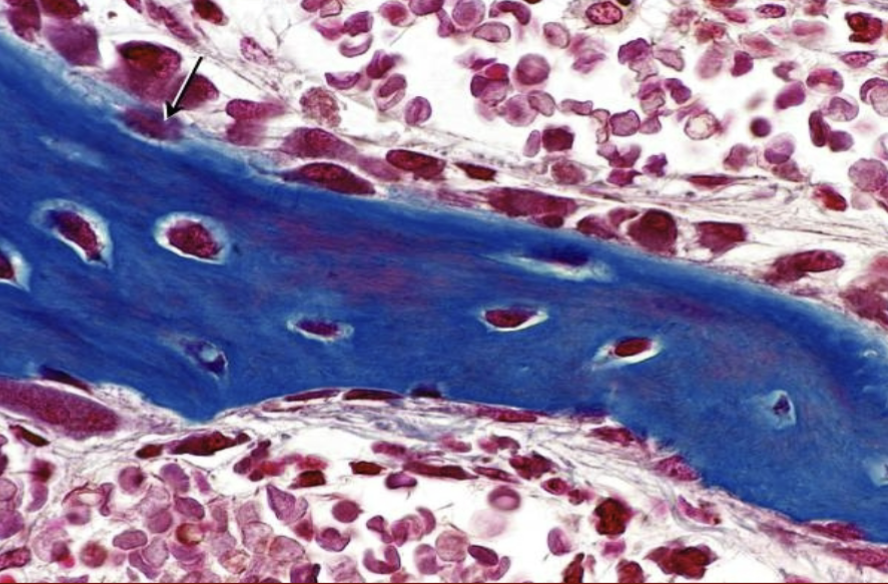
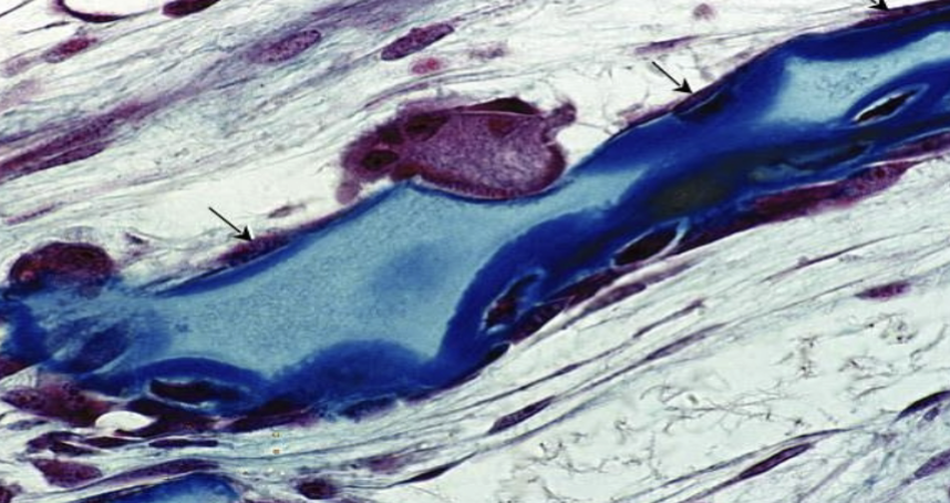
<p>What is this?</p>

What is this?

osteoblast

6
New cards
<p>What is this?</p>

What is this?

Osteoclasts

7
New cards
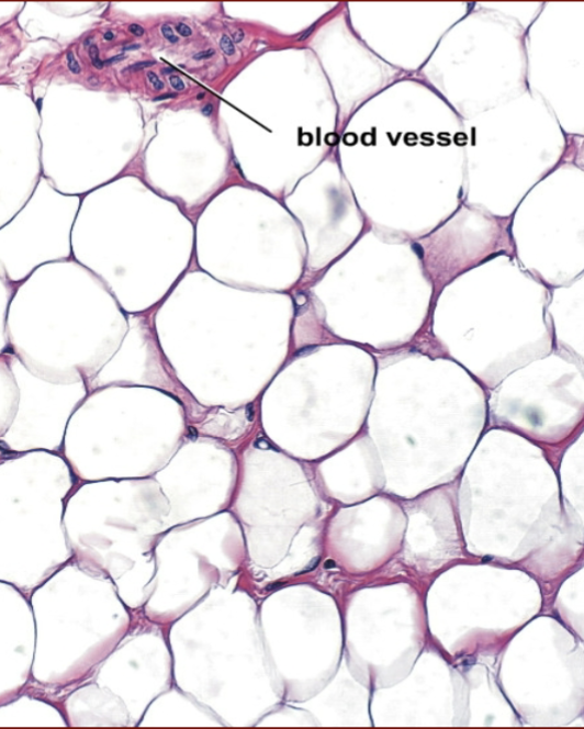
<p>What is this?</p>

What is this?

adipose tissue

8
New cards
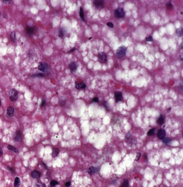
<p>What is this?</p>

What is this?

Brown adipose tissue

9
New cards
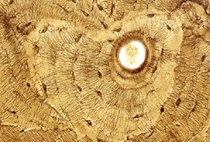
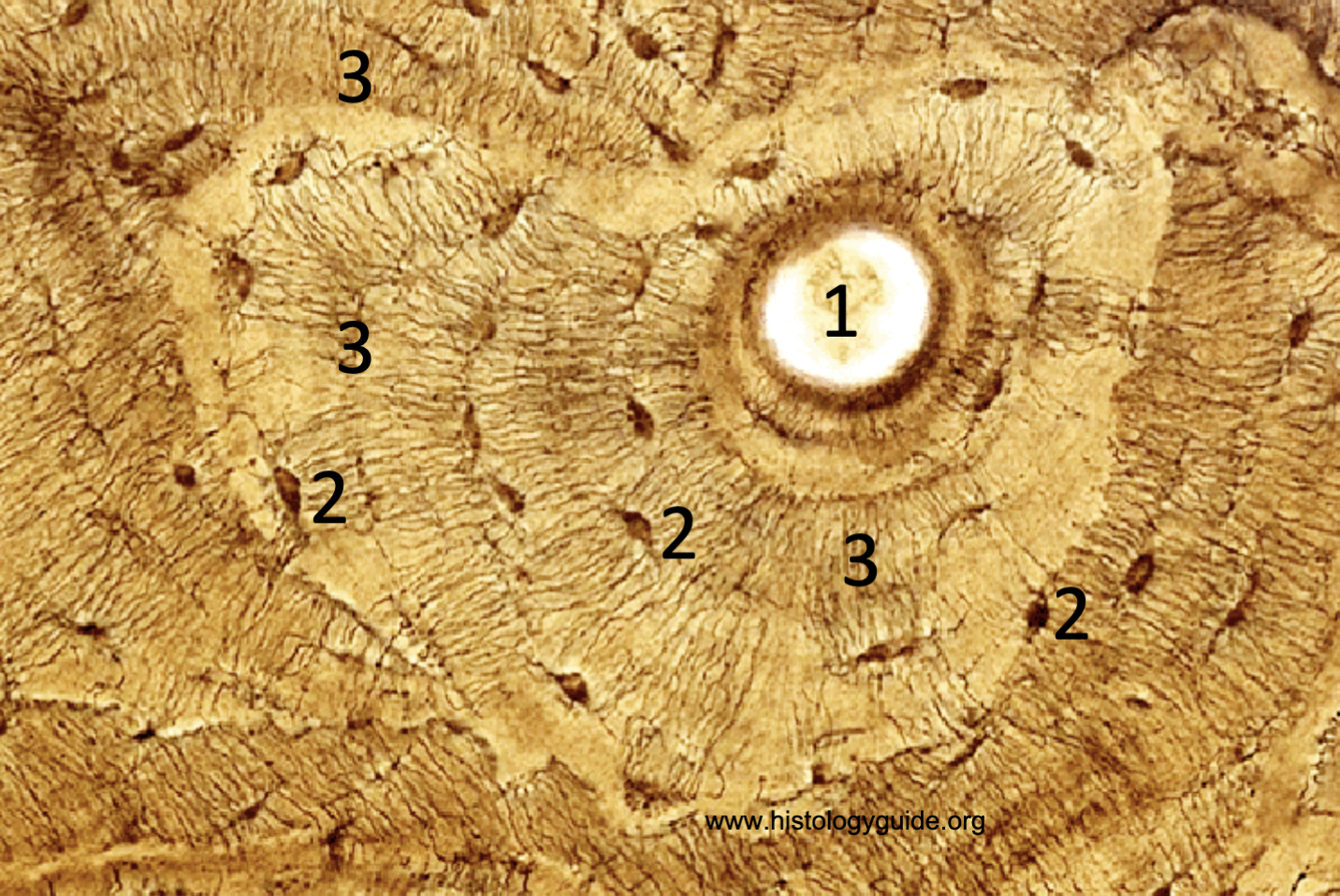
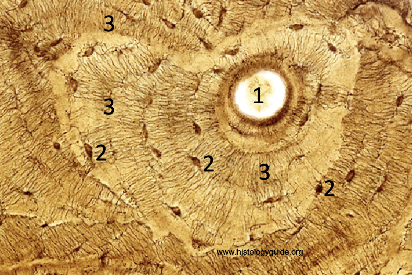
<p>What is 1?</p>

What is 1?

Ostenonal canal

10
New cards
<p>What is 2?</p>

What is 2?

Lacunae

11
New cards

What is 3?

Canaliculi

<p>Canaliculi</p>
12
New cards
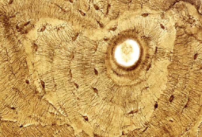
<p>What are the rings in this histology slide of a bone?</p>

What are the rings in this histology slide of a bone?

Lacunae
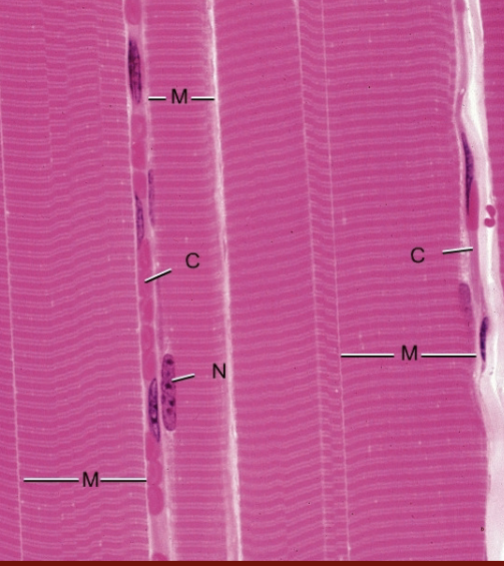
13
New cards
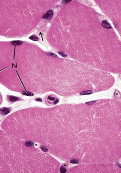
<p>What is this?</p>

What is this?

Skeletal Muscle

14
New cards
<p>What is this?</p>

What is this?

Skeletal Muscle

15
New cards
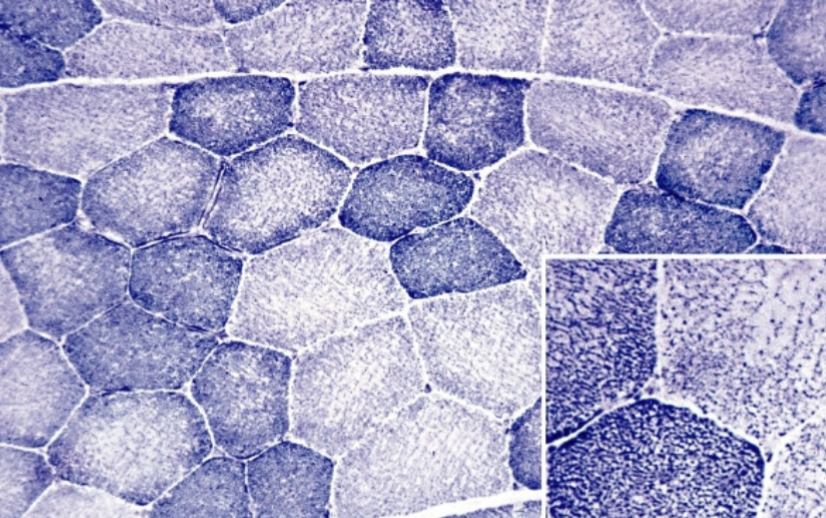
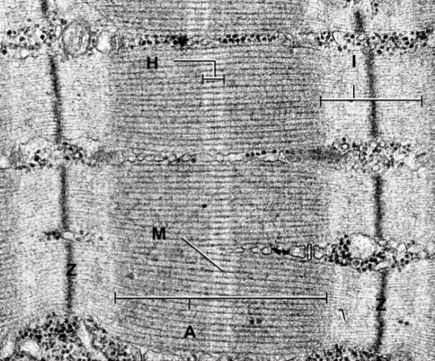
<p>What are the two types of fiber seen in this image?</p>

What are the two types of fiber seen in this image?

Type 1 and type 2
16
New cards
<p>What is this?</p>

What is this?

myofibrils

17
New cards
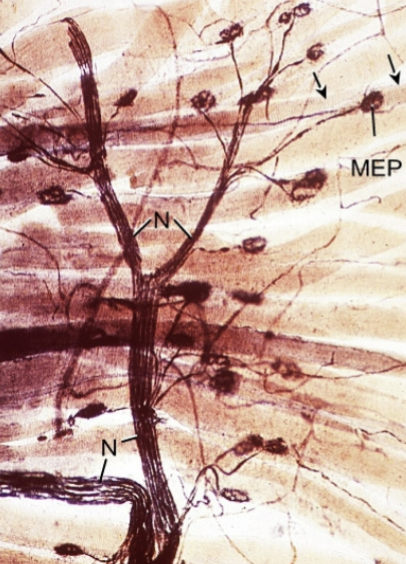
<p>What is this?</p>

What is this?

neuromuscular junction

18
New cards
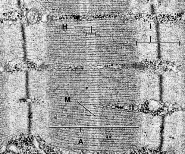
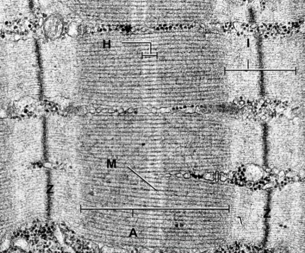
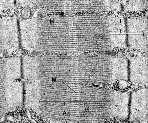
<p>What is the darkened line in the most medial part of this image?</p>

What is the darkened line in the most medial part of this image?

M line
19
New cards
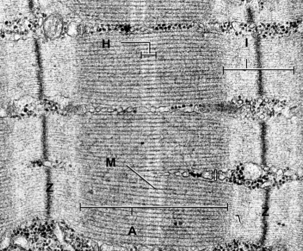
<p>What are the two dark bands on the left and right of the image?</p>

What are the two dark bands on the left and right of the image?

Z line
20
New cards
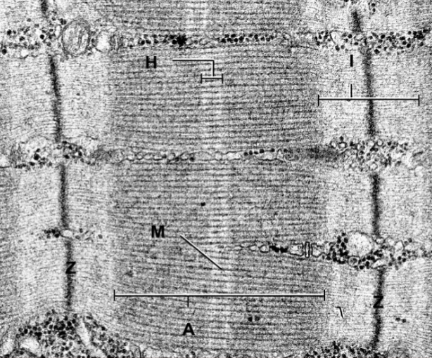
<p>What is the thick dark band on either side of the M-band?</p>

What is the thick dark band on either side of the M-band?

A band
21
New cards
<p>What band is thick and on either side of the z bands?</p>

What band is thick and on either side of the z bands?

I band
22
New cards
<p>What is the light band in the middle of the image that houses the M line?</p>

What is the light band in the middle of the image that houses the M line?

H band
23
New cards
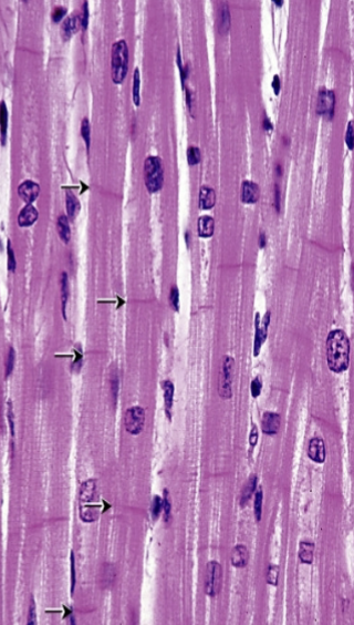
<p>What type of muscle is this?</p>

What type of muscle is this?

Cardiac muscle
24
New cards
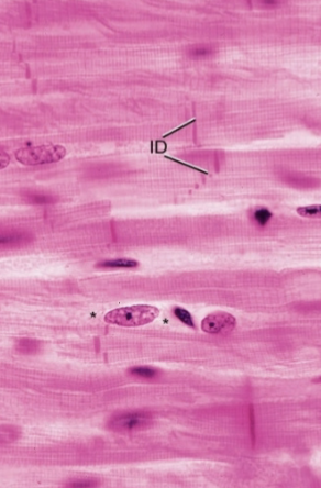
<p>What type of muscle is this?</p>

What type of muscle is this?

Cardiac muscle
25
New cards
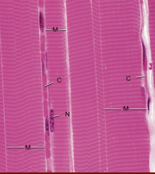
<p>What type of muscle is this?</p>

What type of muscle is this?

skeletal muscle
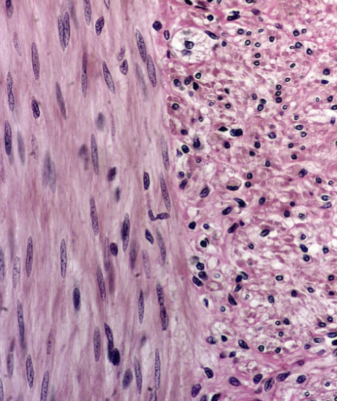
26
New cards
<p>What type of muscle is this?</p>

What type of muscle is this?

smooth muscle
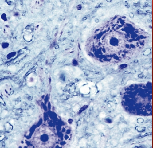
27
New cards
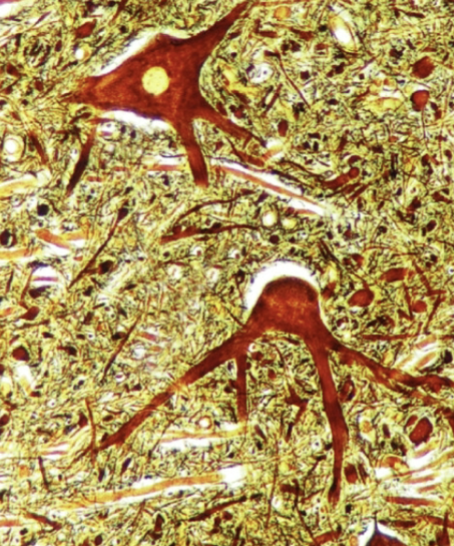
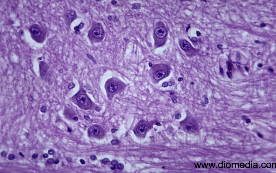
<p>What is this?</p>

What is this?

nerve cell bodies

28
New cards
<p>What are those red things?</p>

What are those red things?

nerve cell bodies
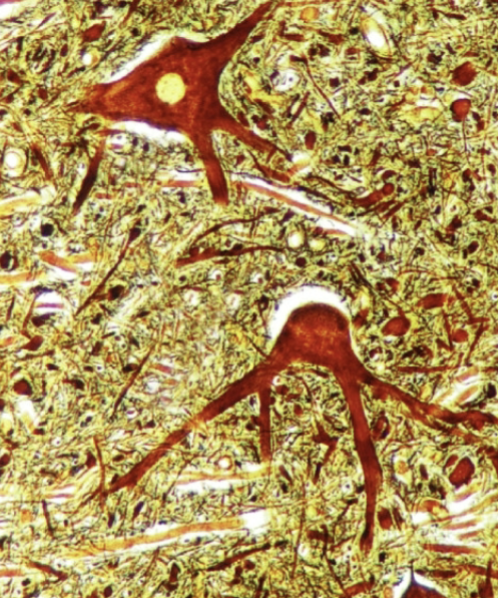
29
New cards
<p>What are those branches?</p>

What are those branches?

axonal hillock
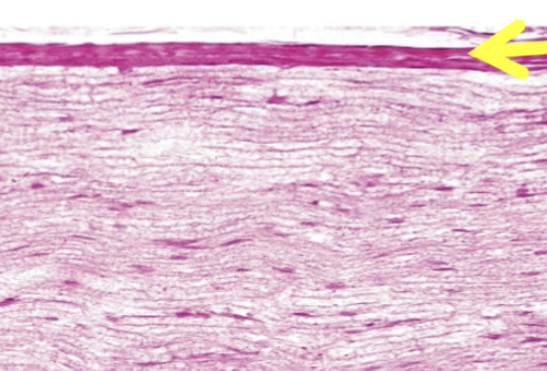
30
New cards
<p>What is this?</p>

What is this?

fascicles

31
New cards
<p>What is this</p>

What is this

fascicles?

32
New cards
<p>What is this (as a whole)?</p>

What is this (as a whole)?

fascicles

33
New cards
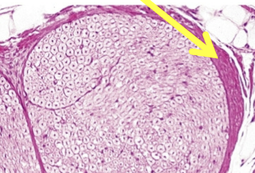
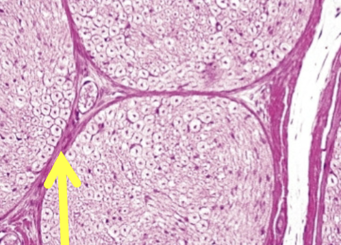
<p>What is the yellow arrow pointing to?</p>

What is the yellow arrow pointing to?

epineurium
34
New cards
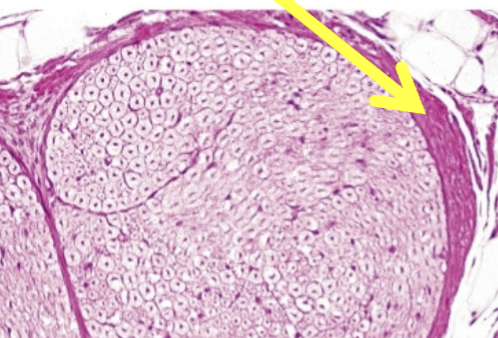
<p>What is the yellow arrow pointing to?</p>

What is the yellow arrow pointing to?

Perineurium
35
New cards
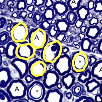
<p>What is the yellow arrow pointing to?</p>

What is the yellow arrow pointing to?

Epineurium
36
New cards
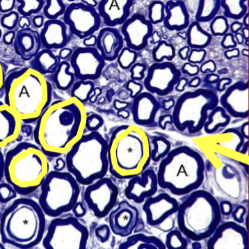
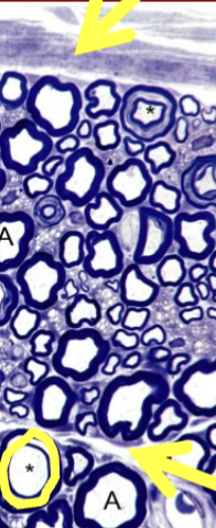
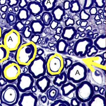
<p>What are the yellow circles pointing out?</p>

What are the yellow circles pointing out?

Endoneurium
37
New cards
<p>What is the arrow at the top pointing to?</p>

What is the arrow at the top pointing to?

Epineurium
38
New cards
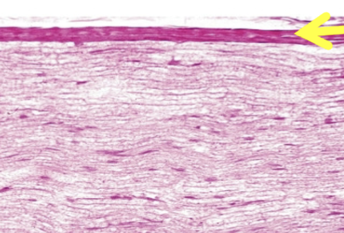
<p>What is the yellow arrow pointing to?</p>

What is the yellow arrow pointing to?

Perineurium
39
New cards
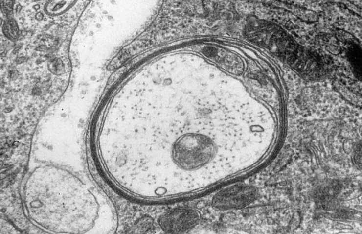
<p>What is this fiber covered by?</p>

What is this fiber covered by?

myelination
40
New cards
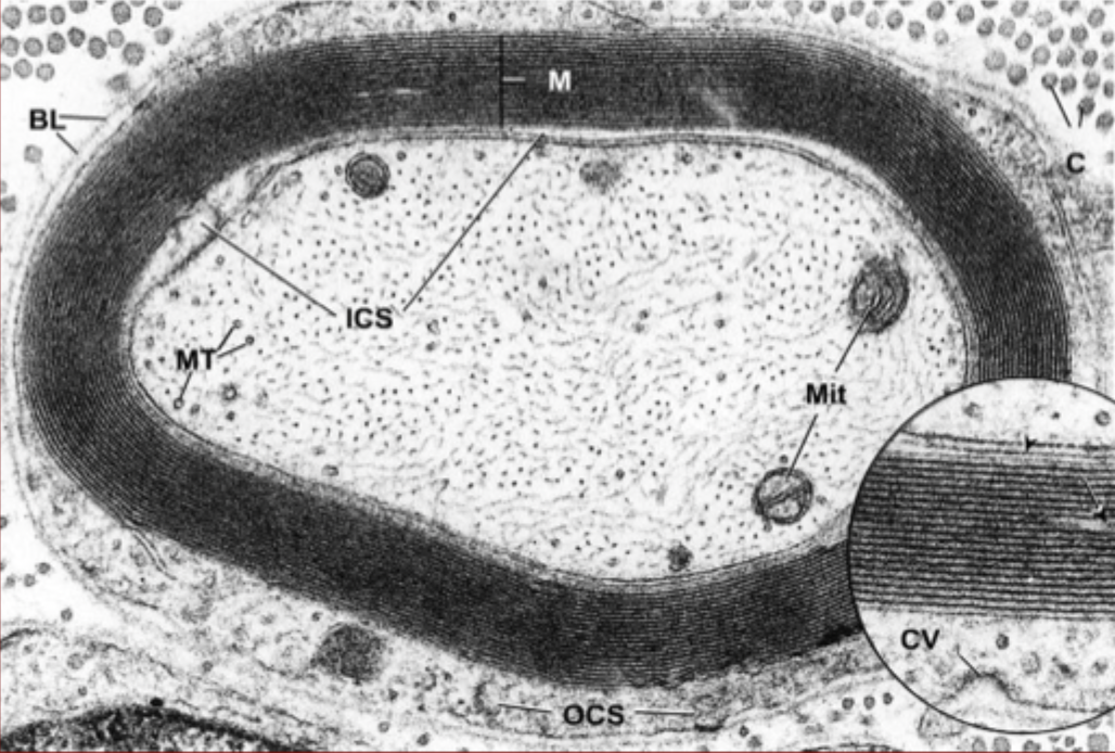
<p>What is that dark layered rings around this fiber?</p>

What is that dark layered rings around this fiber?

Myelination
41
New cards
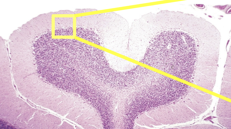
<p>What is this an image of?</p>

What is this an image of?

cerebellum

42
New cards
<p>What is this an image of?</p>

What is this an image of?

Brain stem/medulla oblongata
43
New cards
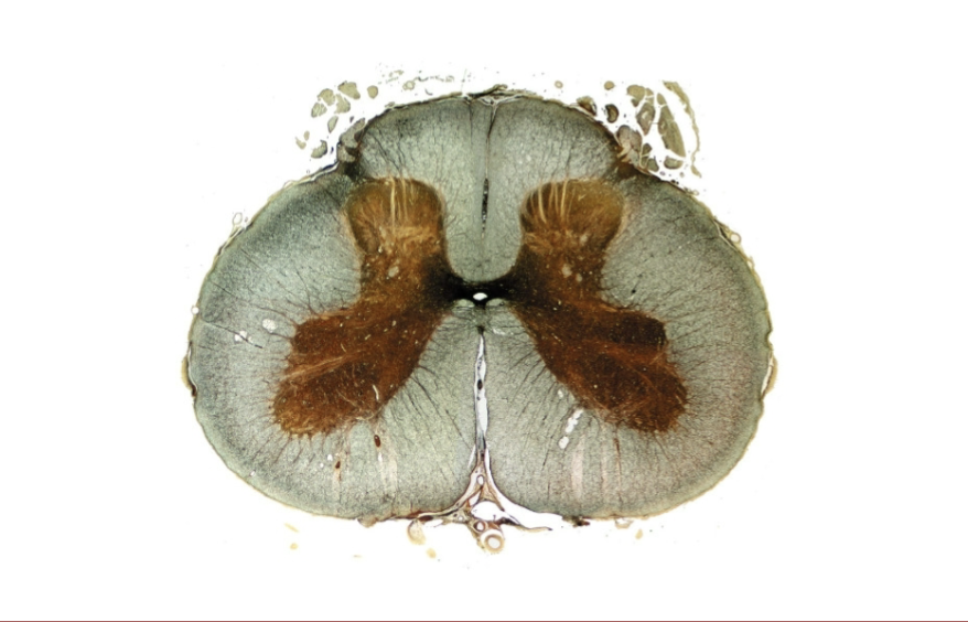
<p>What is this?</p>

What is this?

spinal cord

44
New cards
<p>What is this an image of?</p>

What is this an image of?

unmyelinated fibers
45
New cards
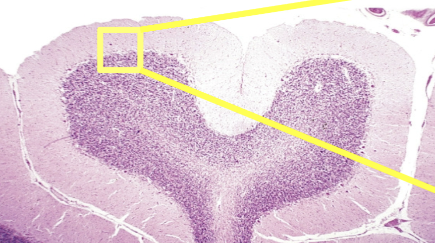
<p>What is the cortex of the cerebellum composed of?</p>

What is the cortex of the cerebellum composed of?

gray matter
46
New cards
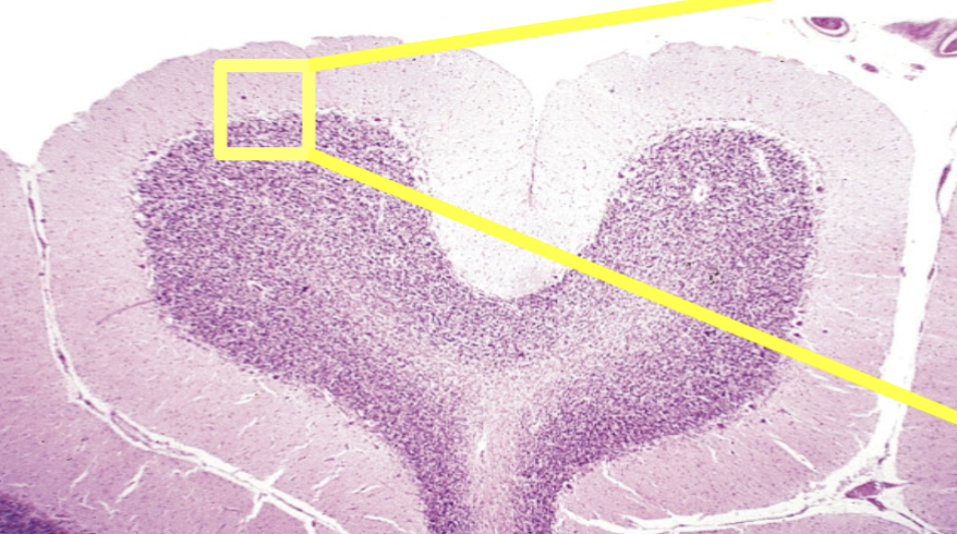
<p>What is the medulla of the cerebellum composed of?</p>

What is the medulla of the cerebellum composed of?

white matter
47
New cards
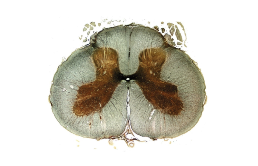
<p>What is the cortex of the spinal cord composed of?</p>

What is the cortex of the spinal cord composed of?

white matter
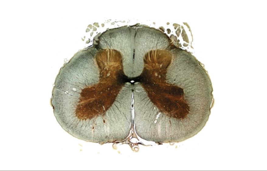
48
New cards
<p>What is the medulla of the spinal cord composed of?</p>

What is the medulla of the spinal cord composed of?

gray matter
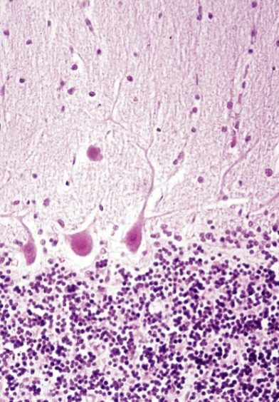
49
New cards
<p>What are the fibers seen in the cerebellum?</p>

What are the fibers seen in the cerebellum?

purkinje fibers
50
New cards
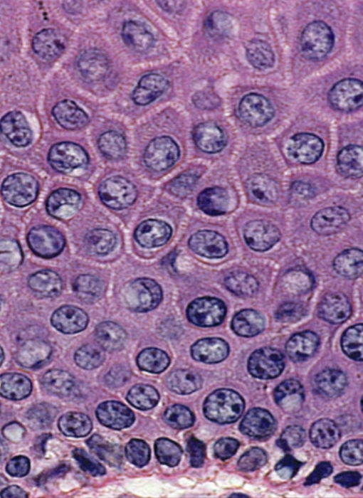
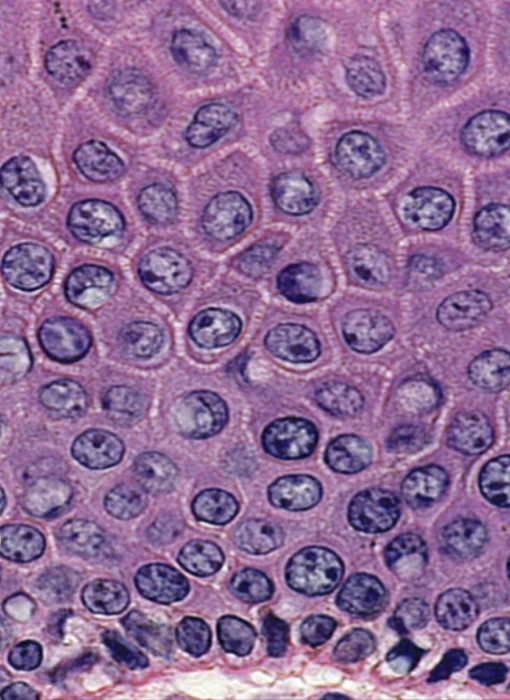
<p>What layer of stratum is at the very top of this image? </p>

What layer of stratum is at the very top of this image?

S. granulosum
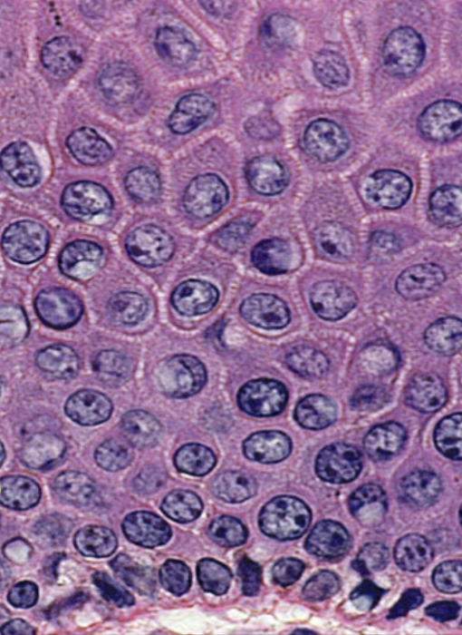
51
New cards
<p>What is the middle section of this image of stratum?</p>

What is the middle section of this image of stratum?

S. spinosum
52
New cards
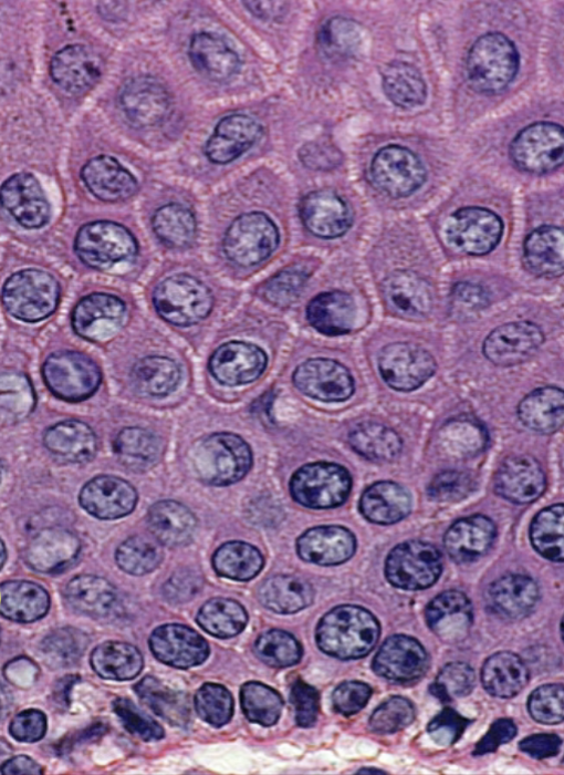
<p>What is the bottom later of cells in this image of stratum?</p>

What is the bottom later of cells in this image of stratum?

s. basale
53
New cards
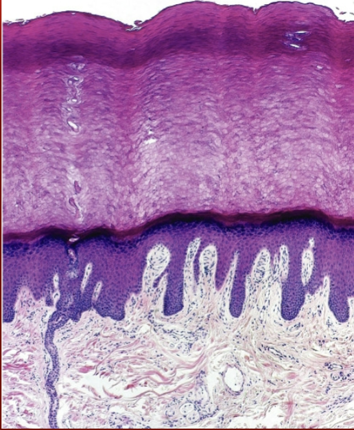
<p>What is this?</p>

What is this?

stratum of the epidermis
54
New cards
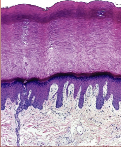
<p>Whats that light pink later called?</p>

Whats that light pink later called?

dermis
55
New cards
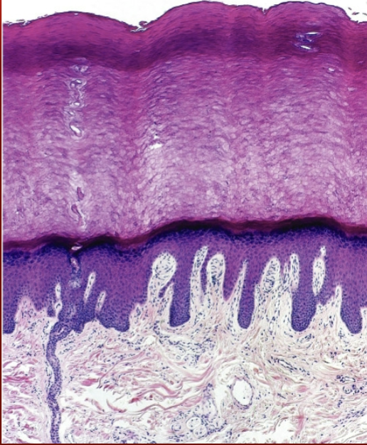
<p>What are those downard projections in the dermis?</p>

What are those downard projections in the dermis?

Dermal papillae
56
New cards
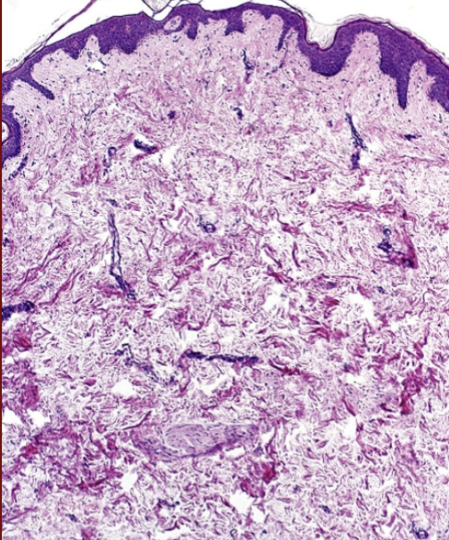
<p>What are the ridges on the dermal papillae (downward projections)?</p>

What are the ridges on the dermal papillae (downward projections)?

Epidermal (rete) ridges
57
New cards
<p>What is the lighter colored stuff of the dermis?</p>

What is the lighter colored stuff of the dermis?

papillary and reticular layers
58
New cards
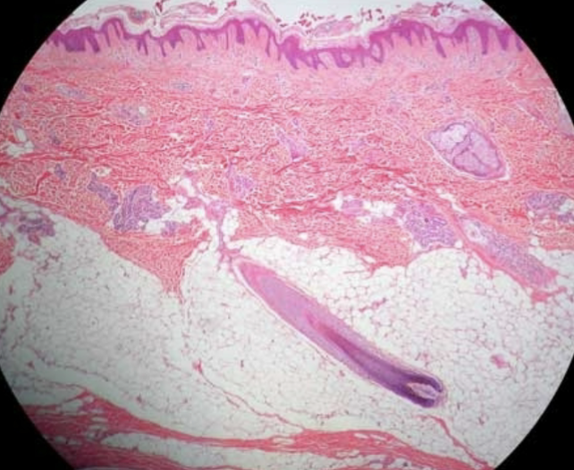
<p>What is that tadpole looking thing neat the bottom left of the image?</p>

What is that tadpole looking thing neat the bottom left of the image?

Hair follicle
59
New cards
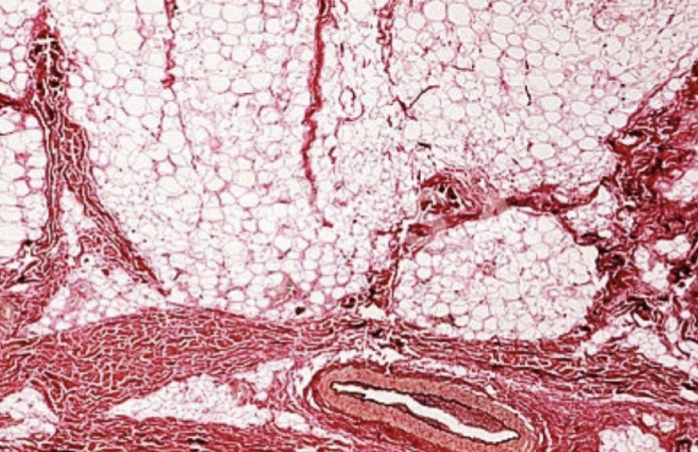
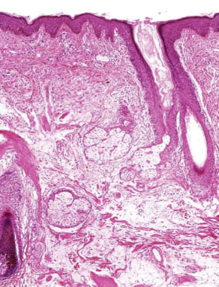
<p>What layer of the skin is this?</p>

What layer of the skin is this?

Subcutis/hypodermis
60
New cards
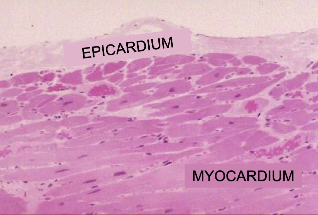
<p>What is the bottom layer of this image?</p>

What is the bottom layer of this image?

Subcutis/hypodermis
61
New cards
<p>What is this?</p>

What is this?

heart

62
New cards
<p>What is this?</p>

What is this?

arteries

63
New cards
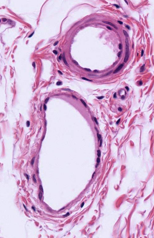
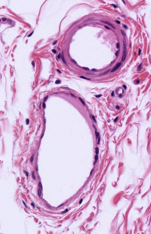
<p>What is the smaller hole with the thick layers around it?</p>

What is the smaller hole with the thick layers around it?

arteries
64
New cards
<p>What is the big hole?</p>

What is the big hole?

vein

65
New cards
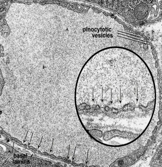
<p>What is this an image of?</p>

What is this an image of?

capillaries
66
New cards
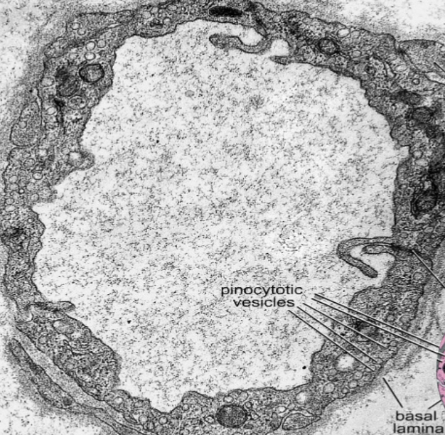
<p>What type of capillary is this?</p>

What type of capillary is this?

Fenestrated
67
New cards
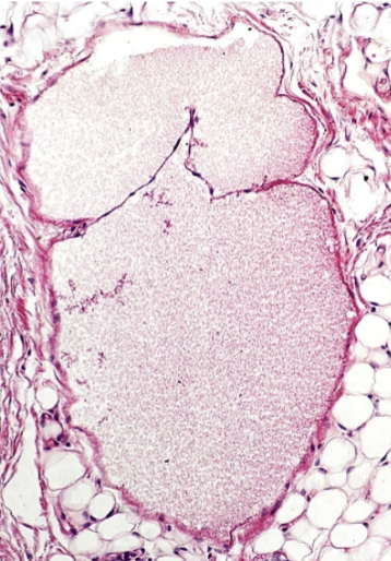
<p>What type of capilary is this?</p>

What type of capilary is this?

continuous
68
New cards
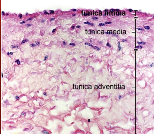
<p>What is this an image of?</p>

What is this an image of?

vein valves
69
New cards
<p>What is this?</p>

What is this?

vein

70
New cards
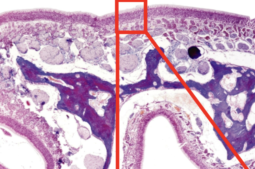
<p>What is this?</p>

What is this?

Nostrils

71
New cards
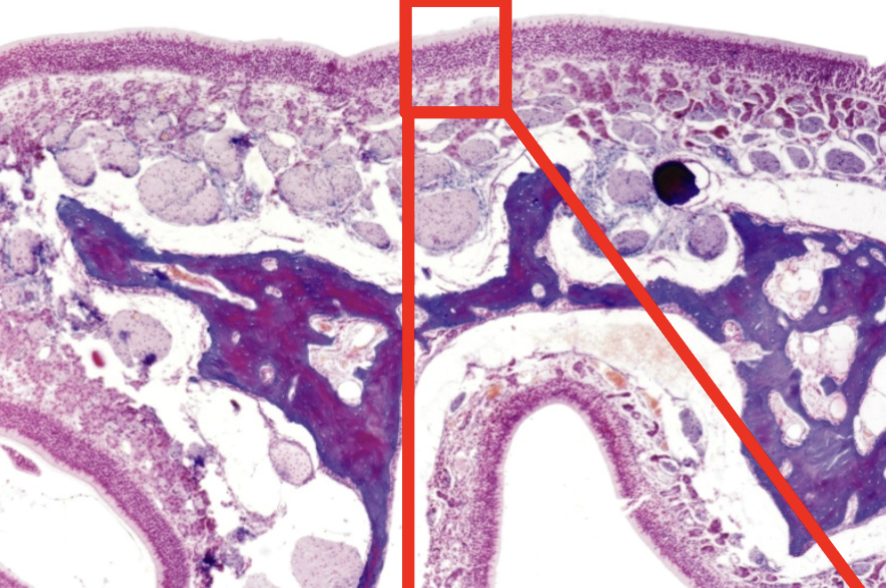
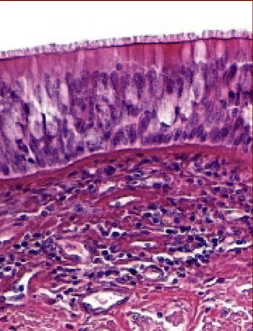
<p>What is the space in the lower area supposed to be?</p>

What is the space in the lower area supposed to be?

nasal cavity
72
New cards
<p>What is this?</p>

What is this?

nostrils

73
New cards
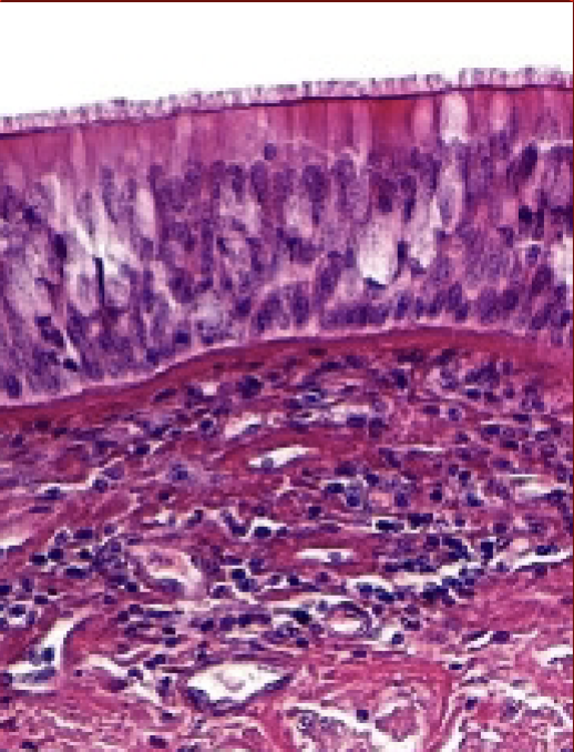
<p>What are those areas of no dye in the top layer of the ciliated columnar epithelium?</p>

What are those areas of no dye in the top layer of the ciliated columnar epithelium?

goblet cells
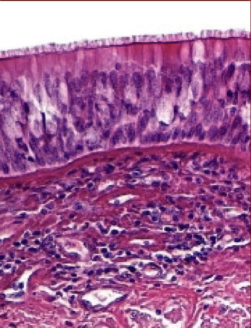
74
New cards
<p>What type of cells is the top layer of these nostrils?</p>

What type of cells is the top layer of these nostrils?

ciliated columnar epithelium?
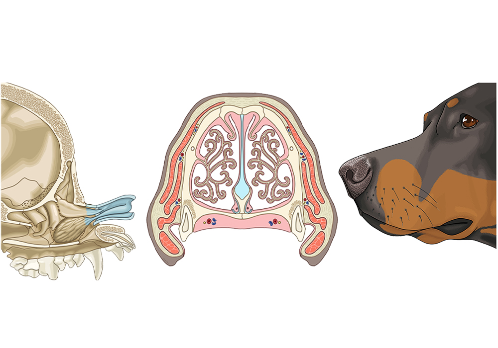
75
New cards
<p>What are those weird swirly things?</p>

What are those weird swirly things?

cartilaginous turbinates
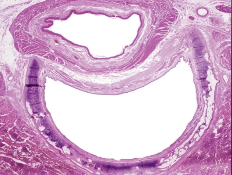
76
New cards
<p>What is that C-shaped hole?</p>

What is that C-shaped hole?

trachea

77
New cards
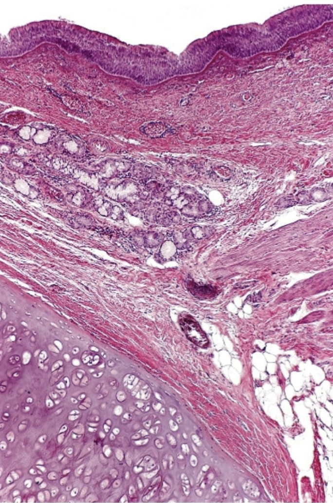
<p>What is this?</p>

What is this?

trachea

78
New cards
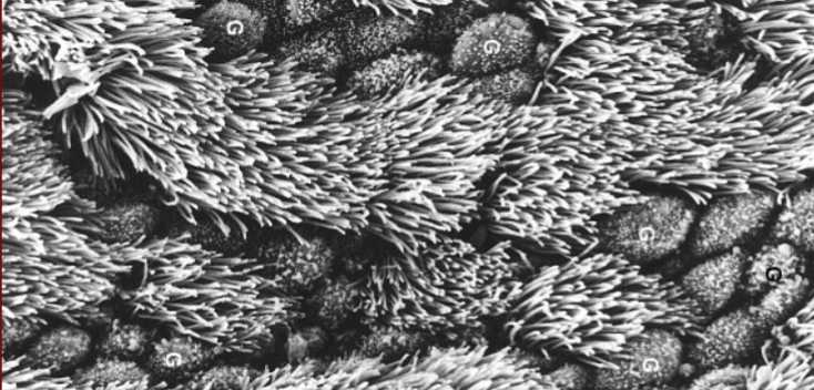
<p>What are these frilly things that can be found in the trachea?</p>

What are these frilly things that can be found in the trachea?

mucociliary elevator
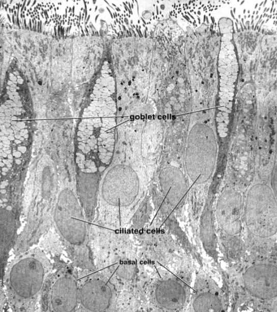
79
New cards
<p>What kind of cells are these that are in the trachea?</p>

What kind of cells are these that are in the trachea?

mucous cells
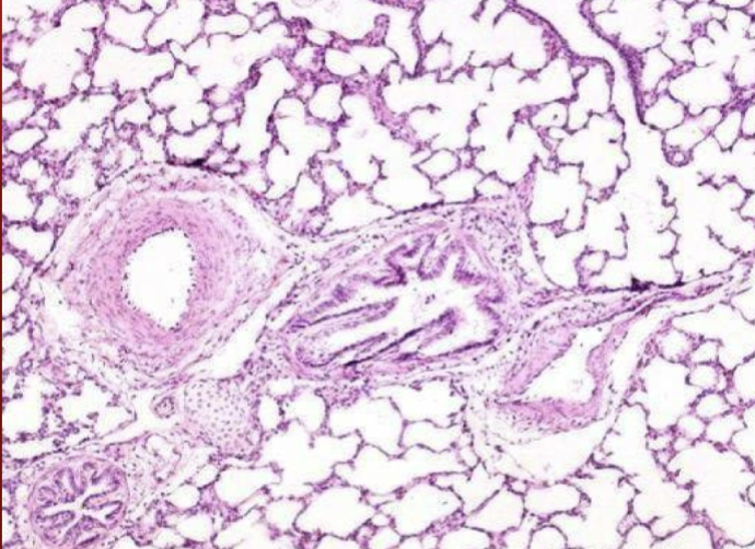
80
New cards
<p>What is the hole all the way to the left of this image?</p>

What is the hole all the way to the left of this image?

artery
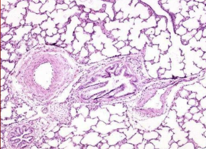
81
New cards
<p>What is the middle hole in this image?</p>

What is the middle hole in this image?

bronchioles
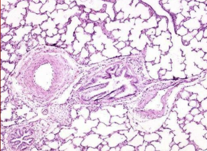
82
New cards
<p>What is the hole all the way to the right?</p>

What is the hole all the way to the right?

vein
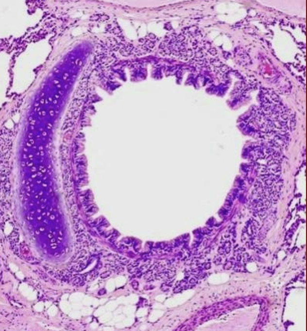
83
New cards
<p>What is this?</p>

What is this?

bronchi

84
New cards
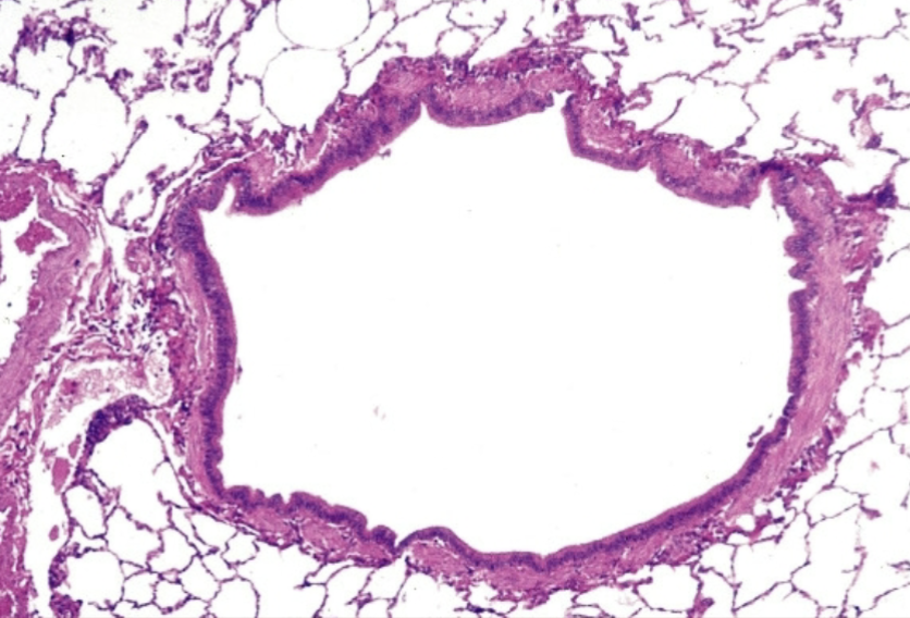
<p>What is this?</p>

What is this?

bronicoles

85
New cards
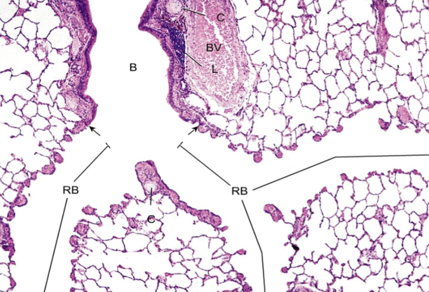
<p>What is this?</p>

What is this?

bronicoles

86
New cards
<p>What is this?</p>

What is this?

alveoli

87
New cards
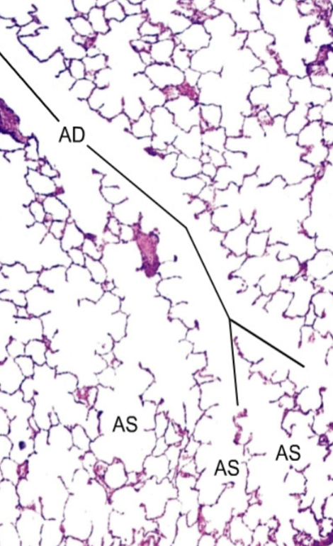
<p>What is this showing?</p>

What is this showing?

bronchioles to alveoli
88
New cards
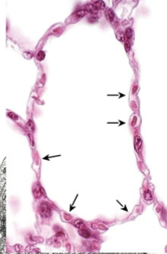
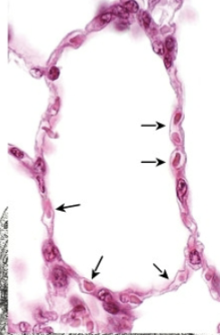
<p>What are those arrows pointing to?</p>

What are those arrows pointing to?

alveolar pores
89
New cards
<p>What is this?</p>

What is this?

alveolus

90
New cards
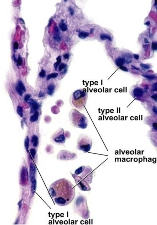
<p>What part of the distal airwats is this showing?</p>

What part of the distal airwats is this showing?

interstitium
91
New cards
<p>What part of the distal airways is this showing?</p>

What part of the distal airways is this showing?

interstitium
92
New cards
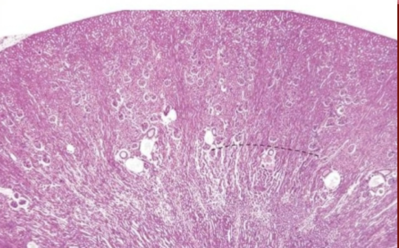
<p>What is this image of?</p>

What is this image of?

kidney

93
New cards

What is that line in the middle?