Nucleic acids hybridization techniques (EXAM 2)
1/24
There's no tags or description
Looks like no tags are added yet.
Name | Mastery | Learn | Test | Matching | Spaced | Call with Kai | Chat |
|---|
No analytics yet
Send a link to your students to track their progress
25 Terms
What is nucleic acid hybridization?
Process in which single stranded nucleic acid form different sources but complementary sequences are form double stranded molecules
What do we use nucleic acid hybridization for?
detect and semi-quantify microbes in a sample
Localize a microbe in a sample
Identify a gene in a sample
Detect gene mutations
Study gene expression
Probes
A fragment of DNA or RNA of variable length which is used to detect the presence of homologous nucleotide sequences (DNA/RNA target) in a sample
Genomic DNA probe
Fragment (by PCR or DNA cloning, whole genomic (long hundreds to thousands nt)
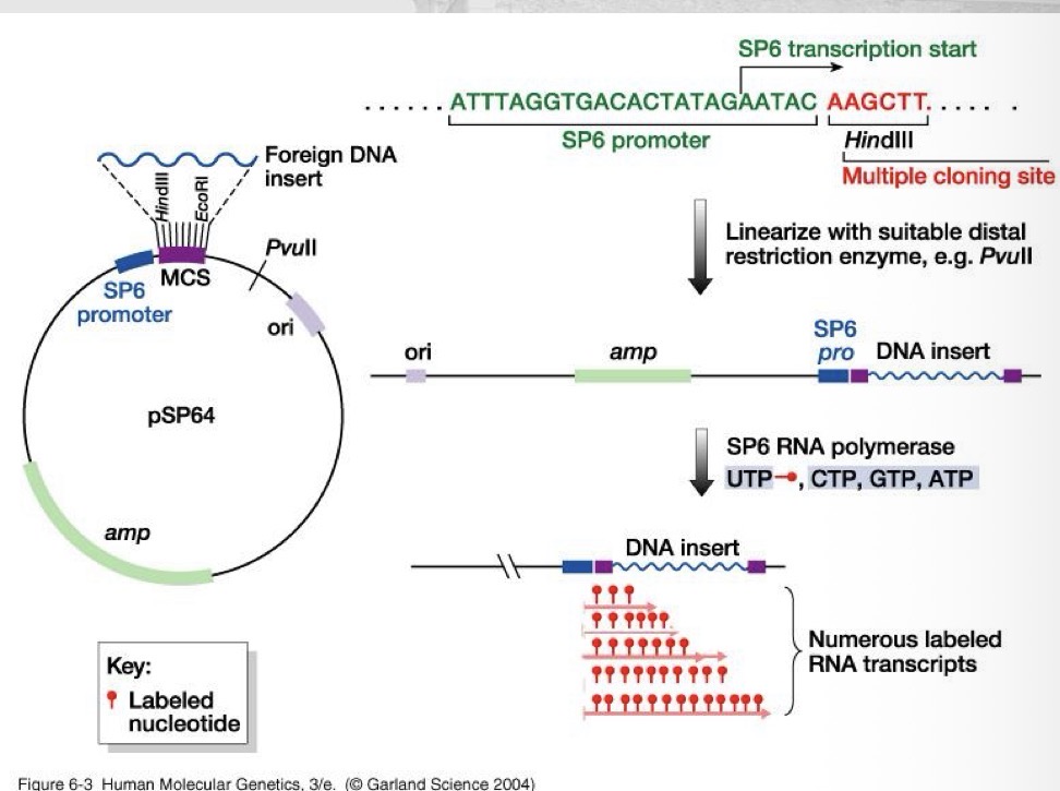
RNA probes (riboprobes)
By transcription of clones DNA (long hundreds to thousands nt)
Oligonucleotide probes
By chemical synthesis (short 15-50 nucleotides)
Method to labeling probes
By strand synthesis: DNA or RNA template; DNA or RNA polymerase and labeled nucleotide
By end labeling: adding a label to the 5’ end of the probe
Label types
Radioactive, non-radioactive (fluorophores, haptens)
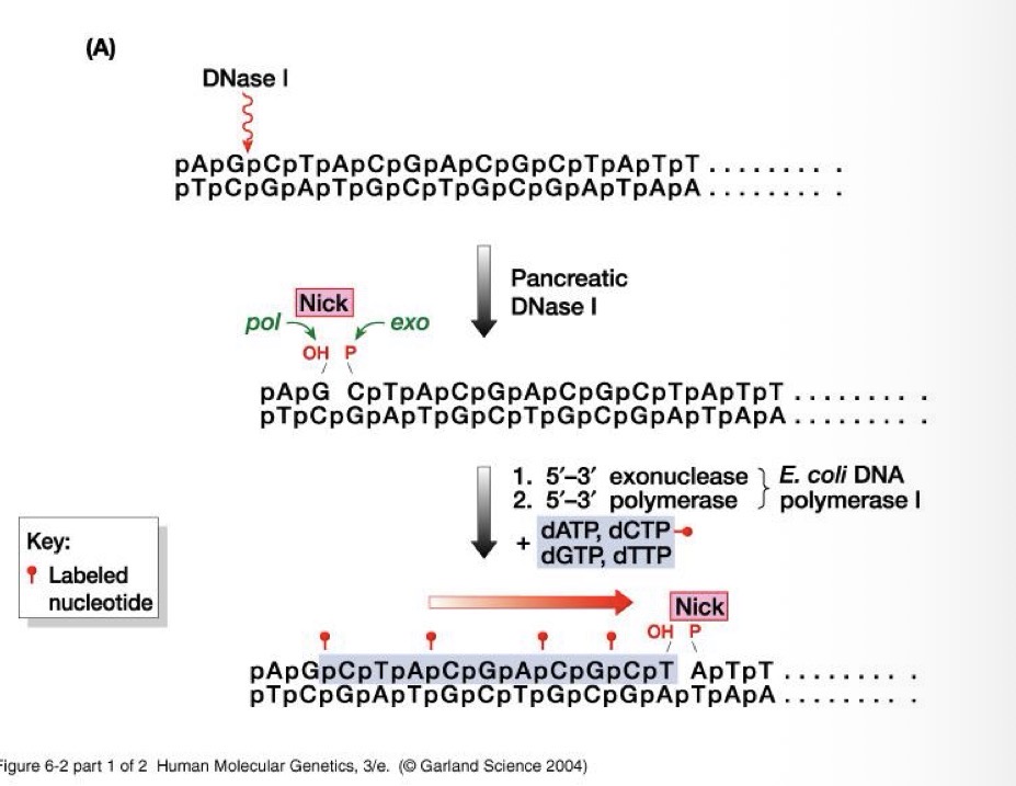
Nick translation - strand synthesis
DNA polymerase removes nucleotides ahead of a nick and replaces them with new nucleotides at the same time
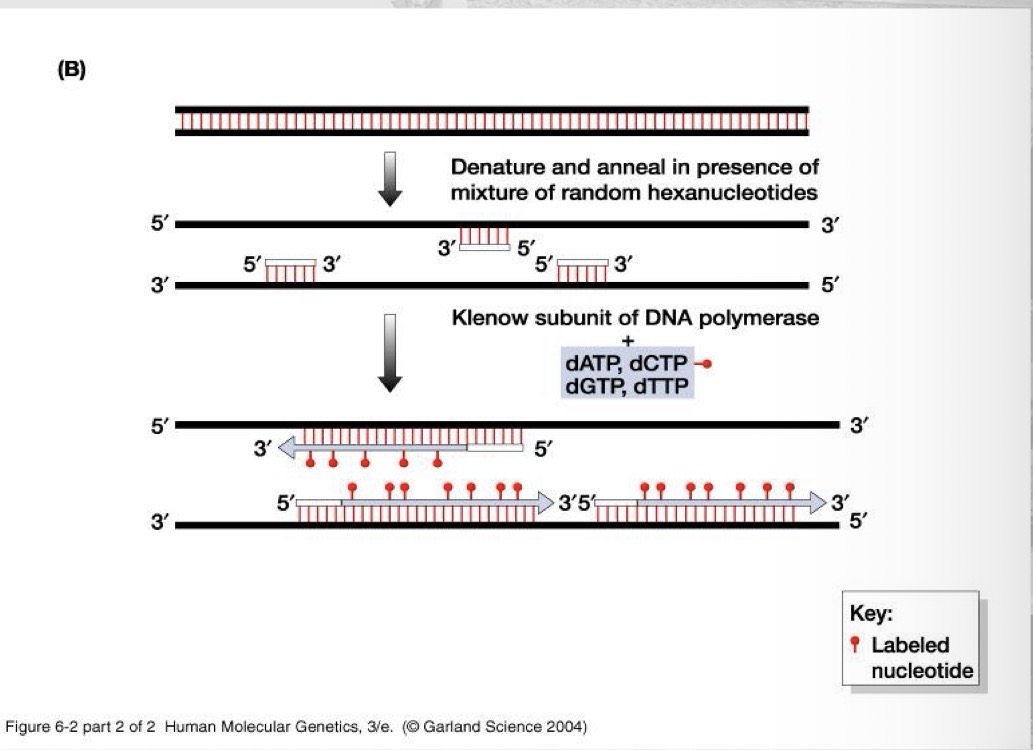
Random primers - strand synthesis
Anneal to multiple locations on a DNA (or RNA derived cDNA) template, providing free 3’-OH ends for DNA polymerase to begin strand synthesis
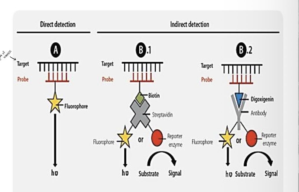
Labeling with fluorophores and haptens
Fluorophores - direct fluorescent label
Haptens - indirect label detected by antibodies
Specific techniques
South blots, northern blot, dot blot, check board DNA-DNA hybridization, in situ hybridization, DNA microarrays
Who discovered southern blotting and when?
Edward souther -1975
Southern blotting
Uses a DNA probe to detect a sequence in a DNA sample and identify the size of the fragment containing it
Southern blot steps
DNA is extracted from cells and cut into fragments by restriction enzymes
Fragments are separated by size using gel electrophoresis
DNA bands are transferred to a nitrocellulose filter by blotting
This produces a nitrocellulose filter w/ DNA fragments positioned exactly on gel
Filter is exposed to a radioactively labeled probe for specific gene
Filer is then exposed to X-ray film. The fragment contains the gene of interest is identified by a band on the developed film
Who founded northern blotting and when?
James Alwine, 1977
Northern blotting
Uses a DNA or RNA probe to assess presence and quantity of a mRNA in a sample (gene expression)
Dot blotting
Simpler form of Southern and Northern blots: detect and semi-quantitate a DNA or mRNA sequence in a sample without separation (electrophoresis)
Add antibody → detect protein
Add probe → detect DNA
Who invented check board DNA-DNA hybridization and when?
Socransky, 1994
Check board DNA-DNA hybridization
A high throughout version of dot blot used for analysis of dental samples: detection and semi-quantification of up to 40 bacterial species in up to 28 dental samples using whole genomic probes
In situ hybridization
Uses a DNA or RNA probe, usually fluorescent, to detect and localize a DNA or mRNA in a tissue or cells
DNA microarrays
A glass or silicon chip with thousands of unlabeled DNA probes spotted on it for hybridization with labeled nucleic acids from samples
DNA microarrays uses
Detection and quantification of microbes in a sample
Study/compare gene expression
Detect gene mutations (SNPs)
Detect gene copy variations (competitive genomic hybridization)
Microarrays principle
Two different cells are being compared
mRNA is extracted - the cell’s active gene produce mRNA (more mRNA = gene is being expressed more)
mRNA → cDNA - an enzyme called reverse transcriptase converts mRNA into cDNA then cDNA is tagged with a fluorescent label
Microarrays chip - each spot contains a DNA probe that matches a specific gene; the labeled cDNA binds to its matching probe through base pairing
Scanner reads fluorescence - bright spots mean a lot of cDNA bound (high gene expression); dim spots mean little cDNA bound (low gene expression)