A&P 1 Lab (Chapter 8-9)
1/33
Earn XP
Description and Tags
The Muscular system
Name | Mastery | Learn | Test | Matching | Spaced | Call with Kai |
|---|
No analytics yet
Send a link to your students to track their progress
34 Terms
What is the function of Muscle Tissue?
generate force and produce movement
What are skeletal muscles?
composed of organized connective tissue wrappings and contractile units with each layer supporting and transmitting force
electrically excitable cells
maintains a resting membrane potential (approx. -70 to -90 mV)
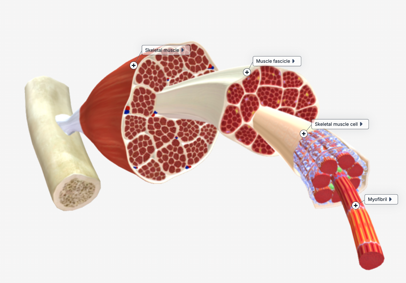
What are the layers of skeletal muscle (4) (in order - from superficial to deep)?
Muscle (the entire organ)
Fascicle
Muscle fiber
Myofibril
What is the first layer of the skeletal muscle? What is it surrounded by and attached to?
muscle
Surrounded by the epimysium
Attached to bone via tendons and supported by the broader sheets of fascia

What is epimysium?
dense connective tissue sheath
What is the second layer of the skeletal muscle? What is it?
fascicle
A bundle of muscle fibers wrapped by the perimysium

What is the third layer of the skeletal muscle? What is it? Function?
muscle fiber
surrounded by the endomysium (the innermost connective tissue layer)
also carries capillaries and nerves

What is the fourth layer of the skeletal muscle? What is it? Function?
myofibril
Cylinder-shaped, contractile organelles composed of bundled specialized proteins that allow the fiber to shorten and generate force

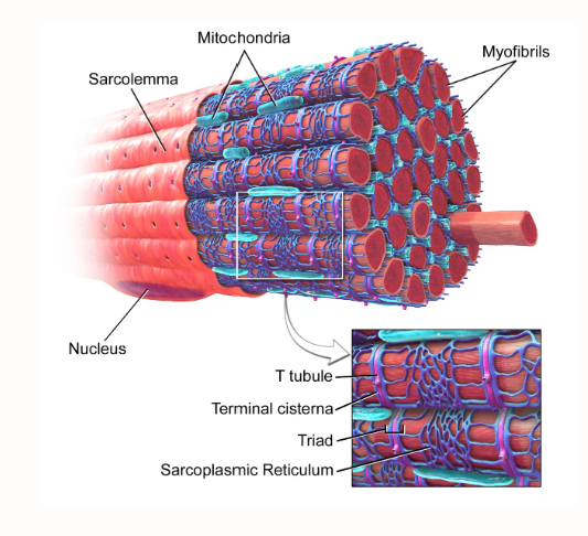
What are the 5 structures of the muscle cells?
myocyte
sarcoplasm
sarcolemma
sarcoplasmic reticulum
myofibrils
What is a myocyte?
The muscle cell itself
Large, multinucleated
Function: force production

What is sarcoplasm?
the cytoplasm of the myocyte
rich in glycogen, myoglobin, enzymes, and the contractile proteins needed for contraction

What is Sarcolemma?
the myocytes plasma membrane
function: electrically capable of transmitting action potentials across the entire fiber surface
What is a sarcoplasmic reticulum?
modified endoplasmic reticulum
Function: stores and releases calcium ions (Ca2+), which is the key trigger for muscle contraction

What are myofibrils?
Cylinder-shaped, contractile organelles composed of bundled specialized proteins that allow the fiber to shorten and generate force

What are T-Tubules? What do they do?
they are the deep invaginations in the sarcolemma that form a tunnel like network that runs throughout the interior of the muscle fiber.
function: carry action potentials inward, making sure all fibers receive the electrical signal and enabling contraction.

What is the Terminal Cisternae? Function?
enlarged bulging sections of the sarcoplasmic reticulum that has one T-tubules between two terminal cisternae, forming the structure triad
Function: when the action potential arrives via T-tubule, the terminal cisternae releases stored Ca2+ in the sarcoplasm to initiate contraction
Sarcomere (description)
the myofibril is made up of sarcomere
at the ends of the sarcomere is the Z disk or Z line
The lines contains thick (myosin) and thin (actin) filaments
the I bands only contain actin (thin filaments)
the A band only contains myosin (thick filaments + overlap zone)
the H zone/band only contains myosin and is in the center
the M line is in the center of the A band; anchors thick filaments
During the contraction, the thin filaments go toward the center, the I band and H zone shorten, BUT the A band STAYS the same length - the sliding filament theory
What is the Sliding Filament Theory?
During the contraction, the thin filaments go toward the center (to the M line), the I band and H zone shorten, BUT the A band STAYS the same length
What are the protein strands in the myofibril called?
myofilaments
What is the function of myofilaments?
produce the striated (striped) appearance of skeletal muscle
produce the sliding motion of contraction
What are the 3 filaments of the myofibril?
thick
thin
elastic
thick filaments are… + function
myosin
function: pull thin filaments during contraction using globular heads
What do the globular heads contain?
actin-binding site
Basically theres active sites on each myosin head that physically attaches to a active site on an actin during contraction, forming a cross bridge
ATPase activity
Basically each myosin head can bind and hydrolyze ATP to release energy, which allows it to ‘cock’ (re-energize) into position like a spring then snap forward to pull the actin filament, creating muscle contraction.
thin filaments are… and composed of… + function
actin
composed of actin, tropomyosin and troponin
function: contain binding sites for myosin head to attach to during contraction
What are the 3 proteins thin filaments composed of?
actin, troponin, and tropomyosin
Actin is…
Bead-shaped subunits arranged in two intertwining strands. Each actin subunit has an active site that can bind to a myosin head.
Tropomyosin is…
A long, rope-like protein that coils around the actin strands and physically covers the active sites at rest
function: prevents myosin from binding and keeping the muscle relaxed.
Troponin is…
a regulatory protein complex that holds tropomyosin in the blocking position; when Ca²⁺ binds to the troponin, it changes shape, pulling tropomyosin away from actin active sites.
elastic filaments are composed of… + function
composed of titin (‘giant’ protein - pronounced like titan)
molecular springs that help return the sarcomere to resting length and prevent overstretching
What is the resting membrane potential of skeletal muscle fibers?
Skeletal muscle fibers maintain a resting membrane potential of approximately –70 to –90 mV.
What is an action potential in a skeletal muscle fiber?
An action potential is a rapid, self-propagating change in membrane voltage triggered by a stimulus (which the sarcolemma genereates), which initiates muscle contraction.
what are the 2 phases of action potentials?
phase 1: depolarization
phase 2: repolarization
Depolarization occurs when…
Na⁺ channels open in response to stimulation.
Sodium ions rush into the cell down their concentration gradient.
The membrane potential rapidly shifts from negative to more positive (approximately +30 mV), reversing the resting charge.
Repolarization occurs when…
Na⁺ channels close and K⁺ channels open.
Potassium ions flow out of the cell, restoring the negative charge inside.
Once resting membrane potential is reached (~–70 mV), K⁺ channels close and the cell is ready for the next stimulus.