UofSC BIO302 Exam 1- Stephanie Ackerson
 Call Kai
 Learn
Learn Practice Test
 Spaced Repetition
 Match
Match Flashcards
 Knowt Play
1/180

There's no tags or description
Looks like no tags are added yet.
Name | Mastery | Learn | Test | Matching | Spaced | Call with Kai | Chat |
|---|
No analytics yet
Send a link to your students to track their progress
181 Terms
Proteins are an extremely diverse classification of macromolecules. What are different examples of functional roles of proteins?
•Structural: Provides the cell with shape and structure (Actin, Lamin A/C, tubulin)
•Enzymes: Catalyze covalent bond breakage or formation (Trypsin, polymerases)
•Transport: Carries other molecules or ions (Hemoglobin)
•Motor: Generates movement in cells and tissues (Myosin)
•Storage: Stores small molecules or ions (Ferritin)
•Signal: Carries signals from cell to cell or within the cell (Insulin)
•Receptor: Detects signals and transmits them to the cell's response machinery (Insulin receptor)
•Gene Regulatory: Binds to DNA to switch genes on or off (p53, p21, lactose repressor)
How are polypeptides formed?
Amino acids are linked together by peptide bonds.
A protein is made of amino acids linked together in a polypeptide chain.
How do we denote the different terminals of a protein? Why are they named this way?
Each polypeptide has a free amino group at one end. This end is called the N terminal, or the amino terminal, and the other end has a free carboxyl group, also known as the C or carboxyl terminal.
D
Aspartic Acid (1 letter)
Asp
Aspartic Acid (3 letter)
E
Glutamic acid (1 letter)
Glu
Glutamic acid (3 letter)
R
Arginine (1 letter)
Arg
Arginine (3 letter)
K
Lysine (1 letter)
Lys
Lysine (3 letter)
H
Histidine (1 letter)
His
Histidine (3 letter)
N
Asparagine (1 letter)
Asn
Asparagine (3 letter)
Q
Glutamine (1 letter)
Gln
Glutamine (3 letter)
S
Serine (1 letter)
Ser
Serine (3 letter)
T
Threonine (1 letter)
Thr
Threonine (3 letter)
Y
Tyrosine (1 letter)
Tyr
Tyrosine (3 letter)
A
Alanine (1 letter)
Ala
Alanine (3 letter)
G
Glycine (1 letter)
Gly
Glycine (3 letter)
V
Valine (1 letter)
Val
Valine (1 letter)
L
Leucine (1 letter)
Leu
Leucine (3 letter)
I
Isoleucine (1 letter)
Ile
Isoleucine (3 letter)
P
Proline (1 letter)
Pro
Proline (3 letter)
F
Phenylalanine (1 letter)
Phe
Phenylalanine (3 letter)
M
Methionine (1 letter)
Met
Methionine (3 letter)
W
Tryptophan (1 letter)
Trp
Tryptophan (3 letter)
C
Cysteine (1 letter)
Cys
Cysteine (3 letter)
Types of Noncovalent Bonds
•Hydrogen bonds
•Electrostatic attractions
•van der Waals attractions
What is the point of noncovalent bonds
they help proteins fold
Hydrogen bonds
Interaction between a positively charged hydrogen atom in one molecule and a negatively charged atom in another.
• help stabilize a protein molecule's folded shape
partial + and partial -,
Electrostatic attractions
Holds ions together in an ionic compound
Van der Waals attractions
• Weak interaction due to fluctuating electrical charges
• Often Hydrophobic interactions
• Minimizing of distribution of hydrophobic molecules within water
Hydrophobic Forces in Proteins
help proteins fold into compact formations
____________ within a protein molecule
help stabilize its folded shape
Hydrogen bonds
Denatured Protein
a protein whose structure has been changed by physical or chemical agents, can be changed back sometimes
Prions
infectious protein particles that do not have a genome, created by a misfolded protein, causes a build up of amyloid fibrils
Chaperone proteins
- can guide the folding of newly synthesized polypeptide chain
- some act as isolation chambers that help a polypeptide fold
Levels of Protein Folding
•Primary
•Secondary
•Tertiary: Folding involving side chains (R groups)
•Quaternary: Multiple polypeptide chains come together to form a complex
Primary Structure
amico acid sequence
Secondary Structure
- the local spatial conformation of the polypeptide backbone excluding the side chains
- α-helices and β-sheets
α-helices
The N-H (amino group) of every peptide bond is hydrogen-bonded to the C=O (carboxyl group) of a neighboring peptide bond located 4 aa away in the same chain
hydrogen bonds in back bone adds stability
r group decides what environment we can interact with
intertwined a helices can create
a stiff coiled-coil
β-sheets
The individual strands in the sheet are held together by hydrogen-bonding between peptide bonds in different strands, and the side chains in each strand project alternately above and below the plane of the sheet
Come in parallel and anti-paralell (c is pointy, n is flat)
Can form amyloid structures
Tertiary Structure
The overall, three-dimensional shape of a polypeptide due to interactions of the R groups of the amino acids making up the chain.
Quaternery Structure
-multiple proteins assemble to form a super-structure
-most biological functions are so complicated that multiple proteins must cooperate in a complex
Disulfide Bonds
-Strong chemical side bonds formed when the sulfur atoms in two adjacent protein chains are joined together.
-help stabilize a favored protein conformation
Serine proteases comprise a
family of proteolytic enzymes, elastase and chemotrypson
An actin filament is composed of
identical protein subunits
Single protein subunits can pack to form
a
filament, tube, or a spherical shell
Scaffold Proteins
- can concentrate interacting proteins in the cell
-Proteins often form large complexes that function as machines. Many interacting proteins are brought together by scaffolds
- increases the probability of complex forming
Collagen is a
triple helix formed by three
protein chains that wrap around one another
Binding sites allow a protein to
interact with specific ligands
An antibody is
Y-shaped and has two identical
binding sites for its antigen, antibody is antigen ligand
Ligands
A molecule that binds specifically to a receptor site of another molecule.
Ex: neutral molecules water (H2O), ammonia (NH3), and carbon monoxide (CO) and the anions cyanide (CN-), chloride (Cl-), and hydroxide (OH-
specific ligand that binds
to enzyme
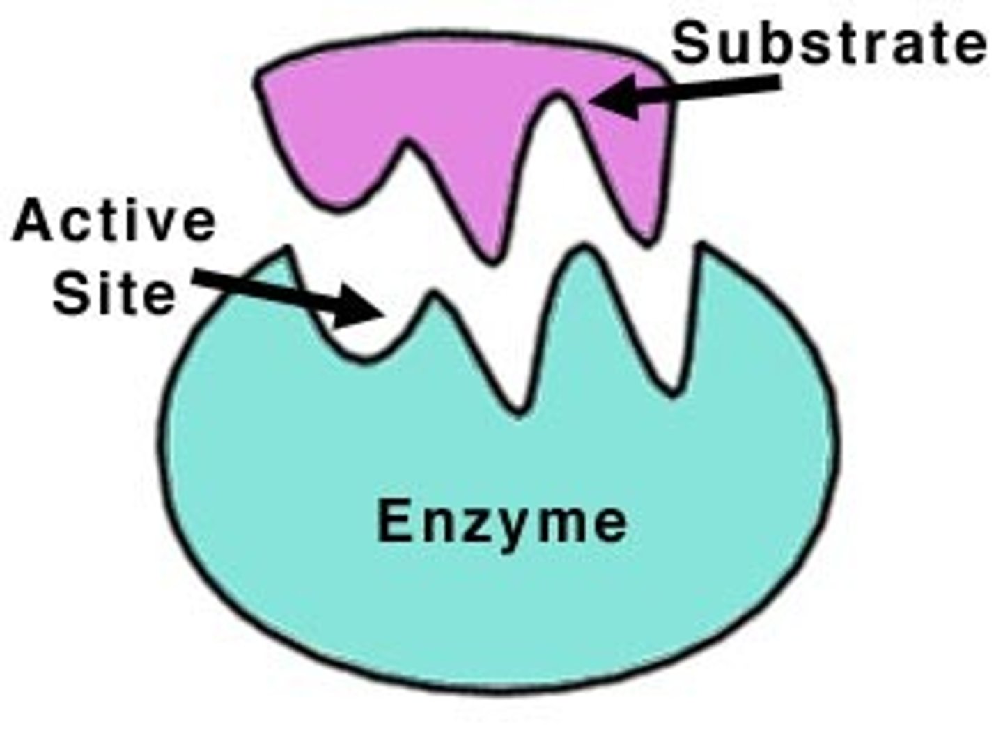
substrate
Binding site
- The folding of the polypeptide chain creates a cavity on the protein surface
kinase
puts a phosphate on something
phosphotase
removes a phosphate from something
Enzyme Catalyst
A type of protein that speeds up a chemical reaction in a living thing
Feedback Inhibition
- regulates the flow through biosynthetic pathways
- triggers a conformational change
- Prevents cells from wasting energy and substrates on chemical reactions that are not necessary at the time
how to regulate protein activity
Gene Expression
• Protein location
• Feedback loops
protein phosphorylation
a very common means of regulating protein activity
The covalent addition of a phosphate group to a side chain of a protein, catalyzed by a protein kinase; serves as a form of regulation that usually alters the activity or properties of the target protein.
(light switch)
Wha activates the enzymatic reaction for oxidation of sugars
increase of ligand (ADP) concentration
Activation of enzymes through ligand binding: why would this be useful?
The binding of a ligand to an allosteric site of a multimeric enzyme often induces positive cooperativity, that is the binding of one substrate induces a favorable conformation change and increases the enzyme's likelihood to bind to a second substrate.
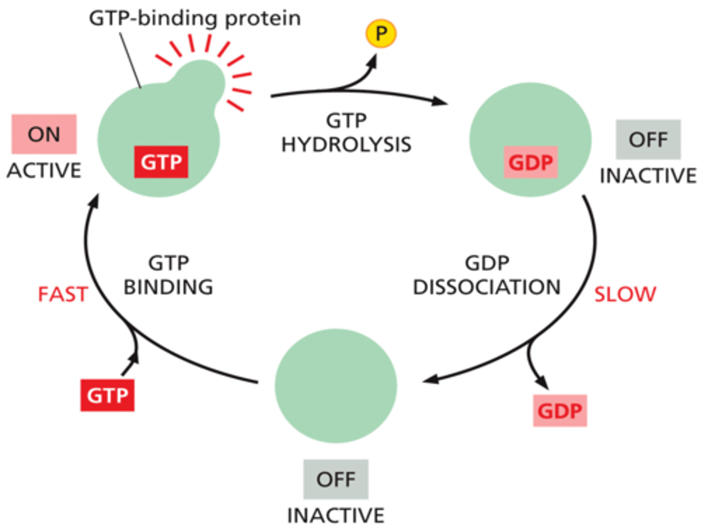
GTP binding proteins form
molecular switch pathway. Examples of these roles include: Signal transduction in response to activation of cell surface receptors, including transmembrane receptors such as those mediating taste, smell and vision.
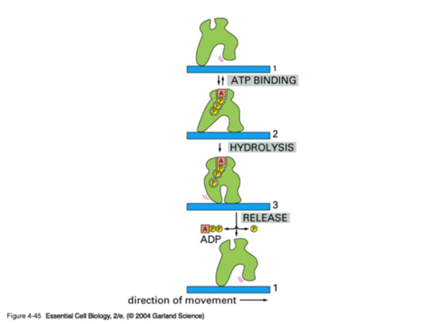
Mechanism of movement by allosteric motor proteins
An orderly transition among three conformations is driven by the hydrolysis of a bound ATP molecule. By repeated cycles of the conformation changes, the protein moves continuously to the right along the thread (in only one direction)
DNA experiment- late 1920s; why is it important
•The bacterium Streptococcus pneumoniae comes in 2 forms that differ in their ability to cause disease
•S strain: Smooth colonies and pathogenic (lethal).
•R strain: Rough colonies and harmless
•In the late 1920s, Fred Griffith found that a substance present in the pathogenic S strain could permanently transform the nonlethal R strain into the deadly S strain
-avery et al used this study for theirs
- Heat-killed bacteria can transform living cells
How to control protein function
The modification of a protein at multiple sites produce a
regulatory code that controls the protein behavior
DNA experiment- 1944; why is it important
•Avery, Macleod, and McCarty prepared an extract from the disease-causing S strain and fractionated it into different fractions including RNA, protein, DNA, lipids, and carbohydrates
•DNA can permanently change the harmless R strain into the pathogenic S strain
•The first evidence that DNA can serve as the genetic material
Evidence in Viruses (1952)
•Hershey and Chase used bacteriophages to demonstrate that DNA rather than protein carries the genetic material
- used seperate markers for DNA and protein and followed the DNA throughout generations
Watson-Crick DNA structure
•1953: James Watson & Francis Crick determined the DNA structure and revealed how DNA might be copied and encode the instructions for making proteins
Rosalind Franklin obtained
excellent
X-ray diffraction patterns of
crystalline B-form DNA
Genes
: store the genetic information which determines the characteristics of a
species as a whole and the individuals within it
parts of a nucleic acid
sugar, phosphate group, nitrogen base
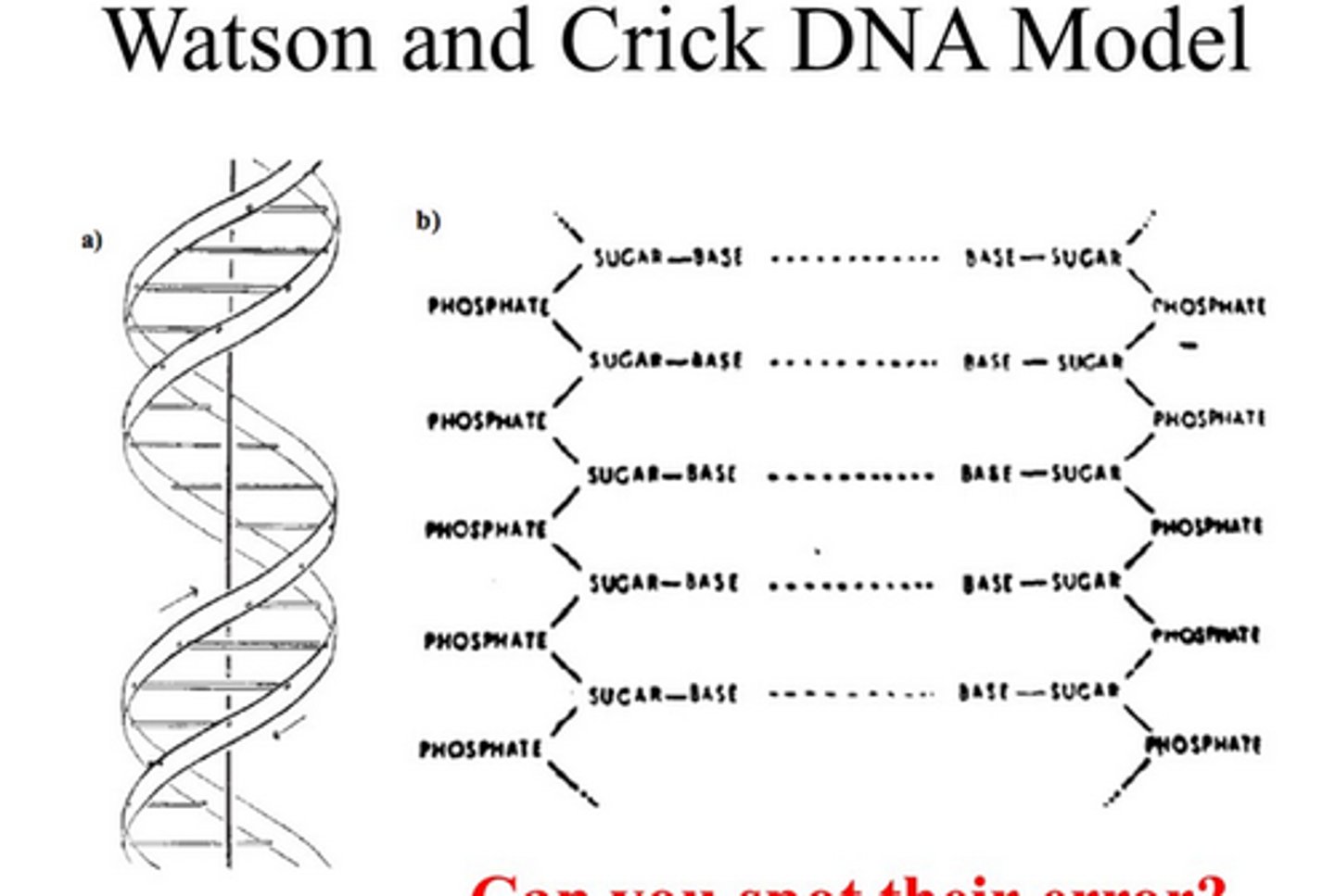
Watson-Crick DNA Structure
•Two polynucleotide strands that wind around each other about a common axis to form a helix
•The two strands run Antiparallel
•The planes of the bases are nearly at right angles to the helical axis
•Each base is base-paired by hydrogen bonding to a base on opposite strand. Complementary base-pairing
10 base pairs per turn
Chromosomes become visible
as cells prepare to
divide
Chromatin:
The combination of DNA and
proteins that make up the contents of the
nucleus of a cell
Purines
Bases with a double-ring structure.
Adenine and Guanine
Pyrimidines
cytosine, thymine, uracil
thymine- DNA or RNA?
DNA
Purines and Pyrimidines are
Nitrogenus Bases
uracil- DNA or RNA?
RNA
Directionality of DNA
-Transcription occurs in the 5' to 3' direction, based on orientation of sugar.
-Directionality: 3' end carries the unlinked -OH group attached to the 3' position on the sugar. 5' carries a free phosphate group attached to the 5' position
What bonds connect the sugar-phosphate backbone?
phosphodiester bonds between the sugar and phosphate