Mock exam wrong answers
 Call Kai
 Learn
Learn Practice Test
 Spaced Repetition
 Match
Match Flashcards
 Knowt Play
1/153

There's no tags or description
Looks like no tags are added yet.
Name | Mastery | Learn | Test | Matching | Spaced | Call with Kai | Chat |
|---|
No analytics yet
Send a link to your students to track their progress
154 Terms
mid diastolic closure of the mitral valve may be seen with what abnormality?
dilated cardiomyopathy
indicates rapid increase of LV diastolic pressures associated with severe AI
in PSAX of the LV, the IVS is flattened in diastole and normal in systole, what is the associated issue with this finding?
ASD
indicitive of right heart volume overload
left to right shunting
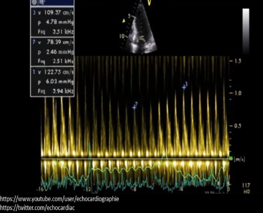
this doppler tracing is consistent with what cardiac abnromality?
contrictive pericarditis
respiratory variation of MV and TV velocities
what best describes the doppler tracing within a PDA obtained in the PSAX view?
continuous forward flow toward the transducer in systole and diastole
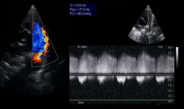
a patient presents for a 6 month FU for a reported aortic peak velicity of 3.3 m/s and a AVA of 1.4. On todays exam, the CW shows an aortic peak velocity of 2.5 m.s with the same AVA. What could explain the variation in measurements?
decrease in EF% from last exam
the early heart tube normally…
loops anterior and to the right
what is an abnormal finding on a treadmill stress echo?
systemic blood pressure drops during exercise and persists after stopping
hypotension
what congenital defect is seen with ebstein anomaly?
secundum ASD
_____ is the preferred method of diagnosis for contrictive pericarditis
echo
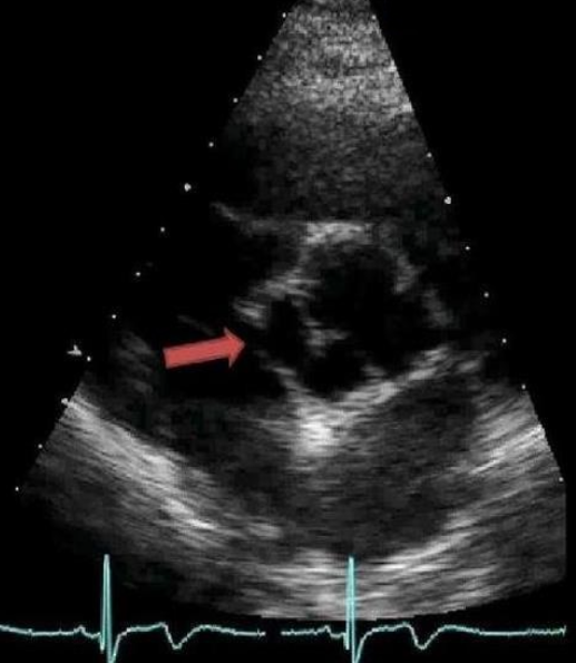
this abnormality is associated with what clinically critical complication?
dissection
this image shows aneurysmal dilation of the sinus of valsalva, the weakening can lead to rupture and dissection

what tubular structure is labeled A?
right main coronary artery
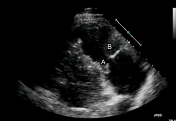
what is indicated by the letter A?
septal tricuspid leaflet
a patient with a history of moderate pulmonary HTN and severe TR. The RVSP is calculated at 30 mmHg on todays exam. What could explain these findings?
development of dilated cardiomyopathy
as ventricles are dilated, pressure gradients would decrease
rupture of the LV free wall usually involves…
the inferolateral wall with the circumflex or LAD occlusion
a lateral scar on a patients chest is suggestive of what type of surgery?
cardiac shunt placement
what is a responsibility of the sonographer during a pericardiocentesis
instruct the patient on breathing techniques
what will lead to pulmonary hypertension?
pulmonary vein stenosis and mitral stenosis
MS causes PV and LA dilation
what grade of DD is most commonly associated with chronic systemic HTN?
grade 1
LV wall thickening and recuced myocardial compliance
what measurement is an acccurate predictor of adverse outcomes in LV DD?
MV deceleration time
<160 ms = bad
what is the necessary info to estimate the systolic pressure in the pulmonary artery in a patient with a PDA?
systolic BP and peak velocity across the PDA
what is the best method to evaluate the severity of MR in a mechanical mitral valve?
A4C PISA method
what describes the effect of dobutime on the LV?
increases oxygen demand and LV contractility
what LA diameter would likely be demonstrated in a patient with chronic mild MR?
3.8 cm
won’t lead to significant changes in size
what echo finding is seen with RV pacing, left bundle branch block and recent cardiac surgery?
paradoxical septal motion

what is this doppler tracing suggestive of?
severe TR
significant systolic flow reversal (above the baseline) caused by flail TV leaflet
when measureing LA volume…
LA length is measured from the center of the mitral annulus to the inner edge of the posterior LA wall
the IVS is normally seen with a mild bowing ____
toward the right ventricle
placing the transducer at the 5th intercostal space on the patients chest can lead to…
an oval appearance of the LV in short axis
what is a characteristic of chronic systemic HTN identified on doppler?
E to A reversal
E/A ratio 1.0
what best describes the valvular abnormalitis seen in a heart with dilated cardiomyopathy?
due to volume overload caused by hypocontractile LV, the presense of multivalvular regurgitation is common
the majority of pulmonary venous flow into the LA occurs during…
early systole
S wave
diastolic flow reversal in the hepatic veins, diminished A velocity and prominent a-wave on the pulmonary vein tracing, with a mitral E/A ratio of 1.9 correlates with what?
constrictive pericarditis

what is the diagnosis if the right side is very big but the left side still looks mostly normal?
severe MR
cause LA dilation, pulmonary HTN and volume overload in the right side causing it to look really big

a 35 yr old complains of an episode of sudden chest pain with SOB that happened last week. what is true about this image
it is normal
what is an indication of right coronary artery disease on a exercise stress echo?
reduced TAPSE
> 4 mm
what is true regarding MR?
the larger the residual opening between the mitral leaflets during ventricular systole the lower the pressure gradient of the MR jet
blood moves through a bigger opening with less resistance/pressure
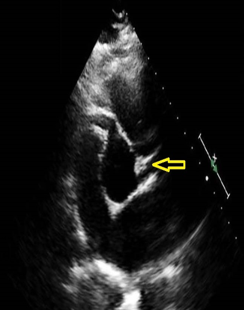
color doppler is applied over the vessel indicated with the arrow. The map demonstrates flow away as blue. what color will fill the the vessel during diastole?
the left coronary artery will display as red flow

what correctly describes the findings of this PSAX view?
LV aneurysm of the inferior wall

what describes the findings of this image?
trace MR with significant AS
this is a native valve with limited mobility and significant atherosclerosis
what would be given to a patient who is having an anaphylactic response caused by contrast injection
hydrocortisone
at what point during an echo using saline UEA will the patient perform provocative maneuvers?
about 5 seconds after the UEA injection
in a patient with a PDA, normal intracardiac pressures can be assumed if the peak pressure gradient across the ductus is equivalent to…
100 mmHg
the PPG between the aorta and the PA is 100, assuming the peak velocity across the PDA is 5
patients with a history of ____ are at high risk of getting acute endocarditis, while patients with a hisotyr of ____ are at a high risk of getting subacute endocarditis
IV drug use, prosthetic valves
acute = staph infection
subacute = bacteria already in the body
severe pulmonic recurgitation:
demonstrates a PHT of <100 msec
causes premature opening of the PV and flow reversal in the MPA
when assessing MR using vena contracta, what adjustement should be made to the machine?
set the color velosiry scale to 50-60 cm/s
PISA is when the baseline is moved
what describes the preferred method when measuring RA volume on 2D imaging?
A4C, with prove tilted upward to view the superior aspect of the chamber, measure at end systole
a 67 year old male presents to the emergency room with sudden onset of cyanosis, fatigue, and oliguria. the systemic BP is 70/40. What is the most likely finding on the echo?
abnormal left ventricular wall motion
a 22 year old patient presents for an echo and you see biventricular wall thickening, mild pericardial effusion and thickening of the leaflets of all 4 valves. What is the most likely cause of these findings?
amyloidosis
infiltrative disease indicated by whole heart affected
how is LVOT diameter measured?
PLAX at its greatest capacity in mid systole
perpendicular to long axis of AO at the AV insertion points
a 20 year old male presents for an echo due to SOB and cyanosis. What is the mostly likely reason for his symptoms?
tetralogy of fallot
MC cardiac cause of cyanosis in adults
when evaluating an adult with suspected congenital heart disease, how do you identify the left atrium?
locate the left atrial appendage
b/c the atra can be switched and not necassarily with their valve
what transducer postion provides the best view of the descending thoracic aorta for assessment of diameter?
apical
a patient presents with AS. LVOT velocity is 1 m/s, LVOT diameter is 2 cm, and AV velocity is 4 m/s. What is the AVA?
0.8 cm²
AVA = 0.785 x (LVOT D)² x LVOT PSV / AV PSV
= 0.785 x (2)² x 1 / 4

what is this video showing?
this is a short axis TEE view of a large ASD
what is recommended for calculating the effective orifice area for prosthetic mitral valve?
continuity equation
planimetry cannot be performed on mechanical prosthetics
what is used to calculate effective orifice area for a native mitral valve?
pressure half time
what correctly describes normal flow in the coronary arteries?
early in ventricular diastole flow moves into the coronary arteries
during systole the AV cusps cover the openings
what is the correct way to change transducer position from the subcostal 4 chamber to view the 4 pulmonary veins emptying into the LA?
angle slightly anterior
prolapse of the right aortic cusp is most commonly seen with what type of VSD?
outlet
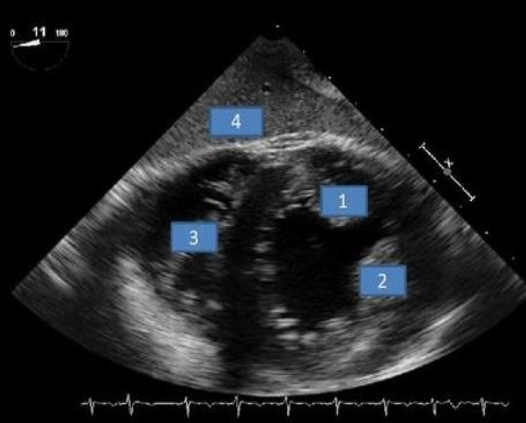
what structure is #2?
anterolateral papillary
doppler evaluation of the aortic valve demonstrates _____ with atrial fibrillation?
beat to bear variation in aortic velocity
what is best evaluated with perflurocarbon ultrasound enhancing agent?
LVOT gradients
what is associated with equal diastolic pressures in the right and left ventricles?
contrictive pericarditis
becuase the pericardium restricts diastolic filling, diastolic pressures in the 2 ventricles will be very similar
what conduction abnormality is commonly seen with chronic mitral stenosis?
atrial fibrillation
due to atrial enlargment

what is letter A?
right atrium
what system setting should be kept the same during resting and stress images for a stress echo?
image depth
to more accuratly evaluate wall motion changes and chamber size
if a patient suffers from a syncopal episode due to a vasovagel reation after the injection of UEA, how should you postion them to help them recover?
trendelenburg position
legs being above the head will increase venous return to the heart and increase volume of oxygenated blood to the body
an inpatient is referred for an echo to identify an embolic sourse for a series of recent “mini strokes”. she was admitted 2 days ago to undergo heparin treatment for DVT in the left leg. the VQ scan was normal with no evidence of PE. what is the most likely reason the study was ordered?
to evaluate the patient for a possible PFO
A 40 year old with a 4 mm VSD will demonstrate dilation of which chamber first?
left atrium and left ventricle
What is best evaluated using a supine bike?
diastolic dysfunction
A patient with trisomy 21 presents for an echo to rule out congenital heart defects. The most common form of congenital heart disease that occurs in patients with this genetic disorder is ____
atrioventricular canal defect
A patient presents for an echo due to recent onset of SOV and an abnormal ekg. The ekg report indicates the prevents of electrical alterans. What is the expected finding?
significant pericardial effusion
Which Doppler flow parameter is most accurate when assessing aortic stenosis in a patient with significant aortic regurgitation?
mean pressure gradient
_____ is a common finding with infiltrative CM, endocarditis, and a recent MI
a pericardial effusion
You begin your echo exam by performing the parasternal long axis view. color Doppler demonstrates severe mitral regurgitation what hard structure should be evaluated for associated dilation before changing views?
left atrium
You identify thickened tethered, mitral leaflets, decreased EF slope on PW Doppler and right ventricular hypertrophy on an echo. These are all signs of ____
mitral stenosis
___ can cause the aortic valve velocity to be overestimated, while ____ can cause the aortic valve velocity to be underestimated.
significant aortic regurgitation, significant mitral regurgitation
When performing an echo on a patient with suspected congenital heart disease. You’re unable to demonstrate two separate vessels exiting the two ventricles. These findings are most suggestive of:
truncus arteiosus
Bilateral distended jugular vans and a knocking sound her during cardiac auscultation are reported at the physical exam. What is the most likely abnormality identified on the echo?
constrictive pericarditis
What correctly describes mitral valve Doppler tracing in most cases of hypertrophic CM?
E/A ratio < 0.8
What is the purpose of the eustachian valve in fatal circulation?
to guide flow through the foramen ovale
Ruptured sinus of valsalva, patent ductus arteriosus, and an aorta pulmonary window are associated with what type of murmur?
continuous murmur
The most common pattern of left ventricular hypertrophy in hypertrophic CM is:
isolated interventricular septal hypertrophy
What transducer can still be used safely?
betadine infiltration of the matching layer
What is not an expected complication caused by mild moderate tricuspid regurgitation?
pulmonary htn
What is a potential cause for underestimated of aortic stenosis when calculating AVA with the continuity equation?
sub optimal parasternal view, causing overestimation of the LVOT diameter
you perform an echo and the only abnormal finding present is mild tricuspid regurgitation. What do you expect the peak pressure gradient to be for the tricuspid regurgitation?
20 mmHg
What heart sound is associated with tricuspid stenosis?
opening snap
You are performing an echo on a patient with an ASD and the PW Doppler assessment of the flow across the defect yields a peak velocity of 1.1 m/s. What does this indicate about the atrial pressures?
the pressures in the right and left atrium are normal
What describes the appearance of the Doppler tracing of the mitral valve in a patient with restrictive cardiomyopathy due to amyloidosis?
higher peak velocity of the E wave and very low peak velocity of the A wave
What commonly requires the use of high PRF Doppler to evaluate normal flow?
LVOT peak systolic velocity
What Doppler measurements are normally used to evaluate a cardiac transplant for rejection?
mitral deceleration time, IVRT, and E velocity
Severe tricuspid stenosis leads to:
hepatic congestion
You suspect a thrombosis present in the left atrium. What should you evaluate as a potential cause for this finding?
mitral valve area
the standard algorithm for grading diastolic dysfunction cannot be used in patient with:
significant mitral annular calcification
What is the most common echo finding in HIV patients?
pericardial effusion
When performing an echo with saline ultrasound enhancing agent to evaluate an eccentric jet of tricuspid regurgitation. Which chamber should you monitor during UEA administration?
right atrium
What correctly describes when to remove personal protective equipment after completing an ultrasound exam on a patient with Covid?
remove everything but the respirator before leaving the patient care area
What level of pulmonary hypertension is present with an acceleration time of 50 msec for the pulmonary valve Doppler tracing?
severe
< 60 = bad