bio 37 midterm 1
1/198
There's no tags or description
Looks like no tags are added yet.
Name | Mastery | Learn | Test | Matching | Spaced | Call with Kai |
|---|
No analytics yet
Send a link to your students to track their progress
199 Terms
how heavy is the average brain?
2.5 lbs
how much cerebrospinal fluid is there on the outside/inside of the brain?
½ of a cup
how many neurons? how many inputs does each have? how many glial cells per neuron? what is the diameter of each one?
1) 10^12 neurons
2) thousands of inputs, up to 150k
3) 20 glial helper cells per neuron
4) diameter = 8-80 micrometers
how many miles of axons in a brain? how fast do they conduct signals?
100k miles of axons conducting at 200 mph
how much of the body’s blood flow and energy does the brain use?
20%
what percentage of human genes are expressed in the brain?
25%
dementia — is it a group of disorders? or just 1
group of disorders that affects memory, thinking, and interferes with daily life
is a majority of dementia disorders reversible or irreversible? stats?
most are irreversible (85%-95%)
around 5%-15% are reversible
examples of irreversible dementia (6)
1) alzheimers
2) dementia with lewy bodies
3) fronto-temporal dementia
4) vascular dementia
5) parkinsons disease dementia
6) traumatic brain injury
examples of reversible dementia (5)
1) depressive pseudodementia
2) metabolic problems
3) medication side effects
4) infections
5) dementia b/c of structural lesions
what is the most common type of dementia?
alzheimers
is dementia increasing in the population?
yes, elderly population growing so more old people = more dementia
hydrocephalus
water on the brain — enlargement of ventricles with CSF
crushes brain tissue causing variety of symptoms
microcephaly — what is it and what issues can it lead to
essentially super small brain — can lead to seizures, developmental delay, intellectual disability, problems with movement and balance, feeding problems, hearing loss, and vision problems
anencephaly
born with no brain — individuals usually die upon birth
what connects the brain to the spinal cord?
medulla (goes through pons first)
the medulla is a part of the….
brainstem
order of communication amongst diff types of neurons in the brain?
sensory neurons (get info) —> interneurons (makes sense of it) —> motor neurons (create movement)
motor neurons go to effector cells in body
sensory neurons — function, monitors what?
receives information from environment (touch, pain, temperature, light) and relays it to the central nervous system
monitors conditions important to bodily homeostasis of blood pH and o2 levels
interneurons — function
associates sensory and motor activity in CNS — makes sense of all the information collected, and does computational stuff to work out what the information is conveying (what does the body need?)
motor neurons — function
comes out from brain/spinal cord and sends signals from these places to the muscles — allows mvmt!
what are the different ways you can cut up the brain?
1) sagittal
2) coronal
3) horizontal
sagittal plane of brain
divides into left and right sections

coronal plane of brain
dorsal ventral split (essentially like sliced into front vs back sides)
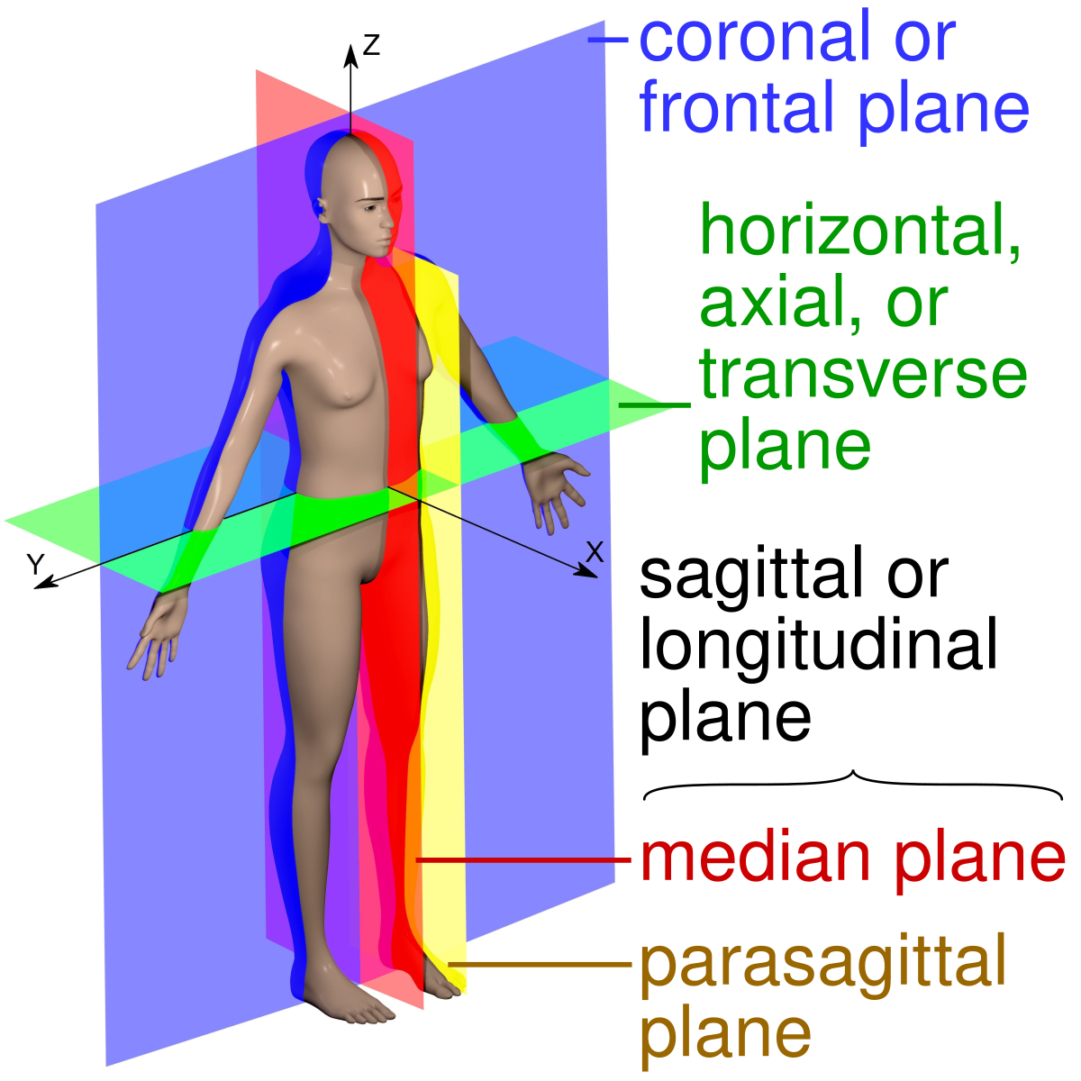
horizontal plane of brain
divided into top and bottom — straight through the middle horizontally

central nervous system composed of?
brain + spinal cord
peripheral nervous system composed of?
nerves and ganglia
nervous system coordinates… and also forms…
movement, touch, pain, senses
also forms emotions, thoughts, consciousness
how does the spinal cord convey info from the brain to the PNS?
spinal nerves
nerves
neurons send info to spinal cord/brain through these — tough/long strands of tissue w/ neurons
effector cells
the cells in glands/muscles that receive signals from motor neurons
spinal cord basic organization
bone surrounding spinal cord to protect the fragile tissue, and spinal nerves stem out from the middle
each vertebrate contains how many clumps of nerves? what are the names of each one and their function?
2 clumps
1) dorsal (back) clump receives sensory information
2) ventral (front) clump sends signals to muscles
different sections of the spinal cord (4)
1) cervical
2) thoracic
3) lumbar
4) sacral regions
cervical part of the spinal cord
top 8 segments that innervate back of head, neck, shoulders and arms
thoracic part of the spinal cord
12 segments below cervical region that innervate thorax and upper abdomen
lumbar part of the spinal cord
5 segments below thoracic region that innervate pelvic girdle and legs
sacral region of the spinal cord
5 segments below lumbar region that innervate urogenital and perianal structures + back of legs
dermatomes
regions of skin that a specific spinal nerve innervates
horizontal slice across body
ex) c2 corresponds to the 2nd cervical nerve
what nerves innervate the face? how many are there?
12 cranial nerves (comes out of the brain)
where do cranial nerves originate mainly?
medulla
locked in syndrome — what is it, what does it result from, damages, what part of the body is usually damaged?
neurological disorder that results in complete paralysis of voluntary muscles except eye mvmt
results from brain injury, strokes, diseases destroying the myelin sheath, or medication overdose
medulla
individuals with locked in syndrome
conscious with thinking/reasoning but unable to speak/move
communication can be possible with possible blinking eye mvmt
jean dominique bauby
man who experienced a stroke and suffered locked in syndrome — only able to blink with left eye
able to write a book by blinking out the letters
how does injecting evans blue into the blood indicate that the brain doesn’t come into contact with blood?
evans blue will dye all other organs but brains remain in similar condition to the control brain w/o dye in the blood
why does blood permeate the brain?
provides oxygen, glucose and removes waste products
why does blood not come in contact with the brain?
brain is sensitive to changes (pH, o2, chemicals), and blood fluctuates dramatically so it would be harmful
also infections would be able to reach the brain and kill neurons — which cant be regenerated
cerebrospinal fluid
fluid bathing the brain, and is separated from blood via blood brain barrier
blood brain barrier — what is it formed by? what is the purpose? can antibiotics cross?
formed by cells in brain capillaries (astrocytes cells form tight junctions that prevent things from move)
stops infections/toxins/drugs from reaching brain
antibiotics too large to cross BBB
why is it difficult to develop drugs for brain disorders?
large molecules don’t cross bbb easily (most drugs (BACE inhibitors) that can treat the pathologies cannot get into brain)
what can cross the BBB and what drugs does this apply to?
small molecules (like oxygen) /LIPID SOLUBLE molecules
applies to barbiturates (sleeping pills), antidepressants
central canal of spinal cord + ventricles of brain — describe them
hollow & filled with cerebrospinal fluid (150 mL)
how is csf made? what is its function?
made by filtering from the blood (area in ventricles takes fluid away from blood)
cushions brain/cord
do our brains float in csf? what would happen if they didnt?
yes — neutrally buoyant — w/o csf and buoyancy it would crush itself b/c of density
lumbar punctures — what happens, what is it used for, where is it done and are there complications?
csf can be drawn from spinal column to diagnose disease (cancer, meningitis, or markers for other diseases like alzheimers (certain proteins can indicate this disease)
also used for delivery of anesthesia to reach cns
needle inserted between L3/L4 or L4/L5
complications are rare but headaches can be caused
what problem of eukaryotic cells do organelles solve?
euka cells relatively large so it’s difficult to properly diffuse molecules across entire cell, so organelles break up the volume
advantages of compartmentalization in euka cells (2)
1) separating incompatible chemical reactions
2) increasing efficiency of these chem reactions
nucleus — structure & function, what does it produce and where?
double membraned nuclear envelope surrounding it + nucleolus
function : info storage/processing, containing chromosomes, and ribosomal RNA (rRNA) synthesis (in nucleolus)
rough endoplasmic reticulum — structure & function, continuous with what? what is its interior called?
network of tubes/sacs studded with ribosomes, with an LUMEN interior — rough ER is continuous with nuclear envelope
protein synthesis (in ribosomes) (MAIN)
modification occurs in lumen (for secretion or membrane use)
smooth endoplasmic reticulum — structure & function— what is stored here? what is made & broken down here?
no ribosomes
enzymes make fatty acids/phospholipids + break down poisonous lipids
reservoir of Calcium ions
golgi apparatus — structure & function, what carries things to this organelle?
series of stacked flat sacs called cisternae
processes, sorts, ships proteins synthesized in rough er
vesicles carry materials to and from here
ribosomes — structure & function, are they membrane bound? where are they found?
non membranous - not technically organelles either… w/ large + small subunit w/ RNA and protein — can be attached to rough er or in cytosol floating
protein synthesis
lysosome— structure & function, how many membranes? in animal or plant cells?
single membrane bound w/ digestive enzymes — in animal cells
digestion + waste processing
tay sachs — malfunction in what organelle
malfunction in lysosome, results in waste buildup in the brain/neurons
mitochondria — structure & function, how many membranes? has its own ___ and makes its own ____?
two membranes, inner membrane folded into sac like cristae, solution inside cristae = mitochondrial matrix. organelle has own DNA + manufactures own ribosomes
atp production (energy)
cytoskeleton— structure & function
holds cell tgth, gives it shape/stability
aids in cell movement and transports materials
organizes cell and other structures
plasma membrane— structure & function
double layer of phospholipids (amphipathic — hydrophilic heads and hydrophobic tails) that separates outside and inside of cell
has a bunch of channels/receptors/proteins that regulate things + receive signals
allows passage of oxygen, nutrients, and waste
animal movements are triggered by electrical signals conducted by…
neurons
neurons — what do they do, how do they do it, and how do muscles respond?
nerve cells transmitting electrical signals in communication — plasma membranes have voltage
muscles respond to signals by contracting
describe the anatomy of a neuron
CELL BODY : contains organelles
DENDRITES : branched extensions connected to cell body that RECEIVE electrical signals from other neurons
AXON : extension that TRANSMITS signals to other cells at synapses
AXON HILLOCK : axon + cell body junction — where the action potential is generated
synapse not a part of the neuron but is the gap between axon and another cell
synaptic terminal — what is it and what happens here?
where axon passes info across the synapse using neurotransmitters
is the plasma membrane of the neuron permeable to charged ions? if not, how do they get charged ions across?
impermeable — uses channels and receptors of proteins that let ions over
why are charged ions important to the function of the neuron?
provides electrical energy through flow of charged ions = able to rapidly signal
electrical potential/voltage — when is it present in a neuron?
diff in charge between 2 points
present in neuron when two sides of plasma membrane do not balance e/o
membrane potential
when electrical potential exists on either side, separation of these charges = membrane potential
Na/K ATPase — how does it work and where are each ion’s concentrations higher?
imports 3 Na+ ions from inside of the cell to outside, using ATP, and then transporting 2 K+ ions from out to in
conc of K+ higher inside, conc of Na+ higher outside cell
ATPase changes shape during the course of this
info is transmitted from a _____ cell to a ______ cell
presynaptic (neuron), postsynaptic (neuron, muscle, gland cell)
what are most neurons nourished/insulated by?
glia
diff types of glia (5)
1) astrocytes
2) ependymal
3) microglia
4) oligodendrocytes
5) schwann cells
astrocytes — how do they support neurons? are they restricted to PNS/CNS or are they in both?
support neurons by giving appropriate chem environment for signaling + form BBB
restricted to CNS, with a star like appearance
ependymal cells
circulate cerebrospinal fluid
microglia
protects nervous system from microorganisms
oligodendrocytes — cns or pns
forms myelin sheaths around axons in CNS
schwann cells — cns or pns
forms myelin sheaths around axons in PNS
which glia share the same function but diff neuron location?
oligodendrocytes + schwann cells
brain tumors — what are they? what are the two most common types? do they occur on glia or neurons? how can secondary tumors arise?
results from uncontrolled cell division and ends in a mass (malignant (cancer) or benign(non-cancer))
most are on glia b/c they can divide, but neurons cannot
main types : gliomas, meningiomas
secondary ones arise from malignant tumors originating in other parts and metastasizing
during action potential a _______ (inflow/outflow) of sodium ions is followed by an _______ (inflow/outflow) of potassium ions
inflow; outflow
resting potential
membrane potential of a neuron not sending signals
describe the general charges of the outside vs inside of the cell membrane?
outside is more positive, inside more negative
at resting potential what are the concentrations of K+ and Na+ like?
K+ = low outside, high inside
Na+ = high outside, low inside
what maintains the concentration gradients? is it powered by anything?
sodium potassium ATPase — uses ATP
what do the gradients represent?
chemical potential energy
describe the workings of the sodium potassium pump
1) 3 Na+ binds to pump —> atp used to change shape of protein = 3 Na+ released to outside
2) new shape = 2 K+ bind and phosphate group detaches = shape changes back to get 2 K+ inside cell
causes net negative charge for inside cell b/c releasing more positive charges than gaining
is a majority of the neuron energy utilized by the Na-K pump?
yes
what is the resting membrane potential #?
-70 mv
name the 3 important ion channels for neurons
1) potassium channel
2) sodium channel
3) calcium channel
all voltage gated
how do voltage gated ion channels open?
opens when depolarization (membrane becomes less negative) occurs
voltage gated potassium channel — purpose? does it open slowly or quickly? depolarization or polarization?
restores membrane potential following depolarization (makes membrane negative again) — opens slowly
releases potassium to the outside
voltage gated sodium channel — does it open slowly or quickly? depolarization or polarization?
depolarizes membrane (more positive) during action potentials — opens rapidly
Na+ into cell
voltage gated calcium channels
opens when membrane depolarizes ( near end of axon) — important for synaptic release
lets calcium INTO cell