Exam 1 Euk
 Call Kai
 Learn
Learn Practice Test
 Spaced Repetition
 Match
Match Flashcards
 Knowt Play
1/65

There's no tags or description
Looks like no tags are added yet.
Name | Mastery | Learn | Test | Matching | Spaced | Call with Kai | Chat |
|---|
No analytics yet
Send a link to your students to track their progress
66 Terms
Cell
Simplest form of life that is capable of living and reproducing in a non-living environment
What are all cells surroded by?
A plasma membrane
Non-living environment
Any environment outside of the cell
What can curved glass surfaces (lens) do?
bend light and form images
two hisotorical scientists who made their own microscope
Robert Hooke (1665)
Anton van Leeuwenhoek (1665-1675)
compound microscope
built by Robert Hooke
two lenses
looked at a wine cork
single-lens microscope
built by Leeuwnhoek
looked at stuff in ponds
three tenets of cell theory
created by Matthias Schleiden (1838) and Theodor Schwann (1839)
the cell is the structural unit of life for all organisms
all organisms are composed of one or more cells
these two scientists thought cells could arise from non-living cell material, but that is not true
created by Rudolf Virchow
cells can arise only by division from a preexisting cell
9 basic properties of cells
highly complex and organized
possess a genetic program and the means to use it
capable of producing more of themselves
acquire and utilize energy
carry out a variety of chemical reactions
engage in mechanical activities
able to respond to stimuli
capable of self-regulation
evolve
three domains of life
bacteria
archaea
eukarya
what is the phylogenetic tree of domains of life based off of?
rRNA
RNA is conserved
two fundamental classes of cells
prokaryotic cells
archaea (archaebacteria)
bacteria (eubacteria)
eukaryotic
protists
fungi
plants
animals
archaea’s relatedness to eukarya and bacteria
archaea and eukarya share similar genes for informational processes
archaea is metabolically and strucurally more like bacteria, that is why they are both prokaryotic
9 features that prokaryotic and eukaryotic cells share
plasma membrane of similar construction
genetic information is encoded in DNA using identical gene code
similar mechs for transcription & translation, including ribosomes
shared metabolic pathways
similar apparatus for conservation of chemical energy
plasma membrane of prokaryotes
mitochondria of eukaryotes
similar mech of photosynthesis
similar mech for synthesizing and inserting membrane proteins
proteasomes of similar construction (archaea and eukarya)
cytoskeletal filaents built of proteins similar to actin and tubulin
12 features in eukaryotic but not prokaryotic cells
division of cells into nucleus and cytoplasm, separated by a nuclear envelope containing complex core structures
complex chromosomes composed of DNA and associated proteins that are capable of compacting into mitotic structures
complex membranous cytoplasmic organelles
specialized cytoplasmic organelles for aerobic respiration and photosynthesis
complex cytoskeletal system (actin filaments, intermediate filaments, and microtubules) and associated motor proteins
complex flagella and cilia
ability to ingest particulate material by enclosure within the plasma membrane vesicles
cellulose-containing cell walls (plants)
cell division using a microtubulue-containing mitotic spingle that separates chromosomes
presence of two copies of genes per cell, one from each parent
presene of three different RNA synthesizing enzymes (RNA polymerases)
sexual reproduction requiring meiosis and fertilization
3 major differences in plant vs animal cells
plants have a cell wall, (central) vacuole, and chloroplast
model organisms
organisms we use to study and experiment on because they are easy to maintain and breed in a lab setting
less ethical to experiment on a human first
experiemntal advantages come with model organisms
viruses
microscopic particles that can infect the cells of biological organisms
viral structure
genetic material surrounded by a protein coat (capsid) and sometimes has a lipid envelope
why is a virus considered non-living?
needs a host to perform function
5 common virus properties
all are obligatory intracellular parasites
outside of a living cell, it exists as a particle or virion, essentially a macromolecular package
genetic material is surrounded by a protein capsule (capsid) usually made up of a specific number of subunits
viruses have surface proteins that bind to particular host cell surface components (specificity)
most viruses have relative narrow host range
two basic types of viral infection
lytic infection: the infecting virus kills the host cell
infects, propogates, kills by breaking membrane to release more virus
lysogenic infection: the infecting virus does not kill the host cell
integrate genetic information into host genome
provirus
integrated viral DNA is called a provirus
3 provirus properties
upon a stimulus, becomes lytic
produces new viral progeny without lysing the host cell
host cell becomes malignant
viroids
small circular RNA infectious agent that lacks a protein coat
240 to 600 nucleotides (10% size of smaller viruses)
do not code for proteins
two technqiues in cell and molecular biology (for this class)
pulse-chase experiment
microscopy
pulse-chase experiment- use of radioisotope
an experiment in which cells are growing radioactive medium for a brief period (the pulse) and then transferred to nonradioactive medium/unlabeled chemicals for a longer period (the chase)
during chase period, samples are taken for analysis
put cells in radioactive medium, wash cells, put them in unlabeled chemical or nonradioactive medium, and take sample during a chosen time interval, then analyze
can follow protein synthesis or other dynamics
purpose is to track the synthesis, movement, processing, and degradation of molecules (like proteins or RNA) inside a cell over time.
light microscope
a microscope that uses visible light and lenses to magnify and resolve small objects, such as cells and microorganisms.
hard to see living organisms
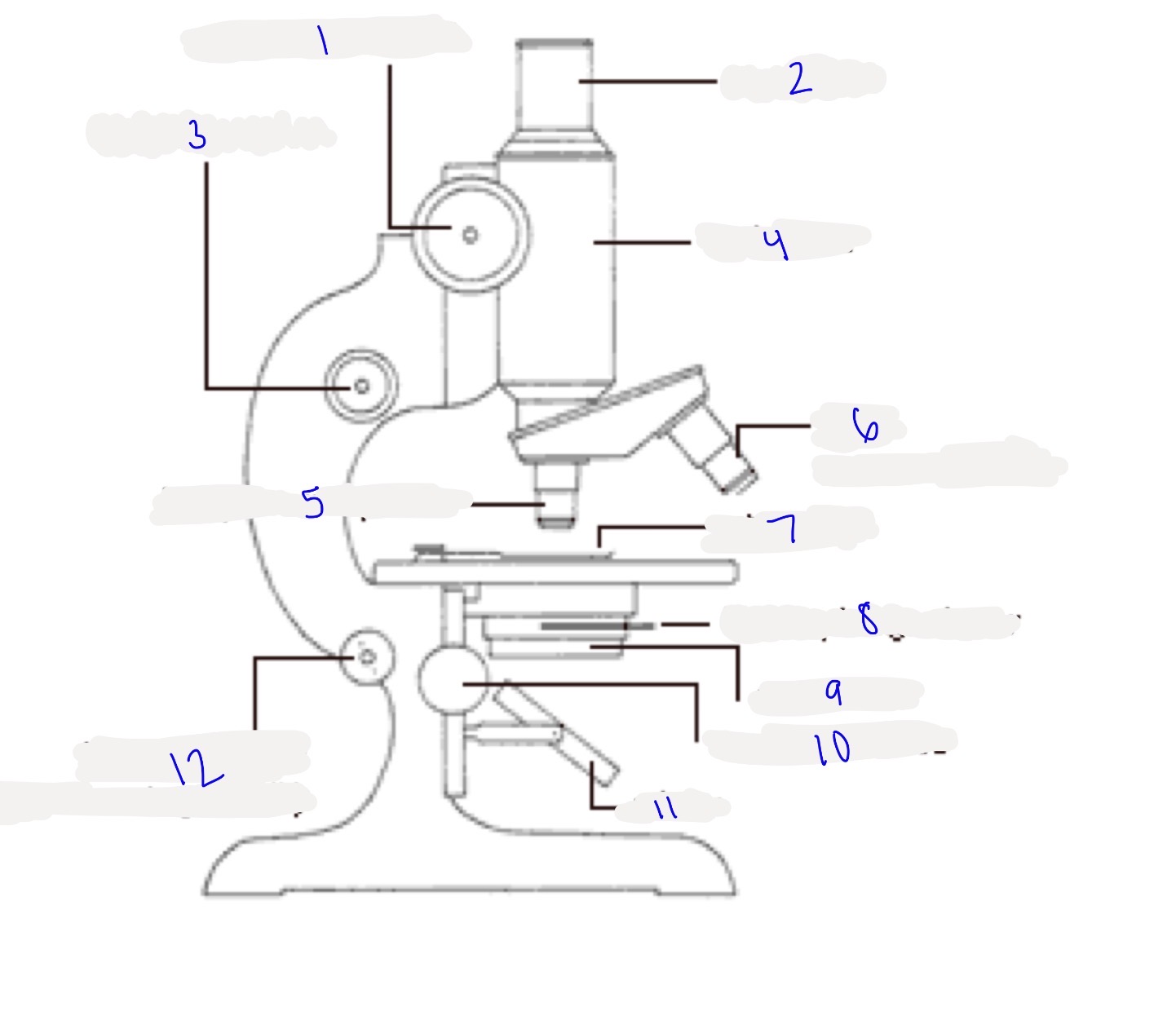
type of microscope, where the light comes from, and parts labeled
light microscope
external light (mirror reflects light from surrounding)
coarse focus knob
eye piece
fine focus knob
body tube
low power objective lens
high power objective lens
stage dip
iris diaphragm lever
condensor
condensor knob
mirror
inclination point (allows the stage to tilt)
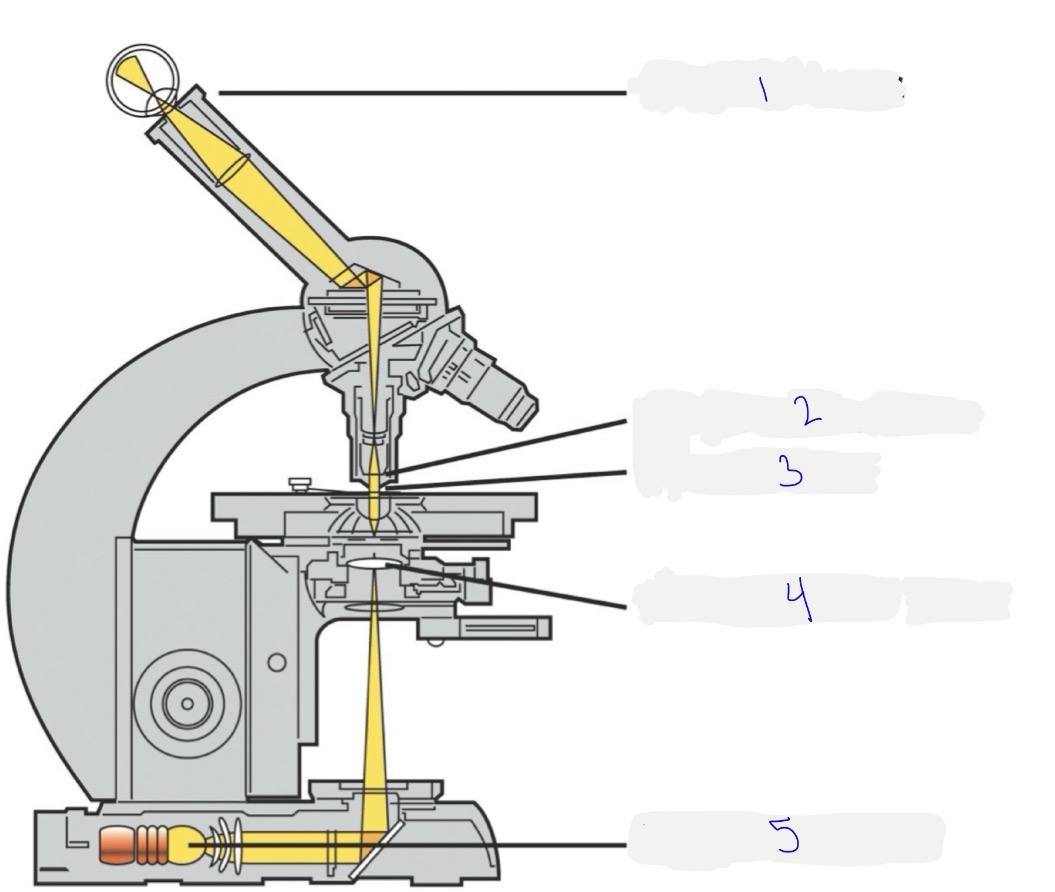
type of microscope, where the light comes from, and parts labeled
light microscope
produces internal light
ocular lens
objective lens
specimen
condensor lens
light source
pathway of light for a light microscope
light is concentrated to the condensor lens
light travels from condensor lens to specimen
light travels to objective lens
light travels to ocular lens
ocular lens function
amplifies image under the microscope
the image inside is reflected by a mirror inside of the microscope, located between the ocular lens and objective lens
what determines the quality of a microscope?
the objective lens
objective lens
amplifies the object/specimen itself
forms the image on the mirror
light path of the objective lens to form the image
distance between objective lens and specimen
lower power objective lens, the distance is bigger
higher power objective lens, the distance is smaller
lens is specific to the specimen: only at a certain distance will you get a clear image
2 alpha angle, objective lens, and speciment
increasing distance between objective lens and specimen decreases 2 alpha angle
decreasing distance between objective lens and speciment increases 2 alpha angle
higher power objective lens will create a larger angle than a lower power lens
empty magnification
a magnification that does not tell you any new details than you already saw
making an image where you can see everything clearly bigger and still seeing everything clearly
an effective magnification will tell you more information about an image
resolution
how small the distance (d) that a lens can distinguish between two neighboring points as two distinct entities
used to measure the optical quality of an objective lens
equation for resolution
lambda is the wavelength of light used
alpha is the angle created between objective lens and specimen
n is refractive index of the medium between the objective lens and the speciemen (air)
determines how much the light bends
resolution and alpha
larger alpha, d will decrease, better resolution
higher power lens
smaller alpha, d will increase, not as good resolution
lower power lens
lens and “n”
each lens is designed for a specific medium/refractive index. you can’t use the same medium for every lens because different lenses require different refractive indices to work properly.
numerical aperture (N.A)
for a particular lens, the refractive index and the angles involved are fixed, so the amount of light the lens/fiber can accept is also fixed.
tells you how much light a lens or optical fiber can collect/accept.
quality of a lens is determined by the numerical aperture of the lens
diffraction
the bending and spreading of waves when they pass through an opening or around an obstacle.
caused by the waves overlapping and interfering with each other
if two spots overlap, it will be seen as one spot instead of two since the waves overlap
no resolution
diffraction limits resolution
visibility and contrast
essentially the same thing
a part of a specimen is visible when it interacts with light differently than the areas around it or the background.
if contrast is too small, then visibility is too small and we cannot see it
visibility is determined by the reflective index of the objective and of the specimen and its surroundings
if the refractive indices of the specimen and its surrounding medium are very similar, there is little difference in how light interacts with them, so the specimen has low contrast and is difficult to see.
if the refractive indices of the specimen and its surrounding medium are very different, visibility and contrast is better
Why is oil used with a high-power objective?
Oil has a refractive index (~1.5) similar to glass (~1.5). This reduces the bending/refraction of light between the slide and objective, allowing more light to enter the objective, increasing numerical aperture (NA) and improving resolution.
reflective index of air, water, oil
air: 1
water: 1.33
oil: 1.5
oil should have best quality, then water, then air
staining
increases visibility and contrast
very difficult to see internal structure of chromosome and cells because all parts have similar refractive indexes
dyes can specifically stain the target structure , giving it a different refractive index
phase-contrast microscope
converting differences in refractive index into differences in intensity
relative brightness and darkness (increase contrast)
allows us to avoid killing a cell by staining it
function of a phase-contrast microscope
separating the direct light that enters the objective lens from the diffracted light emanating from the specimen
causing light rays from these two sources to interfere with one another (brings them back together after separated to create differences in brightness)
summary: separate the two light sources (direct and diffracted) and let them interfere with each other
fluorescence microscope
light enters a filter that only allows a certain wavelength of light to go through
light reaches beam splitting mirror
beam splitting mirror reflects the incoming light through the objective lens
objective lens focuses on the specimen
light comes back from the specimen, go to the objective lens, which will amplify the image
the light will pass through the beam splitting mirror, go through the filter to the ocular lens to form an image
how does light hit the specimen and then come back
a specimen has some atoms or something which can absorb light, called excitation light
after absorption, it will emit another light for a short period of time
the shorter remnant light will become a longer remnant light called emission light
excitation light is shorter remnant light (shorter wavelength) while emission light is longer remnant light (longer wavelength)
fluorochrome/fluorophore
a fluorescent chemical molecule that absorbs light at a specific wavelength and re-emits it at a longer wavelength
energy is absorbed by the atom, which becomes excited
electron jumps to higher energy level
electron will drop back to its ground state, emitting a photon, causing the atom to fluoresce
this is what creates shorter remnant light to longer remnant light
light absorbed has higher energy than when emitted later because energy is lost
the energy lost is light called fluorescence
uses dyes or the fluorochromes/fluoropheres
what kind of light is excitation light? why is this bad?
UV light
looking at a specimen for a long time using fluorescence microscope can damage it, making it difficult to get a detailed structure
laser scanning confocal microscope
scans sample so it will not damage the specimen
uses laser as the light source
laser uses different wavelengths
light can go over specimen in any different directions so you can observe the whole area rather than just one space
stack images together to create a 3D image
why is it advantageous to change the wavelength/energy of light with a laser scanning confocal microscope?
fluorochrome in the specimens have a requirement for what kind of light they can emit
wavelengths are fixed for a specific fluorescence dye, so we need to be able to change the wavelength of light to best view the specimen
laser scanning confocal microscope structure/light pathway
dichroic mirror is the same as the beam splitting mirror in a fluoroscence microscope
works similar to fluorescence microscope
laser, dichomic mirror, scanner, objective lens, specimen, objective lens, pinhole, computer
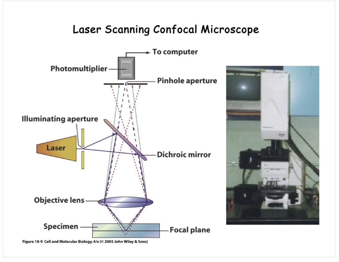
how images are created using laser scanning confocal microscope
light comes from focal plane and will go through the pinhole to form image on the computer
light that comes from the below or above the focal plane will be blocked by the pinhole aperture, creating a clear image
without the pinhole, light from the lower or higher plane will go through to form the image, making it blurred
dichromic mirror/beam splitting mirror
reflects incoming shorter wavelengths light
lets longer wavelengths of light to pass through
focal plane
illuminated plane
the imaginary flat surface where a lens or mirror bends light rays to form a sharp, clear image
specimen does not have to be very thin
pinhole aperture
confocal with the focal plane
light coming from the focal plane will be fixed in the pinhole, (will go through the pinhole) to form an image
relative resolution of our eyes and different microscopes
naked eye is about 100 micrometers
light microscope is 0.2 micrometers, which is sufficient enough to see larger cellular organelles like nuclei and mitochondira
the fine details of the interior structures of organic cells cannot be resolved by light microscope
electron microscopes have better resolution and were created so we can see smaller structures
the amplification of electron microscope to a light one is significantly better
transmission electron microscope vs light microscope pathway of light
light microscopes use light as a light source, while TEM uses a beam of electrons
light microscopes use glass lenses, but electron microscopes cannot because glass woud block the electrons.
electron microscopes use electromagnetic lenses because it can change the path of the electrons, just like glass changes the path of light
light microscopes use different wavelengths of light, beam of electrons use voltage to calculate their wavelength
lambda (wavelength of electrons) is the square root of 150/V (voltage)
increase in voltage makes electron wavelength shorter
condensor lens condenses the electron to the specimen, then objective lens, then ocular lens/projector lens to further amplify the image

preparing specimens for electron microscopes
electrons will damage specimen and dyes
negative stain: stain speciment with heavy metal (uranial acetate or lead citrate).the heavy metal stain interfere or block the electrons so when an electron goes there, that part of the specimen higher exposure (brighter). the parts not stained will appear darker when the electron goes there
shadow casting: evaporate heavy metal (platinum wire) so it becomes vapor. the vapor will deposit onto the specimen
the less deposition, the more electrons will go through, making it brighter
the more deposition, the less electrons will go through, making it darker
freeze-fracture replication/freeze etching: freeze sample, use a sharp knife to heat the sample
a preparation method that splits frozen biological samples and evaporates surface ice to expose internal cell structures in three dimensions
TEM vs SEM
TEM looks at internal structures of cell, SEM looks at surfaces of objects
TEM specimens need to be sliced and stained with solutions of heavy metals while SEM requires critical-point drying and coating with metals
TEM involves an electron beam that is focused to simultaneously illuminate the entire viewing field
SEM involves electrons that are accelerated as a fine beam to scan the specimen
TEM functions by electrons passing through the specimen to form the image
SEM functions by forming the image by electrons reflecting back from the specimen or by secondary electrons given off by the specimen after being struck by the primary electron beam
comparison of microscope (bright-field, fluorescence, confocal, electron)
