L2- COPD, smoke inhalation, EVALI
 Call Kai
 Learn
Learn Practice Test
 Spaced Repetition
 Match
Match Flashcards
 Knowt Play
1/86

There's no tags or description
Looks like no tags are added yet.
Name | Mastery | Learn | Test | Matching | Spaced | Call with Kai | Chat |
|---|
No analytics yet
Send a link to your students to track their progress
87 Terms
characterization of COPD
persistent respiratory sx
airflow obstruction
hx of significant exposure to noxious particles or gases
largely irreversible and progressive
COPD- epidemiology
predisposing factors
inhaled pollutants
cigarette smoke
biomass fuel for cooking and heating
occupation al dusts and chemical
alpha 1 antitrypsin def
main patterns of COPD
1) chronic bromchitis
a mucus problem
blue bloaters
2) emphysema
structural problem
pink puffers
chronic bronchitis: pathogenesis
repeated noxious environmental exposure → chronic inflammation and irritation of bronchioles→ increase mucus gland size and volume + ciliary damage→ bronchial endothelial thickening + excessive mucus production and retention → airway obstruction + chronic cough and phlegm production
chronic bronchitis: physiologic consequences
1) airway obstruction disrupts ventilation
decrease tidal vol due to
increase resistance to air flow in and out
air trapping leads to hyperinflation and reduced capacity for inhalation
2) damage to the mucociiary defenses
reduces clearance of pathogens
increase risk of pulmonary infections (ex. pneumonia)
emphysema: pathogenesis
resident alveolar macrophages phagocytes pathogens adn irritants and release protease
proteases destroy pathogens and break down collagen and elastin→ structural issue
chronic irritant exposure leads to increase protease production/activity and decreased antiprotease activity
results in destruction of capillaries and alveolar wal with air space enlargement (dilation)
alpha 1 antitrypsin
protease inhibitor
synthesized by hepatocytes
alpha 1 antitrypsin deficiency
genetic disorder
deficiency at AATT= protease > antiprotease
results in increased risk of emphysema, even with limited exposure to air pollutants
in which pts might you suspect alpha 1 antitrypsin deficiency
young pt with no smoke exposure
physiologic consequences- emphysema
alveolar wall and capillary destruction
septal destruction reduces alveolar surface area→ decreased area for gas exchange→ increased dead space
elastin destruction impairs ability to recoil→ air trapping→ decreased tidal volume
clinical presentation- chronic COPD
productive cough (bronichitis)
sputum production
SOB (emphysema)
clinical presentation: acute exacerbation COPD
increased cough
freq and severity
increased sputum
vol or change in character
increased SOB
general physical exam- COPD
± cachexia
vital signs- COPD
± hypoxemia and tachypnea
pulmonary exam findings- chronic bronchitis
noisy lungs- rales, rhonchi, wheezing
signs of RHF
cyanosis
peripheral edema
physical exam findings- emphysema
lung sounds are absent or distant
hyperresonance to percussion
barrel chest
dx criteria- COPD
1) respiratory sx consistent with COPD
chronic cough
sputum production
dyspnea
2) persistent airflow limitation
post- bronchodilator FEV1/FVC <70%
obstructive pattern
3) no alternative explanation fro sx and airflow obstruction
DDx- COPD
1) asthma
complete or near complete reversibility of airflow obstruction
2) CHF
dyspnea
dilated heart
3) bronchiectasis
recurrent pneumonia
hemoptysis
Digital clubbing
characteristics imaging abnormalities
initial assessment- COPD
1) spirometry
2) assess fro severity
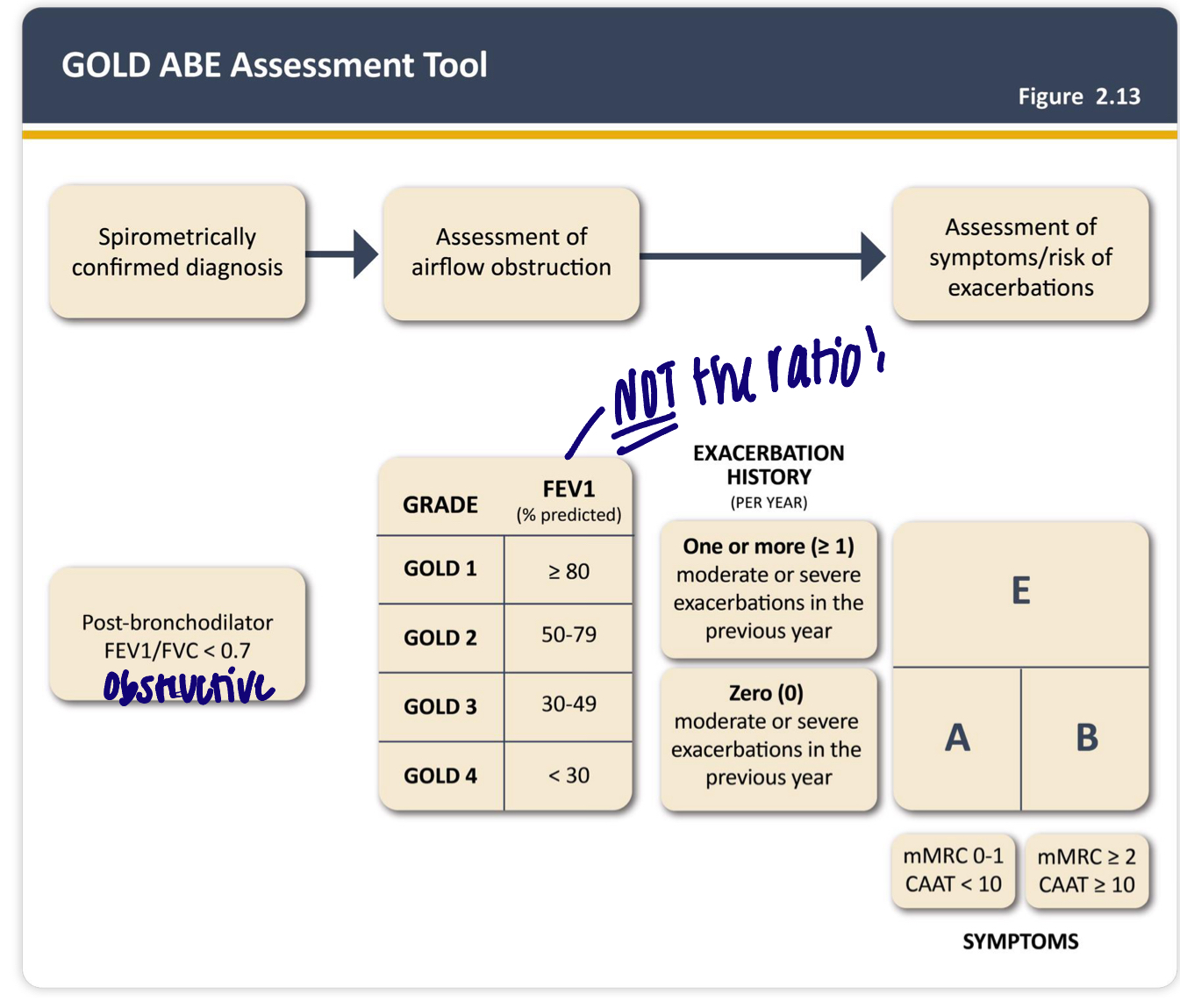
GOLD ABE
3) CBC with differntial
4) CXR
5) test fro alpha 1 antitrypsin deficiency
what conforms obstructive pattern
FEV1/FVC <70%
FEV1 is disproportionately reduced relative to FVC
GOLD ABE spirometry
pre and post bronchodilator spirometry
COPD is irreversible
remains <0.7 with bronchodilator
use FEV1 to assess COPD severity
degree of airflow obstruction is prognostic factor
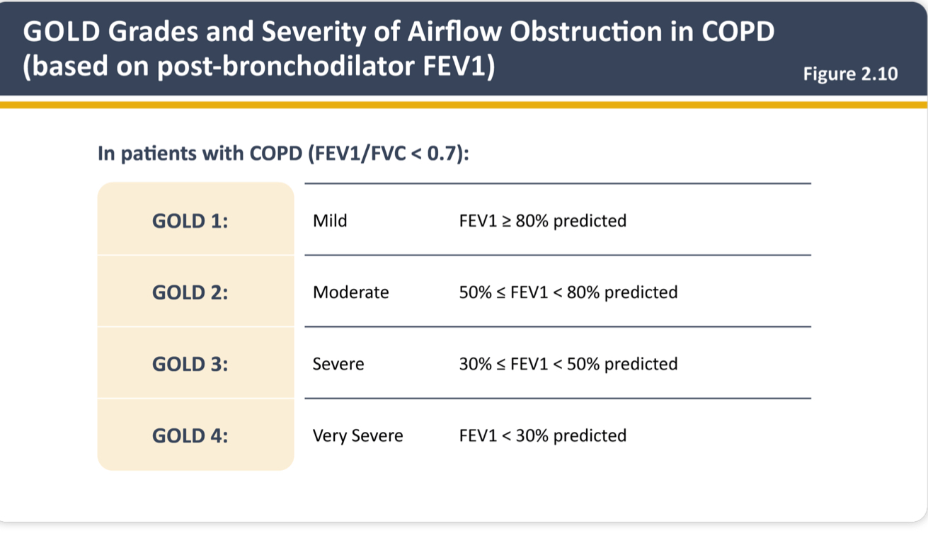
GOLD ABE- sx severity
subjectively how severe is pt airflow obstruction
mMRC
CAAT
GOLD ABE assessment
CBC results- COPD
secondary polycythemia (erythrocytosis)- physiologic adaptation to chronic hypoxemia, more. ommon in chronic bronchitis
anemia
likely anemia of chronic disease
leukocytosis, neutrophilia, elevated eosinophils
eosinophil count- used to guide therapy
start ICS if eos >300 cells
most reliable radiographic findings of COPD
1) hyperinflation
2) flattening of the diaphragms
additional radiographic findings of COPD
increase in the retrosternal clear space
hyperlucency of lungs
prominent pulm arteries
may contain bullae
radiographic findings- chronic bronchitis
increased pulm vasculature
right heart enlargement
increased AP diameter
radiographic findings- emphysema
decreased pulm vasculature
hyperinflation, flattened diaphragms
hyperlucency
bullae/blebs
increased AP diameter
stable COPD management
1) non pharm tx
2) oxygen therapy if needed
3) pharm management
rescue therapy fro EVERYONE- short acting bronchodilator (SABA + SAMA). + therapy based on ABE placement
non pharm tx- COPD
stop smoking
vaccinations
avoid precipitating environmental exposures
pulm rehabilitation
physical activity
adequate sleep/healthy diet
oxygen therapy
supplemental O2 via nasal cannula 1-3L/minute
pts with
resting O2 saturation <88% OR
ABG with PaO2 <55mmHg OR
cor pulmonae
titrate to O2 sat of 92%
why do you not want to shoot for 100% oxygen therapy
pts with COPD have chronic hypercapnia→ central chemoreceptors become “numb” to hypercapnia → rely on relative hypoxia to maintain respiratory drive
must find balance btwn providing adequate oxygenation and maintaining respiratory respiratory drive
too much O2= loss of respiratory drive
pharmacological tx- COPD
aim to reduce obstruction of airflow→ improve sx
1) inhaled bronchodilators
provide sx relief and reduce exacerbation risk
do not improve mortality
2) corticosteroids
reduce exacerbations + treat exacerbations
3) human alpha 1 antitrypsin
replacement therapy- if applicable
anticholinergics MOA
prevent Ach from binding to muscarinic receptors on smooth muscle leading to less bronchoconstriction and decrease secretions
anticholinergics- SE
dry mouth
thirst
blurred vision
urinary retention
difficulty swallowing
SAMAs
anticholinergic
iprtropium bromide
LAMAs
anticholinergic
tiotropium
aclidnium
umeclidinium
glycopyrrolate
beta 2 agonist- MOA
bind to B2 adrenergic receptors
beta 2 agonist - SE
beta 1 cross reactivity
CNS stimulation
tremor
tachycardia
restlessness
hypokalemia
SABAs
B2 agonist
albuterol
LABAs
beta 2 agonist
formoterol
salmeterol
indacaterol
arformoterol
vilanterol
oldaterol
steroids for COPD- ICS
helps reduce exacerbations
recommend if blood eos >300
steroids fro COPD- oral corticosteroids
only used to tx exacerbations of COPD
long term use not recommended
side effects of oral corticosteroid use
osteoporosis
weight gain
cataracts
glucose intolerance
increased risk of infection
adrenal suppression
initial pharmacological tx- COPD
rescue inhaler (albuterol ± ipratropium) + ABE category
initial pharm tx- COPD group A
minimum sx, ,low risk of exacerbation
long acting bronchodilator
LAMA preferred (due to decrease mucus prod)
initial pharm tx- COPD group B
more sx, low risk of exacerbation
LABA + LAMA
initial pharm tx- COPD group E
high sx, high risk of exacerbation
LABA + LAMA ± ICS
COPD exacerbation
episodic acute worsening of cardinal sx of COPD over <14 days
often caused by airway infection or pollution
characteristic of COPD exacerbation
increased cough
frequency and severity
increased sputum
volume or change in character
increased dyspnea
COPD exacerbation management
oxygen therapy- titrate to resting O2 sat 88-92%
nebulized SABA/SAMA fro bronchodilation
duoneb (albuterol and ipratropium)
systemic corticosteroids for airway inflammation
ex. prednisone burst
IV solumedrol
empiric abx fro 5 days
if requiring hospitalization or if >2 or 3 cardinal sx present
COPD exacerbation abx- no RF
macrolide (azythromycin) OR
2nd/3rd cephalosporin (cefpodoxime)
COPD exacerbation abx- with RF
amoxicillin- clavulanate OR
respiratory FQ (levofloxacin)
smoke inhalation assessment
1) thermal injury to upper airway (UA)
2) injury to lower airways ad lung parenchyma
3) impaired tissue oxygenation
UA- smoke inhalation
thermal injury to UA
often results in upper airway obstruction
cause:
inhalation of superheated gases → immediate injury results in erythema, ulcerationsm and edema
UA smoke inhalation- S/Sx
skin:
soot in the mouth/nose
burns on the face/neck
ENT/pulm
mucosal edema
impaired ability to clear oral secretions
inspiratory stridor
black sputum
UA smoke inhalation- exam
fiberoptic laryngoscope or bronchoscope
UA smoke inhalation- tx
airway management
endotracheal intubation for most pts
low threshold fro intubation
tracheostomy is necessary
high humidity face mask with supplemental O2
suctioning (if applicable)
elevation of the heas 30 degrees
promotes secretion clearing
topical epinepherine
can be used to resuce edema of the oropharyngeal mucous membrane
lower airway smoke inhalation
injury to the lower airways and lung parenchyma
thermal burns are usually limited to UA
causes:
inhalation of chemicals in the smoke
toxic gases and products of combustion
aldehydes and organic acids
LA smoke inhalation- early exam findings
dyspnea, tachypnea, tachycardia
wheezing, rhonchi
LA smoke inhalation- 1-2 days exam findings
can develop pulmonary alveolar edema
LA smoke inhalation- 2-3 days exam findings
sloughing of bronchiolar mucosa
leads to atelectasis, airway obstructio, worsening hypoxemia
LA smoke inhalation- 5-7 days exam findings
bacterial colonization and pneumonia
LA smoke inhalation tx
protect airway- likely will need endotracheal intubation
suctioning of debris and secretions
supplemental O2
humidification of inspired gases
bronchodilators
chest PT to help clear secretions
NOT recommended tx for LA smoke inhalation
corticosteroids
ineffective and possibly harmful
prophylactic abx
smoke inhalation- impaired tissue oxygenation
carbon monoxide and cyanide poisoning should be suspected in all pts
diffusion and perfusion are interrupted
CO poisoning source
fire related smoke = MC cause
poorly functioning or improperly vented heating systems
car exhaust
CO poisoning- patho
1) impaired O2 delivery
CO has 240x greater affinity for binding hemoglobin than O2
carboxyhemoglobin = CO bound to heme
2) impaired O2 utilization
impairment of oxidative phosphorylation at the mitochondrial level
injury to myocytes
3) reactive O2 species
superoxide generation and oxidative stress
CO poisoning- Sx
mild-moderate
headache
nausea
dizziness
drowsiness
vomiting
severe
seizures, syncope, coma
cardiac: angina, dyspnea, cardiac dysrhythmia, hypotension
CO poisoning physical exam
bright red retinal vessels on fundoscopy
cherry-red skin/lips
classic but uncommon
CO poisoning workup
serum carboxyhemoglobin levels increase
do not correlate with severity!
ABG
falsely elevated PO2
lactic acidosis from cellular hypoxia and cytochrome oxidase inhibition
CBC, CMP, UA, CXR
troponin
MI is common among moderately to severe CO poisoning pt
masimo pulse CO-oximeter
regular pulse oximters and ABG cannot differentiate oxyhemoglobin vs carboxyhemoglobin
goal of CO poisoning tx
remove from CP source and provide high flow O2
high flow → competitive binding of heme by O2
tx for mild CO poisoning
100% O2 via nonrebreather
continue until COHb <10%
tx for severe CO poisoning
hyperbaric O2chamber (if available)
goal= reduce neurological sequelae
cyanide
mitochondrial toxin that is among most rapidly lethal poisons
hydrogen cyanide
colorless gas with odor of bitter almonds
hydrogen cyanide poisoning source
in industrialized countries, the most common cause= domestic fires
other sources include industrial, medical, diet, and cyanide salt ingestion
cyanide poisoning patho
disrupts the aerobic metabolism in mitochondria via inhibiting cytochrome oxidase
despite ample oxygen, cells cannot utilize bc of poisoned electron transport chain
leads to anaerobic metabolism
damaging to systemic tissues, especially cardiovascular system and brain (bc lactic acid)
cyanide poisoning sx and physical exam
similar to CO poisoning
may detect bitter almond odor
cyanide poisoning labs
venous O2 (SpO2) increase
tissues failed to uptake arterial O2
ABG
increased anion gap acidosis
increased serum lactate
cyanide levels- unreliable adn untimely
cyanide poisoning- tx
hydroxocobalamin
injectable form of B12
causes red discoloration of skin and body fluids fro 2-3 days
EVALI
e-cigarette or vaping product associated lung injury
EVALI contributing factors
diacetyl
THC/adulteration of THC
adulteration of delivery devices
vitamin E acetate
strongly linked
EVALI clinical presentation
respiratory sx
cough, SOB, CP
constitutional sx
fever, weight loss, chills
GI sx
n/v/d
continued sx
fatigue, dyspnea, decreased exercse capacity may linger
tachycardia
tachypnea
hypoxemia
EVALI- workup
no specific labs
could see leukocytosis, elevated CRP and ESR
imaging- may look similar to pneumonia
CXR→ bilateral opacities
chest CT→ nonspecific
EVALI - dx
dx of exclusion
reported use of e-cig or vapign product in last 3 months
pulmonary infiltrates on chest imaging
negative infectious workup
no alternative plausible dx
EVALI- dispossition
admit if sus and have following:
O2 sat <95 on RA
are in respiratory distress
or have comorbidities
EVALI- tx
stop vaping
systemic steroids
empiric abx
cover for PNA
supplemental O2