Anatomy Module 2: The Skeletal System
 Call Kai
 Learn
Learn Practice Test
 Spaced Repetition
 Match
Match Flashcards
 Knowt Play
1/72

There's no tags or description
Looks like no tags are added yet.
Name | Mastery | Learn | Test | Matching | Spaced | Call with Kai | Chat |
|---|
No analytics yet
Send a link to your students to track their progress
73 Terms
functions of the skeletal system
Structural support — without bone, the body would be an unsupported mass of soft tissue.
Protection of internal organs
Skull → protects the brain
Vertebral column → protects the spinal cord
Rib cage → protects the heart and lungs
Mineral storage — reservoir for calcium, phosphorus, and other minerals
Thyroid & parathyroid glands (endocrine system) regulate reabsorption of minerals from bone into the bloodstream when levels are low
Blood cell formation (hematopoiesis)
Occurs in bone marrow (inside long bones)
In early childhood, the spleen plays a larger role in red blood cell formation before the skeletal system fully matures (skeletal maturity generally reached by late teens, ~17–19 years old)
After maturity, the spleen's hematopoietic role diminishes (it still filters blood, but no longer significantly produces RBCs)
Motion / Mobility & Balance
Achieved through joints (the "articular system," a subdivision of the skeletal system)
Joints absorb ground reaction forces and enable balance and movement
epiphysis
Small junction region between the diaphysis and metaphysis; site of the growth plate; most visible in children (still cartilaginous) — less distinct in adults
metaphysis
The flared ends of a long bone
diaphysis
The long central shaft — the largest part of the bone
articular cartilage
Covers the epiphyseal ends of bones at joints to reduce friction
anatomy of a long bone
ex. humerus
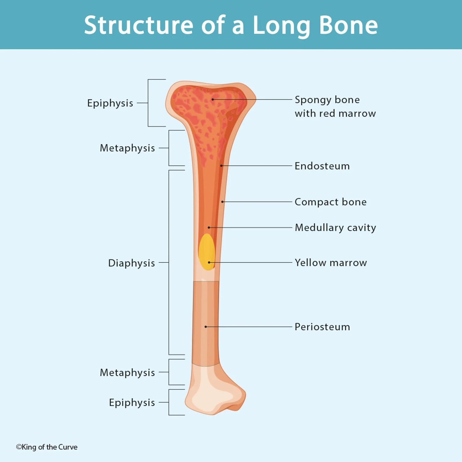
cortical bone
dense outer layer; gives structural rigidity
spongy (cancellous) bone
porous inner bone; absorbs shock and mechanical stress, preventing fractures from concentrated forces
medullar cavity
hollow cavity inside spongy bone containing bone marrow, the site of red blood cell formation
periosteum
covers the outside of bone
Latin: peri = "on top of"
Function: provides attachment sites for tendons/ligaments (soft tissue "blends in" with the periosteum at attachment points)
endosteum
lines the inside of the medullary cavity
Latin: endo = "inside"
Function: provides a smooth lining so blood cells can move through the marrow cavity without friction
composition of bone tissue
~50% minerals (calcium, phosphorus — provide strength)
~25% protein
~25% water (mostly interstitial fluid, not "drinking water")
Haversian canal
Central vertical canal running through bone; carries blood vessels/nutrients
Volkmann’s canal
Horizontal canals connecting Haversian canals
lamellae
Concentric rings of bone tissue (osteocytes) surrounding the Haversian canal
lacunae
Small spaces between lamellae containing interstitial fluid (a mix of lymph, proteins, blood vessels, WBCs, RBCs)
osteocytes
Mature bone cells arranged in the lamellar rings
osteogenic cells
"Blueprint"/stem-like precursor cells present from birth; differentiate into osteoblasts (and are associated with chondrocytes in early cartilage)
chondrocytes
Cartilage cells — present throughout the initially cartilaginous skeleton
osteoblasts
Bone-forming cells; combine with calcium (delivered via blood vessels) to form calcified matrix → become osteocytes
osteoclasts
Bone-destroying/resorbing cells; break down bone to release calcium/minerals into the bloodstream
osteocytes
Mature bone cells that make up the lamellar rings of formed bone
osteoporosis
caused by increased osteoclastic activity relative to bone formation → brittle bones due to low calcium levels. Treatment includes calcium supplementation.
osteophytes
(bone spurs) — form when arthritis causes chronic bone erosion; osteoblasts respond by forming new bone haphazardly (not in an organized fashion), creating bony outgrowths.
bone remodeling
(post-fracture) — osteoblasts initially form bone in a disorganized/bulging fashion; osteoclasts then reshape it back toward the original contour. This remodeling phase takes roughly 1 year, even though initial fracture union takes only ~3–6 weeks.
bone width growth
as osteoblasts add bone on the outside, osteoclasts resorb bone from the inside, allowing the medullary cavity to widen proportionally as the bone thickens.
embryological origin of bone
Fertilization → single cell → rapid division
Gastrulation: cells organize into 3 germ (dermal) layers:
Ectoderm (outer layer) → skin/epidermis, nervous system
Mesoderm (middle layer) → bone, muscle, cartilage, ligaments
Endoderm (inner layer) → GI tract, respiratory tract (internal "tube" structures)
Bone-forming cells originate from mesenchymal cells of the mesoderm, which differentiate into osteogenic cells and chondroblasts.
intramembranous ossification
Used for: flat bones (e.g., skull bones)
Process: osteogenic cells become trapped between two membranous layers; compression + blood supply → calcium delivery → osteoblasts form a single concentric ring of bone → compact, calcified flat bone
Weaker than endochondral bone — appropriate because flat bones (like the skull) are built for protection, not for withstanding heavy mechanical load
Begins in the embryo around 6–8 weeks gestation
In infants, skull bones are not yet fully fused (sutures remain cartilaginous) — full calcification of the skull occurs within about the first 2 years of life
endochondral ossification
Used for: most bones of the body, especially long bones
Process: cartilage is replaced by bone (not converted into bone)
Two ossification centers per long bone:
Primary ossification center — located in the diaphysis; ossification proceeds outward, this is the main growth driver in length
Secondary ossification center — located in the epiphysis
Mechanism:
Blood vessels proliferate near the cartilaginous template
Osteoblasts form rings around blood vessels (providing entry points into the cartilage)
Blood vessels deliver calcium → osteoblasts combine with calcium → form calcified matrix (concentric rings, similar to Haversian system)
Cartilage is progressively pushed toward the epiphysis/metaphysis as new bone forms at the diaphysis
Growth in length: occurs at the diaphyseal side, pushing the cartilage further toward the epiphysis over time (correction noted: bone length grows on the diaphyseal side, not "toward" the diaphysis)
Growth in width: occurs via the same peripheral osteoblast/calcification process, expanding outward
primary ossification center
located in the diaphysis; ossification proceeds outward, this is the main growth driver in length
endochondral ossification
secondary ossification center
located in the epiphysis
endochondral ossification
endochondral ossification mechanism
Blood vessels proliferate near the cartilaginous template
Osteoblasts form rings around blood vessels (providing entry points into the cartilage)
Blood vessels deliver calcium → osteoblasts combine with calcium → form calcified matrix (concentric rings, similar to Haversian system)
Cartilage is progressively pushed toward the epiphysis/metaphysis as new bone forms at the diaphysis
The Epiphyseal (Growth) Plate — 4 Zones (conceptual, not required to memorize by name)
Zone of proliferation — osteoblasts proliferating from the diaphyseal side
Two transitional zones — osteoblasts receiving blood supply and calcium, becoming calcified matrix
Cartilaginous zone (near metaphysis) — remains cartilaginous; the "incentive" driving continued bone growth
Once growth is complete, all 4 zones fuse into a single epiphyseal line.
Articular cartilage remains cartilaginous for life (never fully ossifies) — reduces joint friction.
"Double-jointedness" = a colloquial term referring to persistent cartilage at the epiphyseal line, allowing extra mobility/flexibility at that joint (not a fully fused, rigid union).
fracture healing
(Same General Process as Ossification)
hematoma formation
cartilaginous callus formation
bony union
remodeling
hematoma formation
~6-8 hours post-fracture
Ruptured blood vessels bleed → clot forms around fracture site for protection; brings in phagocytes (WBCs that clear debris) and osteoblasts
cartilaginous callus formation
following hematoma
Osteogenic cells → osteoblasts form a cartilaginous matrix around the fracture site
bony union
~6 weeks
Blood vessels deliver calcium; osteoblasts form calcified matrix; bone is united across the fracture — but the healed area may look bulkier/thicker than the original bone (osteoblasts lack "organizational" precision)
remodeling
~1 year
Osteoclasts reshape and smooth the newly formed bone back toward its original structure
growth plate fractures
pediatric concern
In children/pre-pubertal individuals, fracture of the epiphysis/growth plate is a serious concern because it can disrupt future bone growth.
Potential consequence: limb length discrepancy (e.g., femur growth plate fracture → leg length difference)
Once the growth plate is fully ossified (post-puberty), fractures there heal like any other fracture, with no further growth-related concerns
bony landmarks
Soft tissue (muscles, tendons, blood vessels, nerves) exerts force on bone even while it is still cartilaginous in early development, creating characteristic surface markings
condyle
Rounded bony prominence
Medial/lateral femoral condyles
tuberosity
Elevated bump (often muscle/tendon attachment)
Tibial tuberosity (quadriceps tendon attachment)
foramen
An opening/hole (for vessels/nerves)
Obturator foramen (pelvis)
groove
Depression/gutter (often for a tendon or vessel)
facet
Flat surface formed where two bones articulate
Patellar facets (articulate with femur); vertebral facet joints
head/neck of bone
Rounded proximal end / narrowed region below the head
Head of femur, head of humerus, neck of femur
tendons
Connect muscle to bone (muscle fibers do NOT attach directly to bone)
ligaments
Connect bone to bone; often layered on top of the articular capsule
articular capsule
Surrounds each synovial joint
cartilage
Covers articular (joint) surfaces to reduce friction
hyaline cartilage
Thin, bluish tinge; found on the surface of virtually every bone; relatively weaker/less tough
General bone surfaces (jaw, clavicle, etc.)
fibrocartilage
Strongest type; fibrous + cartilaginous — built to withstand significant joint forces
Articular cartilage (joint surfaces), menisci (knee), intervertebral discs
elastic cartilage
Highly elastic, flexible
External ear, nose (nares), epiglottis, trachea
long bone
Length > width
Most extremity bones (humerus, femur, etc.) — but NOT carpals/tarsals
flat bones
Flat, plate-like structure
Skull bones — frontal bone, parietal bones, occipital bone (cranial vault)
irregular bones
No consistent/standard shape; adapted to surrounding structures
Maxilla, nasal bone, ethmoid bone, sphenoid bone, vertebrae
short bones
Width > length; small, compact
Carpal bones (wrist), tarsal bones (foot)
Sesamoid bones
Small, pea/sesame-shaped; embedded within a tendon/soft tissue
Patella (largest example), sesamoid bones at base of the great toe and thumb
Acts like a pulley within a tendon, improving the mechanical efficiency of the associated muscle.
Example: The patella, embedded in the quadriceps tendon, provides mechanical advantage during knee extension.
Clinical note: Without a patella (e.g., after removal due to fracture or tumor), the quadriceps must generate ~60% more effort to produce the same knee extension force.
synarthrosis
immobile
Skull sutures (parietal-parietal, parietal-frontal)
functional joint classification
diarthrosis
Highly mobile — most common joint type in the body
Shoulder, elbow, hip, knee, AC joint, sternoclavicular joint, costovertebral joints
functional joint classification
amphiarthrosis
Slightly mobile (mobility + stability)
Symphysis pubis (junction of the two pubic bones); allows slight alternating pelvic motion during gait
functional joint classification
fibrous joints
Bones connected by fibrous connective tissue
Generally synarthrotic (immobile)
Skull sutures
synovial joints
Joint enclosed by an articular capsule + synovial membrane, which secretes synovial fluid to reduce friction
Generally diarthrotic (highly mobile)
Hip, knee, shoulder
cartilaginous
Bones connected by cartilage
Generally amphiarthrotic
Two subtypes of cartilaginous joints:
Primary cartilaginous joints — temporary; cartilage present only until full ossification occurs
Example: Epiphyseal plate (cartilage between diaphysis and epiphysis during growth)
Secondary cartilaginous joints — cartilaginous for life
Example: Intervertebral discs (fibrocartilaginous discs between vertebral bodies); slightly mobile
primary cartilaginous joints
temporary; cartilage present only until full ossification occurs
Example: Epiphyseal plate (cartilage between diaphysis and epiphysis during growth)
secondary cartilaginous joints
cartilaginous for life
Example: Intervertebral discs (fibrocartilaginous discs between vertebral bodies); slightly mobile