Looks like no one added any tags here yet for you.
T or F, the basement membrane of the corneal epithelium is an ECM
T
ECM can be divided into two categories:
pericellular matrix and interstitial matrix
ECM is a dynamic, 3D network of molecules that…
provide structural support for cells and tissues
The basement membrane of epithelia and glycocalyx are what category of ECM?
pericellular matrix
Stroma of cornea (and cartilage/bone) is what category of ECM?
interstitial matrix
What cell/tissue processes are regulated by ECM?
proliferation, migration, differentiation, angiogenesis, immune function, autophagy, and tissue separation/shaping
Major components of ECM (just to see them)
proteoglycans, collagens, MMPs, elastic fibers, lysyl oxidase, laminin, tenascin, fibronectin
Significance of proteoglycans having negative charge in ECM
binds to water
MMPs
matrix metalloproteinases, break down components of ECM
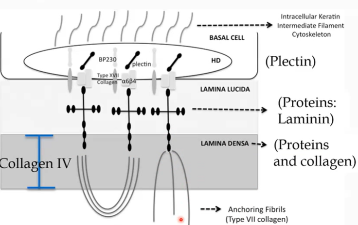
Two layers of any basement membrane
lamina lucida, lamina densa
What is the principal component of lamina lucida of any basement membrane?
Laminin (protein)
Principal component of lamina densa
Collagen IV
What are the anchoring fibrils between lamina densa to sublamina densa?
Collagen type VII
What adheres BM (ECM) to cells?
hemidesmosomes
Hemidesomosomes are in the __ membrane of corneal epithelial cells
basal membrane
What are the components of hemidesmosomes?
intermediate filaments, plectin, integrins
What are intermediate filaments?
A structural cytoskeletal component that strengthens hemidesmosomes
What is plectin?
forms intracellular plaque in hemidesmosomes, connects intermediate filament with integrins
What are integrins?
transmembrane proteins in PM, bind to laminin extracellularly (in lamina lucida), and Plectin (plaque) intracellularly
Describe molecular structure of Collagen IV
Molecular subunit of 4 proteins; chicken wire-like network → strong BM
What might cornea erosions indicate is wrong?
failure of adherence of corneal epithelial cells to BM/Bowman’s via hemidesmosomes (EMBD)
T or F, there is no pain associated with cornea erosions
F, exposed nerve endings induces pain
What is the most common cause of cornea erosions?
corneal injury (trauma)
What diseases come with genetic predisposition to cornea erosions?
Alport’s syndrome, Epidermolysis bullosa
Alport’s syndrome
mutations in Collagen IV → epithelial cell death → cataracts
Epidermolysis bullosa
disruptions in other hemidesmosomal/BM genes
EBMD names
Epithelial BM dystrophy aka Cogan’s microcystic epithelial dystrophy aka map-dot-fingerprint dystrophy
EMBD presentation
map-like lines and subepithelial microcysts where BM has intruded into more superficial layers of cornea
In EBMD, does BM have abnormal thickened or thinned region?
thickened, extra deposition of ECM
2 characteristics of stem cells
1) unspecialized/undifferentiated capable of renewing themselves through cell division
2) have potential to differentiate into a specific cell type
Stem cells can be grouped into two kinds:
1) Embryonic stem cells (pluripotent or totipotent)
2) Somatic “adult” stem cells (multipotent)
pluripotent
can give rise to all tissue cell types
multipotent
can give rise to multiple cell types
unipotent
gives rise to a single cell time
All ocular tissues are derived from what pluripotent stem cell line?
ectoderm (ocular structures) and mesoderm (vasculature, muscles)
Transit Amplifying Cell
highly proliferative cell derived from stem cell, after it committed to developing towards a specific line
Which has a higher proliferation rate, stem cells or transit amplifying cells?
TACs, don’t want stem cells to proliferate uncontrollably
Describe X+Y=Z
X: basal cell proliferation anteriorly (mitosis)
Y: centripetal movement of the epithelial cells from limbus towards center
Z: cells being shed from surface
Describe the conflicting hypotheses of LESC vs CESC
LESC: SC found exclusively in limbus, and TACs move/divide centripetally to refresh corneal epithelium during homeostasis and injury
CESC: SC in limbus AND basal cornea epithelium, TACs move/divide centrifugally. CESCs contribute during homeostasis and injury, while LESCs contribute only during injury
What causes exfoliation/desquamation of corneal epithelial cells?
constant cell loss (sloughing off)
Blinking
Minor abrasions
Eye rubbing
Centripetal movement of cells (~10-15mm/day) is likely due to __ and causes entire corneal epithelium to be renewed in __ months
growth pressure; 9-12 months
Where are limbal epithelial thickenings (source of epithelial cells)?
between palisades of Vogt
What is the source of epithelial cells?
limbal epithelial thickenings between palisades of Vogt
There is more or less proliferation in the posterior/peripheral cornea
More proliferation
LSC location
Limbal stem cells are in the limbal basal cells
Do LSCs (limbal basal cells) have high or low mitotic activity? Short or long life span?
Low; long
LSCs are __potent
multipotent
When needed, cell division of LSC produces what 2 cells?
1 remains a multipotent LSC
1 becomes a TAC and differentiates
What happens if the LSC becomes a TAC?
TAC has increased mitosis rate, migrates centripetally toward center and anteriorly (become wing and superficial cells)
Do TACs become more or less multipotent with each division?
less
When do TACs become post-mitotic (terminally differentiated)?
stop dividing once anterior?
Stem cell “niche”
region that protects stem cell population and helps maintain their multipotency
What is the stem cell niche of limbal stem cells?
epithelial thickenings in limbus
What factors aid multipotency in niche?
Thickness of epithelium
Amount of melanin pigments
Array of BM proteins different from central cornea to promote stemness and proliferation
Presence of nearby blood vessels for O2, GFs, antioxidants
What do blood vessels provide for stem cells in a niche?
O2, growth factors, antioxidants
Where are stromal stem cells relative to the limbal basal cells?
subjacent to limbal basal cells
What SC niche likely nurtures stromal stem cells?
limbal stem cell niche
What is the definition of stem cells that stromal SCs fit?
can self renew
Are multipotent
What do stromal stem cells differentiate into?
keratocytes
Conjunctival stem cells are concentrated in __ conjunctiva or spread throughout conj, not sure
fornicial conj
What do conjunctival stem cells differentiate into (they are multipotent)?
epithelial and goblet cells
How are conjunctival stem cells different from limbal stem cells?
Conj SCs do not help repopulate cornea (don’t express corneal specific proteins), and limbal don’t supply the conj(?)
What happens if limbal stem cells can’t repopulate corneal epithelium?
conjunctival stem cells spread into cornea
What would patient complain of with a limbal stem cell deficiency?
blurry vision, foreign body sensation, photophobia, tearing, pain
What can be observed with slit-lamp with limbal stem cell deficiency?
Loss of palisades of Vogt
Would corneal epithelium thin or thicken with limbal stem cell deficiency?
thin, limbal SCs not repopulating cornea
What can limbal SC deficiency be caused by?
autoimmune disorders, chemical/thermal injury, contact lens wear, surgical damage, infections, congenital malformations…
OSSN general definition and presentation
Ocular Surface Squamous Neoplasia: stem cells divide inappropriately → range from mild dysplasia to invasive squamous cell carcinoma
What areas are more susceptible to OSSN?
conj of limbus (from basal limbal stem cells); also affects cornea
Risk factors for OSSN
UV exposure, HIV, HPV
Pterygium
encroachment of bulbar conj onto the cornea
What resembles an aberrant wound healing response?
pterygium
2 potential etiologies of pterygium
Mutation of limbal stem/epithelial cells → proliferate/differentiate into conj cells
Destruction of limbal stem/epithelial cells (usually a barrier to conj proliferation into cornea) = local limbal stem cell deficiency
Which involves zigzag vessel patterns, OSSN or pterygium?
OSSN
Which involves straight vessel patterns of neovascularization, OSSN or pterygium?
pterygium
Which involves surface keratinization, OSSN or pterygium?
OSSN
Which can become invasive/malignant, OSSN or pterygium?
OSSN
Which involves growth of subepithelial conjunctival fibroblastic tissue over cornea, OSSN or pterygium?
pterygium
Which variably involves growth of conj, limbal/cornea epithelia, OSSN or pterygium?
OSSN
Which involves underlying stroma of activated fibroblasts and ECM remodeling, OSSN or pterygium?
pterygium
Which involves goblet cell hyperplasia, OSSN or pterygium?
pterygium
What is a leading edge of altered limbal epithelial cells characteristic of?
pterygium
Pinguecula
UV damage of fibroblasts beneath epithelium → inappropriate (excess) production of EM proteins (elastin) and possible increase in fibroblast cell number
Where do pinguecula occur?
bulbar conj and limbus
What is a creamy-colored, chalky growth on conjunctival surface characteristic of?
pinguecula
Pinguecula vs pterygium
Pinguecula is more extracellular mass from some damage of cells (no proliferation), pterygium is mutation in p53 (tumor supressant) → fibroblasts proliferate
How can pingueculas affect limbal stem/epithelial cells?
extracellular mass beneath epithelium pushes epi superficially, disrupting protective mechanisms (more exposed to UV, pushed away from vasculature of niche) → can cause mutation of DNA in limbal stem/epithelial cells → pterygium and pinguecula can occur together
Stages of epithelial wound repair
Stage 1. Latent phase, 4-6 hrs after wound - recruit immune cells, change molecular pathways, secreting EGF
Stage 2: Cell migration - cover cells
Stage 3: Epithelial mitosis - make up for loss of cells, activating stem cell pathway
Stage 4: Reassembly of adhesive contacts (hemidesmosomes)
What morphology changes occur in the epithelial cells in the latent phase of epithelial wound repair?
Loss of surface microplicae/glycocalyx
Retraction of epithelial cells at wound edge
Basal cells flatten
In what stage of epithelial wound repair is there signaling/chemotaxis? What is released?
Stage 1, latent phase; cellular stress causes release of Ca2+, H2O2, and nerve damage releases cytokines and substance P
What happens to the basement membrane components (hemidesmosomes, ECM…) in latent phase of epithelial wound repair?
They degrade
What facilitates degradation of hemidesmosomes and ECM in latent phase?
PMNs phagocytize damaged cells and signal for more PMNs
T or F, mitosis is stimulated in latent phase
F, mitosis is inhibited
PMNs infiltrate tears from…
lymphoid follicles and diffuse lymphoid tissue beneath conj
PMNs infiltrate stroma from…
limbal blood vessels upon corneal abrasion
How many cells thick is the leading edge of migrating cells during stage II of epithelial wound repair?
one cell thick
Focal adhesions in migration in wound repair are attachments of what molecules?
actomyosin and transmembrane integrins
What do hemidesmosomes attach to in ECM?
intermediate filaments
What cellular processes help epithelial cells sense the environment in cell migration, and extend from margin of epithelial wound?
lamellipodia (broad) and filopodia (narrow)
What are lamellipodia and filopodia (or cell migration?) driven by?
cytoskeleton polymerization - actin, myosin