Chapter 11: Cranium Long: Merrill’s Atlas of Radiographic Positioning and Procedures, 14th Edition
1/251
There's no tags or description
Looks like no tags are added yet.
Name | Mastery | Learn | Test | Matching | Spaced | Call with Kai |
|---|
No analytics yet
Send a link to your students to track their progress
252 Terms
How many bones make up the cranium?
a. 4
b. 6
c. 8
d. 10
c. 8
How many bones make up the face?
a. 4
b. 10
c. 12
d. 14
d. 14
The cranial bones are rigidly jointed together by articulations called:
a. joints.
b. bursae.
c. sutures.
d. cartilage.
c. sutures.
All of the following are cranial bones except the:
a. maxillae.
b. frontal.
c. sphenoid.
d. occipital.
a. maxillae.
All of the following are facial bones except the:
a. ethmoid.
b. maxillae.
c. mandible.
d. zygomatic bones.
a. ethmoid.
Which skull suture is found between the frontal and parietal bones?
a. Sagittal
b. Coronal
c. Squamosal
d. Lambdoidal
b. Coronal
Which skull suture is located between the parietal bones?
a. Hyoid
b. Coronal
c. Sagittal
d. Squamosal
c. Sagittal
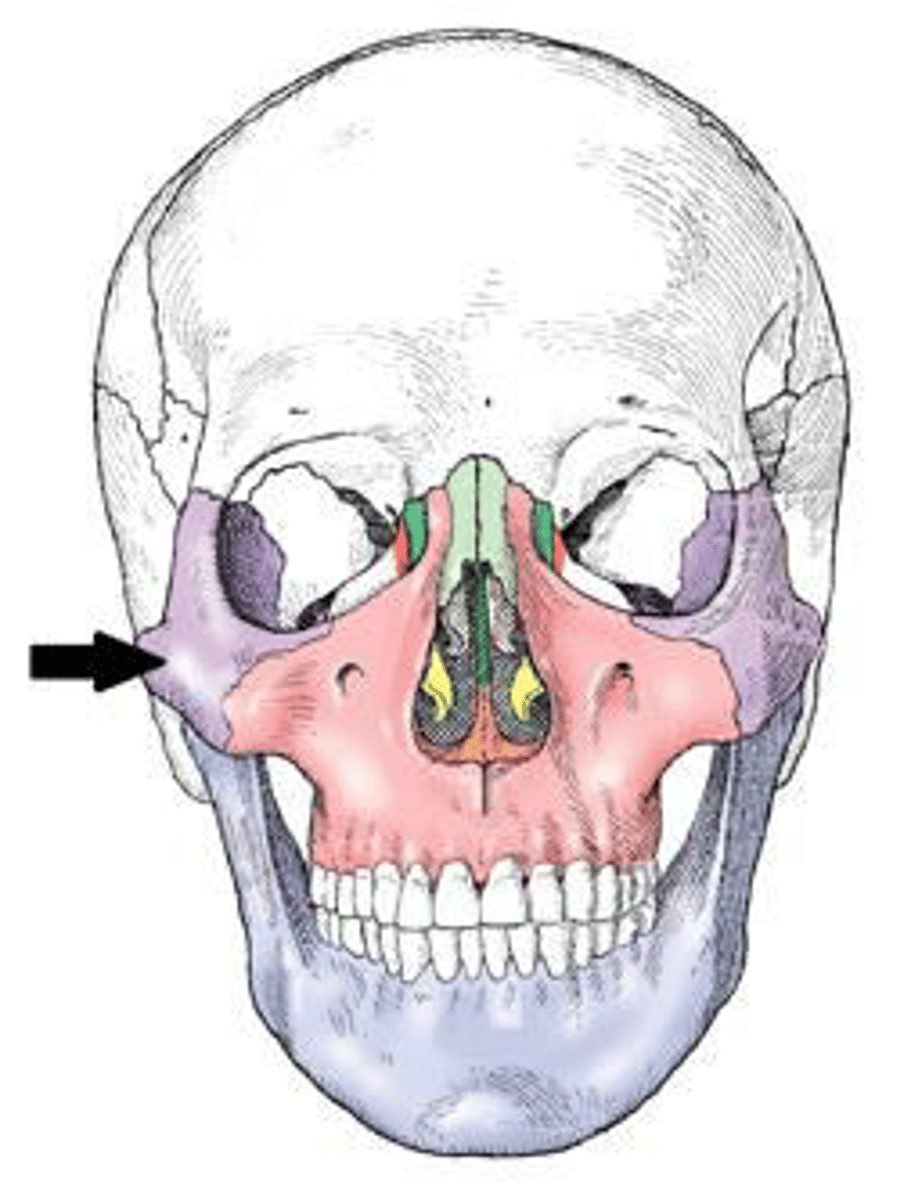
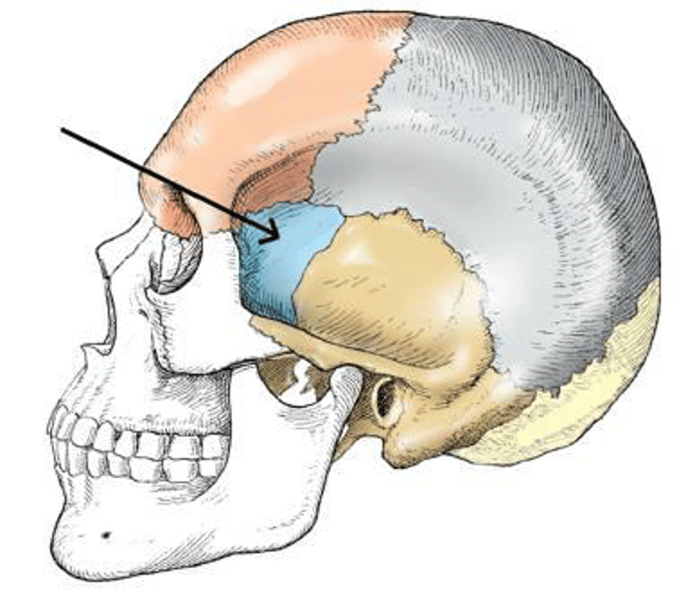
The bone indicated in the figure below is the:
a. parietal.
b. orbital.
c. temporal.
d. zygoma
d. zygoma
3 multiple choice options
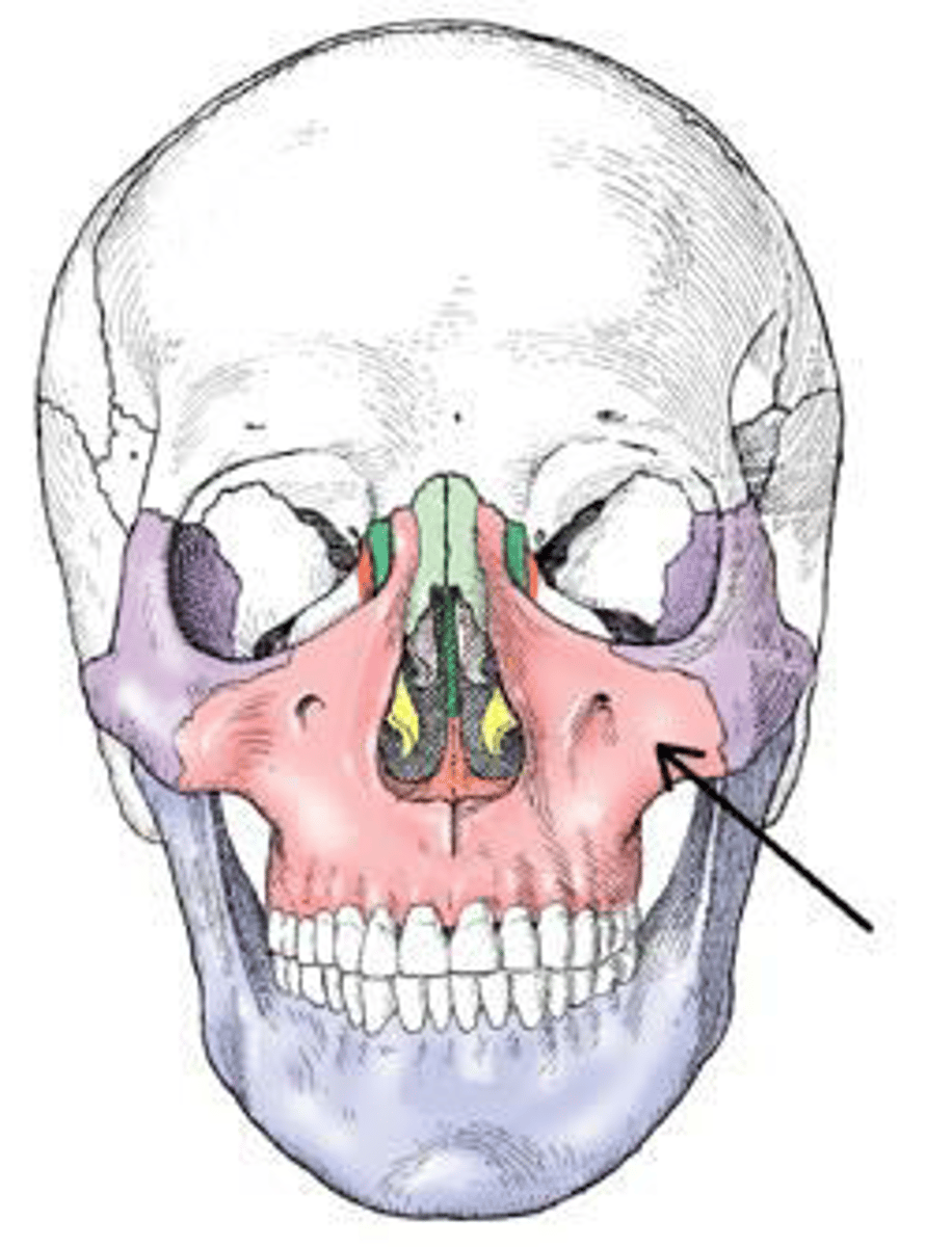
The bone identified in the figure below is the:
a. maxilla.
b. frontal.
c. mandible.
d. ethmoid.
a. maxilla.
3 multiple choice options
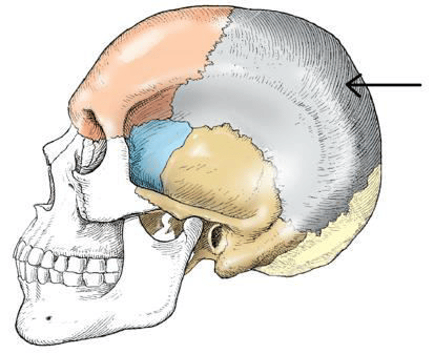
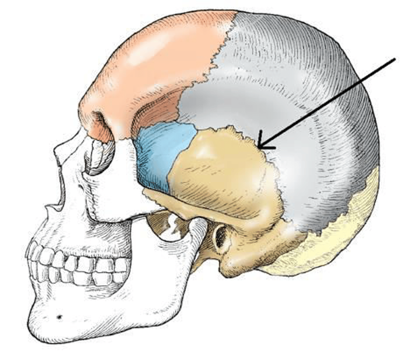
The bone indicated in the figure below is the:
a. temporal.
b. parietal.
c. occipital.
d. sphenoid.
b. parietal.
3 multiple choice options
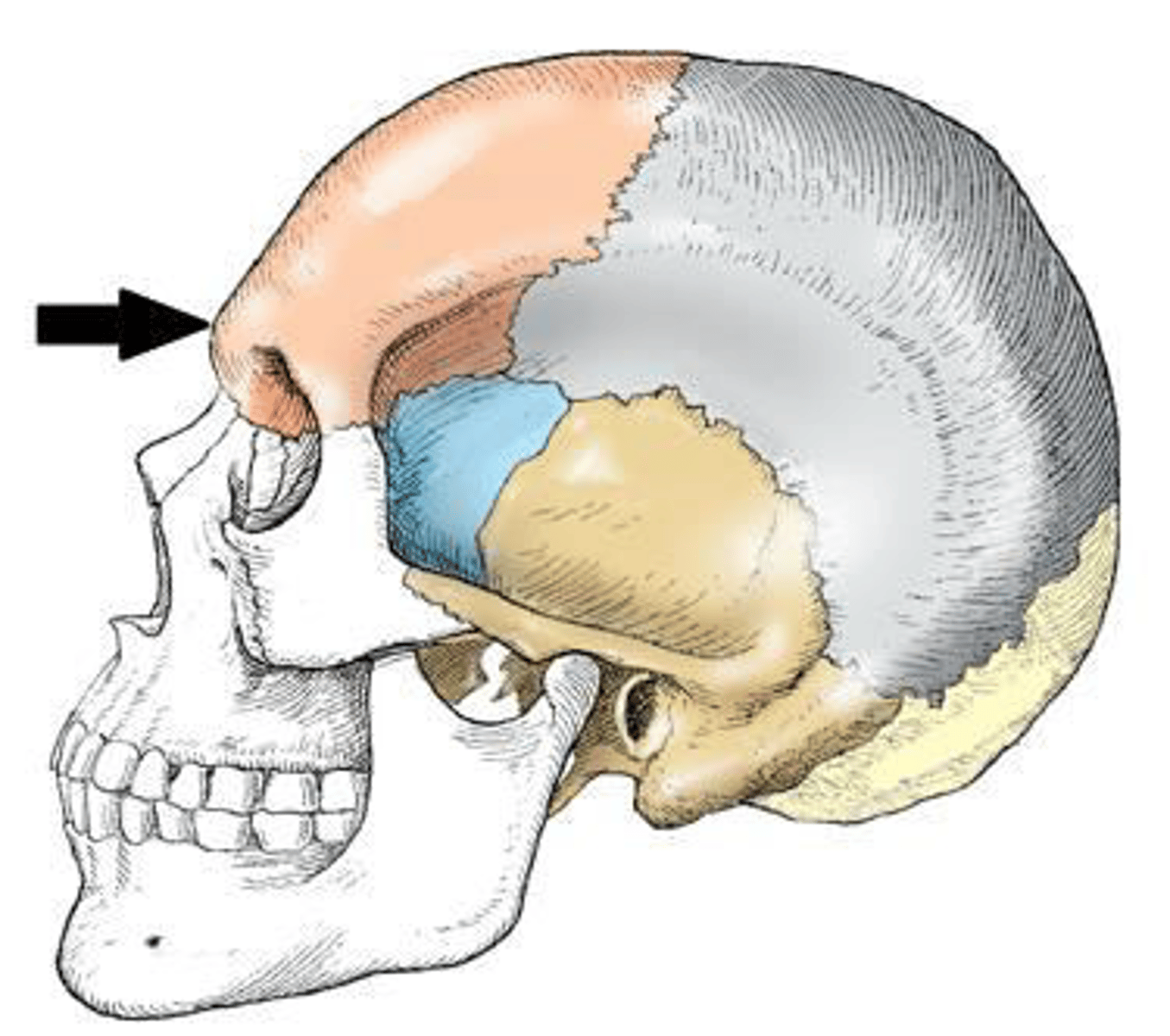
The part of the frontal bone indicated in the figure below is the:
a. bregma.
b. lambda.
c. glabella.
d. acanthion
c. glabella.
3 multiple choice options
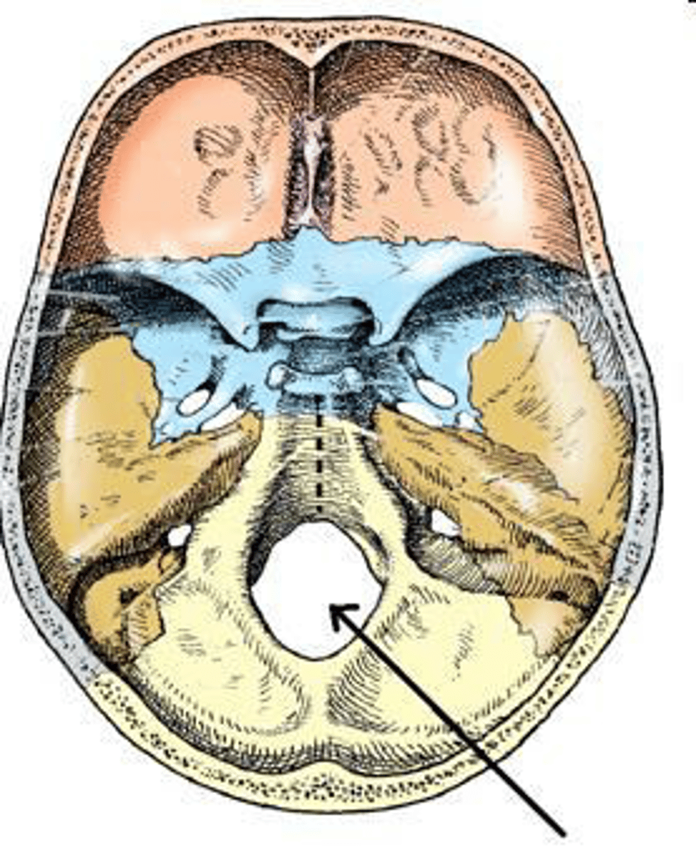
The part of the cranial base identified in the figure below is the:
a. sella turcica.
b. foramen ovale.
c. hypoglossal canal.
d. foramen magnum.
d. foramen magnum.
3 multiple choice options
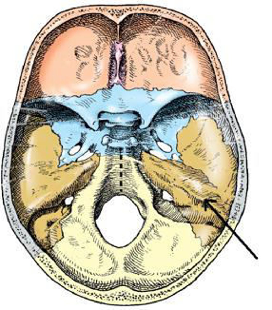
The part of the cranial base identified in the figure below is the:
a. auditory canal.
b. dorsum sellae.
c. greater wing.
d. petrous portion.
d. petrous portion.
3 multiple choice options
All of the following bones contain air sinuses, except:
a. frontal.
b. parietal.
c. ethmoid.
d. sphenoid.
b. parietal.
Which bone has condyles that articulate with the atlas of the cervical spine?
a. Temporal
b. Occipital
c. Parietal
d. Foramen magnum
b. Occipital
The zygomatic arches are a part of which bone?
a. Frontal
b. Parietal
c. Temporal
d. Sphenoid
c. Temporal
Which bone in the skull contains the auditory organs and the organs of hearing?
a. Temporal
b. Sphenoid
c. Occipital
d. Ethmoid
a. Temporal
The petromastoid portion is a part of which bone?
a. Temporal
b. Sphenoid
c. Occipital
d. Ethmoid
a. Temporal
The vestibulocochlear organ is the organ of:
1. hearing.
2. sensation.
3. balance.
a. 1 and 2
b. 1 and 3
c. 2 and 3
d. 1, 2, and 3
b. 1 and 3
Which of the following is located in the middle ear?
a. Cochlea
b. Bony labyrinth
c. Tympanic membrane
d. External acoustic meatus
c. Tympanic membrane
Which of the following is located in the internal ear?
a. Concha
b. Auditory tube
c. Tympanic membrane
d. Semicircular canals
d. Semicircular canals
The maxillary sinus is located in which bone?
a. Temporal
b. Sphenoid
c. Maxilla
d. Ethmoid
c. Maxilla
The largest and most dense bone of the face is the:
a. maxilla.
b. mandible.
c. frontal.
d. sphenoid
b. mandible.
The small bone situated at the base of the tongue is the:
a. hyoid.
b. alveolar.
c. cornu.
d. styloid.
a. hyoid.

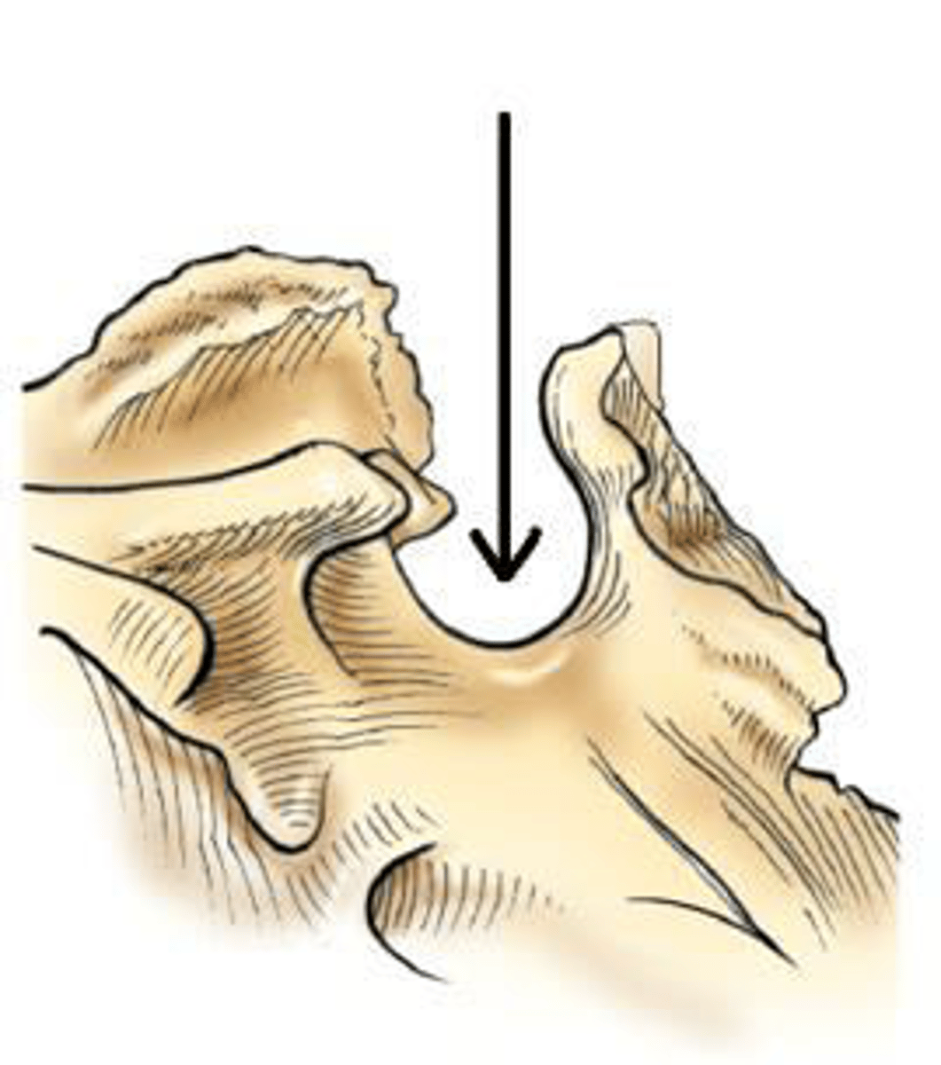
The part of the mandible identified in the figure below is the:
a. body.
b. ramus.
c. symphysis.
d. alveolar portion
a. body.
3 multiple choice options
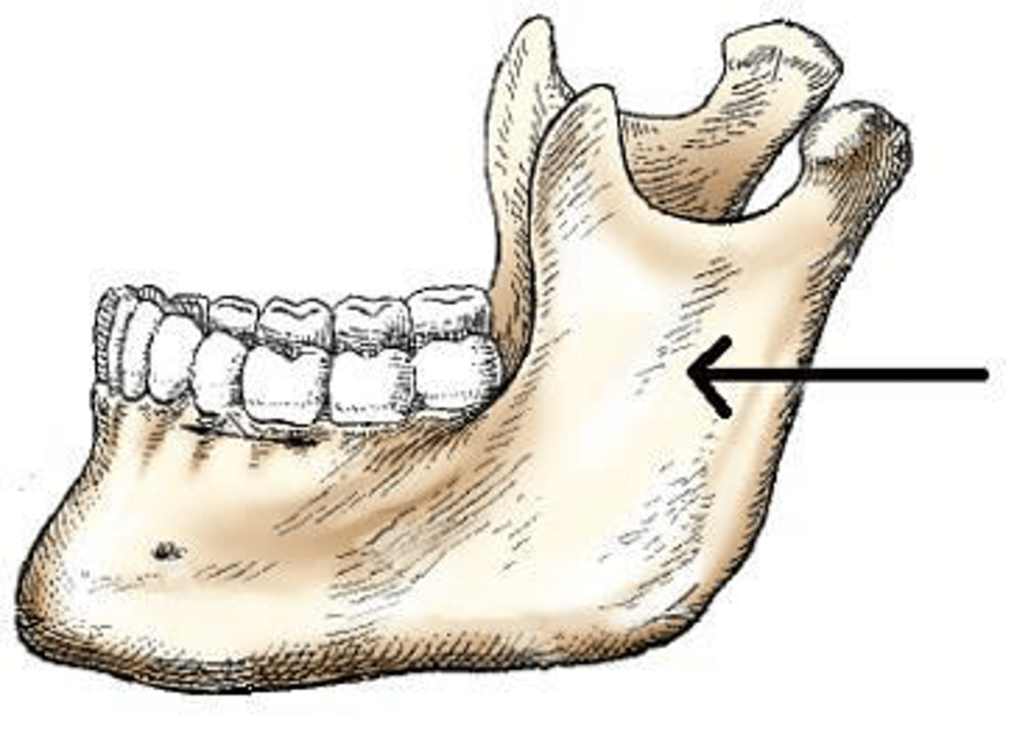
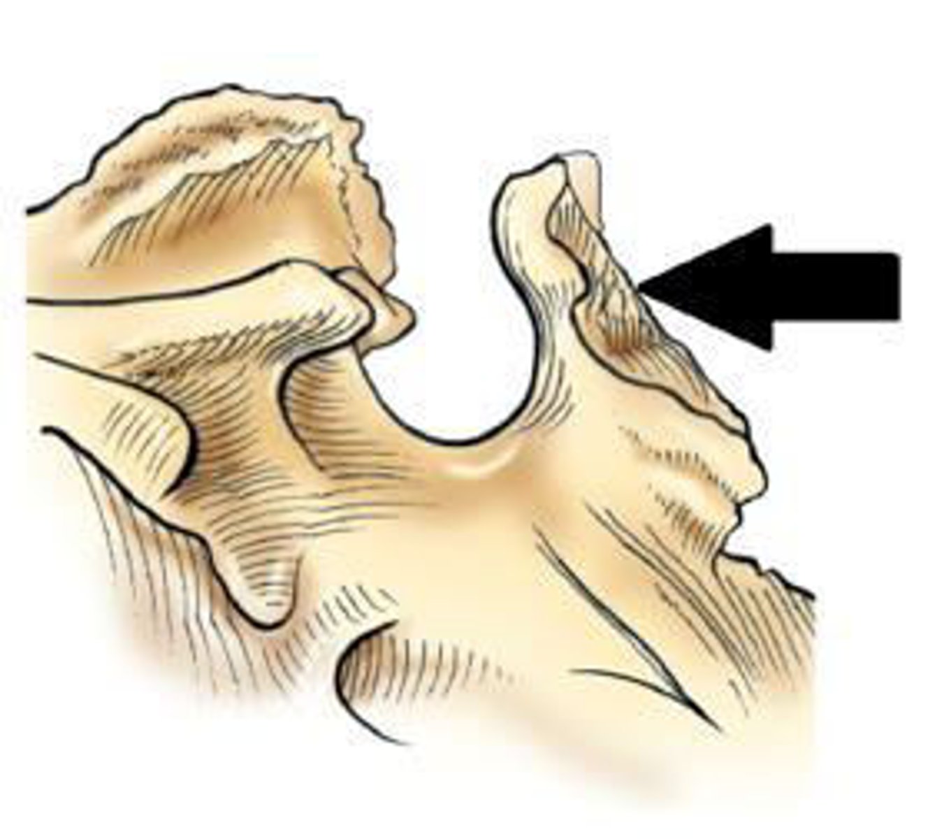
The portion of the mandible identified in the figure below is the:
a. body.
b. ramus.
c. symphysis.
d. alveolar portion.
b. ramus.
3 multiple choice options
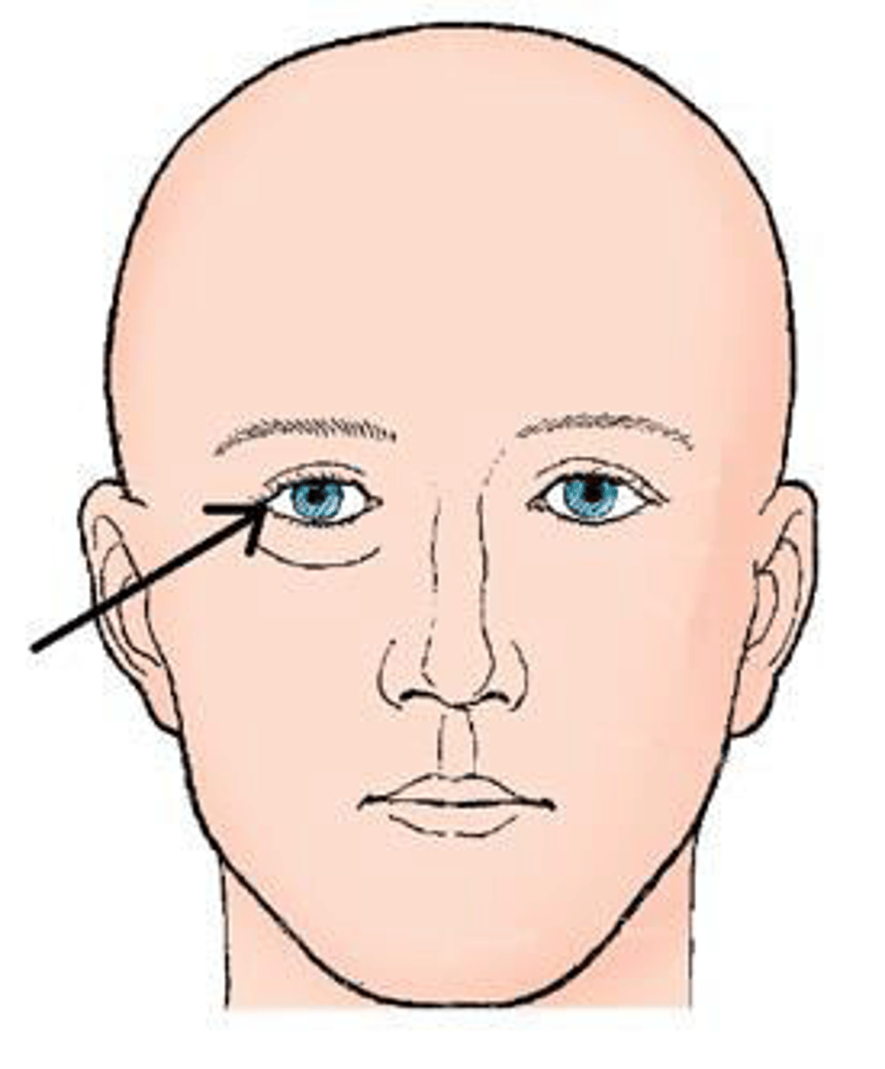
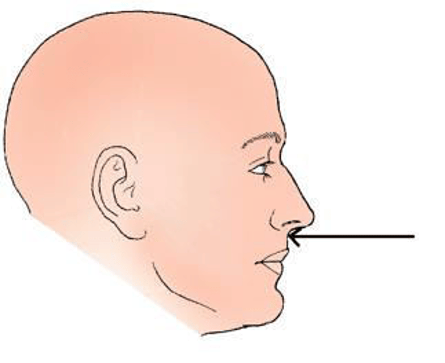
The external landmark identified in the figure below is the:
a. glabella.
b. acanthion.
c. outer canthus.
d. infraorbital margin
c. outer canthus.
3 multiple choice options
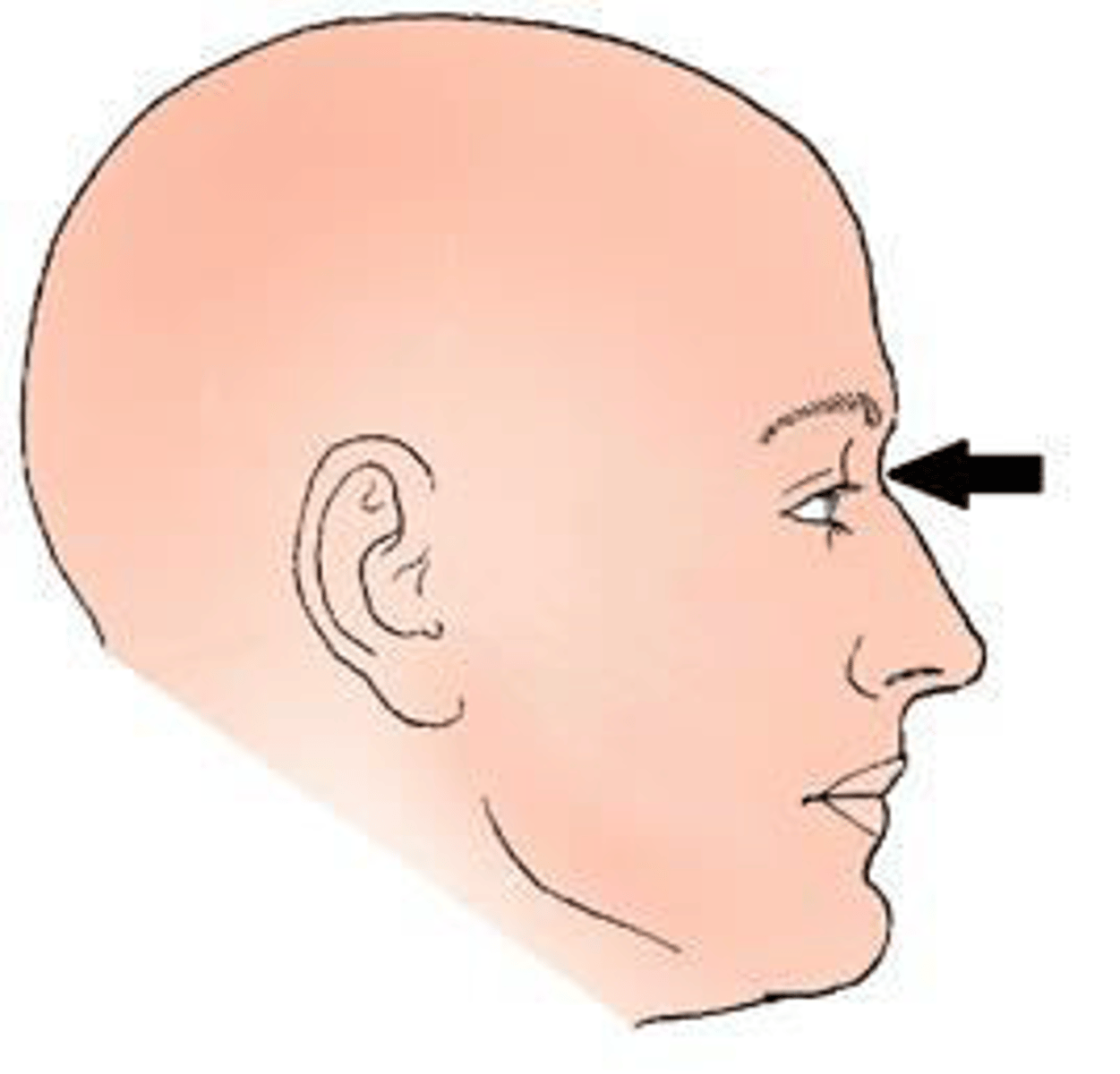
The landmark identified in the figure below is the:
a. nasion.
b. glabella.
c. acanthion.
d. auricular point
c. acanthion.
3 multiple choice options
The landmark identified in the figure below is termed the:
a. nasion.
b. glabella.
c. acanthion.
d. inner canthus
a. nasion.
3 multiple choice options
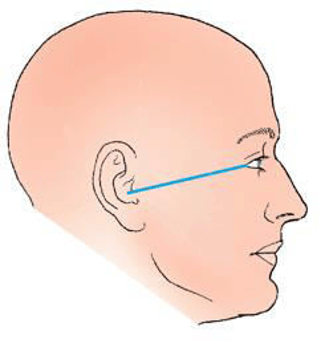
The topographic line shown in the figure below is the:
a. glabellomeatal.
b. orbitomeatal.
c. acanthiomeatal.
d. infraorbitomeatal.
b. orbitomeatal.
3 multiple choice options
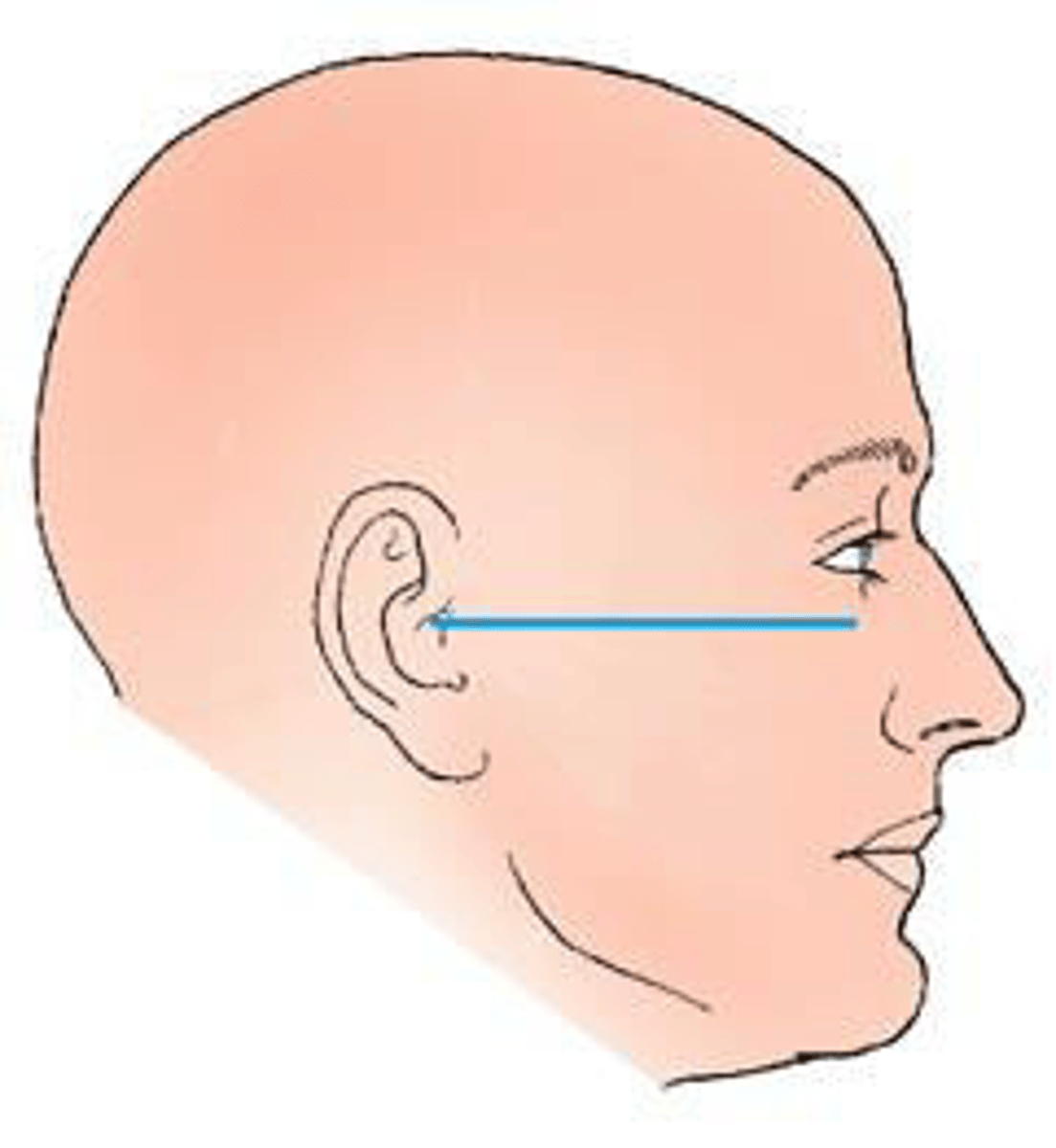
The topographic line identified in the figure below is the:
a. orbitomeatal.
b. mentomeatal.
c. acanthiomeatal.
d. infraorbitomeatal.
d. infraorbitomeatal.
3 multiple choice options
Which of the following skull types is considered average in size and shape?
a. Mesocephalic
b. Brachycephalic
c. Dolichocephalic
a. Mesocephalic
Which skull type is narrow from side to side?
a. Mesocephalic
b. Dolichocephalic
c. Brachycephalic
b. Dolichocephalic
In a typically shaped head, the petrous pyramids project anteriorly and medially at what
angle?
a. 37 degrees
b. 40 degrees
c. 47 degrees
d. 54 degrees
c. 47 degrees
Which plane of the head is placed parallel to the plane of the image receptor for a lateral
projection of the skull?
a. Interpupillary
b. Transverse
c. Midsagittal
d. Midcoronal
c. Midsagittal
The central ray and center of the image receptor position for a lateral projection of the skull is
_____ inch(es) _____ the EAM.
a. 1; below
b. 2; below
c. 1; above
d. 2; above
d. 2; above
Which projection of the skull requires MSP be positioned parallel and interpupillary line
perpendicular to the IR plane?
a. Lateral
b. PA axial (Caldwell)
c. AP axial (Towne)
d. SMV (Schüller)
a. Lateral
All of the following should be seen on a lateral image of the skull, except:
a. superimposed orbital roofs.
b. sella turcica in profile.
c. temporomandibular joints superimposed.
d. mandible overlapping cervical spine.
d. mandible overlapping cervical spine.
Which method of examining the skull will demonstrate the petrous ridges in the orbits, the ethmoid and frontal sinuses, and the crista galli?
a. Towne
b. Caldwell
c. Schüller
d. Waters
b. Caldwell
The central-ray angle for the PA axial (Caldwell) projection of the skull is:
a. 5 degrees cephalad.
b. 10 degrees cephalad.
c. 12 degrees caudad.
d. 15 degrees caudad.
d. 15 degrees caudad.
Which of the following is perpendicular to the image receptor plane for a Caldwell projection of the skull?
a. Glabellomeatal line
b. Acanthiomeatal line
c. Orbitomeatal line
d. Mentomeatal line
c. Orbitomeatal line
Often a patient cannot be turned into the prone position for a PA axial projection of the skull (Caldwell method). What central-ray angle would be used if the AP axial projection is used instead?
a. 10 degrees caudad
b. 15 degrees cephalad
c. 10 to 15 degrees caudad
d. 10 to 15 degrees cephalad
b. 15 degrees cephalad
Which of the following lines is placed perpendicular to the image receptor plane for the AP axial (Towne) projection?
a. Orbitomeatal line
b. Supraorbitomeatal line
c. Glabellomeatal line
d. Acanthiomeatal line
a. Orbitomeatal line
If the patient cannot flex the neck to place the orbitomeatal line perpendicular to the image receptor for an AP axial (Towne) projection, which line should be placed perpendicular?
a. Acanthiomeatal line
b. Infraorbitomeatal line
c. Glabellomeatal line
d. Mentomeatal line
b. Infraorbitomeatal line
For an AP axial (Towne) projection of the skull, the center of the IR is at or near the level of the:
a. foramen magnum.
b. EAM.
c. glabella.
d. acanthion.
a. foramen magnum.
Which method of examining the skull is identified in the figure below?
a. Haas
b. Towne
c. Shüller
d. Caldwell
b. Towne
3 multiple choice options
If the infraorbitomeatal line is placed perpendicular to the image receptor during an AP axial
(Towne) projection of the skull, how much is the central ray angled?
a. 15 degrees caudad
b. 30 degrees caudad
c. 37 degrees caudad
d. 45 degrees caudad
c. 37 degrees caudad
What is the central-ray angulation for demonstration of the entire foramen magnum during an AP axial (Towne) projection?
a. 37 degrees caudad
b. 40 degrees caudad
c. 60 degrees caudad
d. 40 to 60 degrees caudad
d. 40 to 60 degrees caudad
What is the average central-ray angulation for the PA axial (Haas) projection of the skull?
a. 25 degrees caudad
b. 25 degrees cephalad
c. 30 degrees caudad
d. 30 degrees cephalad
b. 25 degrees cephalad
Which line should be placed parallel to the plane of the image receptor for the SMV
projection of the cranial base?
a. Acanthiomeatal line
b. Orbitomeatal line
c. Infraorbitomeatal line
d. Mentomeatal line
c. Infraorbitomeatal line
Radiographic demonstration of the cranial base is performed by which method?
a. Haas
b. Rhese
c. Towne
d. Schüller
d. Schüller
What is the central-ray angulation for the SMV projection?
a. 0 degrees
b. 5 degrees caudad
c. 5 degrees cephalad
d. 5 to 7 degrees cephalad
a. 0 degrees
The x-ray projection demonstrated in the figure below is the:
a. SMV.
a. SMV.
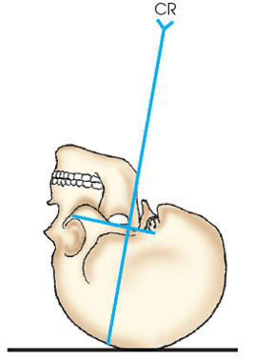
For an SMV projection of the cranial base, the central ray should always be perpendicular to the _____ line.
a. mentomeatal
b. orbitomeatal
c. infraorbitomeatal
d. acanthiomeatal
c. infraorbitomeatal
All are clearly demonstrated on an SMV projection of the cranial base, except:
a. carotid canals.
b. sphenoid sinuses.
c. mastoid process.
d. frontal sinuses.
d. frontal sinuses.
How many bones are contained in the skull?
a. 8
b. 14
c. 22
d. 24
c. 22
The bones of the cranium are joined together by fibrous joints called:
a. diploë.
b. sulci.
c. sutures.
d. cartilage.
c. sutures.
The suture located between the occipital bone and the parietal bones is the:
a. lambdoidal.
b. squamosal.
c. sagittal.
d. coronal.
a. lambdoidal.
The suture identified on the figure below is the:
a. coronal.
b. squamosal.
c. sagittal.
d. lambdoidal.
b. squamosal.
The bone identified on the skull below is the:
a. temporal.
b. parietal.
c. zygoma.
d. sphenoid.
d. sphenoid.
The six areas of incomplete ossification in a newborn infant's skull are called the:
a. sulci.
b. sutures.
c. diploë.
d. fontanels.
d. fontanels.
The opening into the apex of the orbit for the transmission of the optic nerve and ophthalmic artery is called the:
a. optic canal.
b. optic foramen.
c. foramen ovale.
d. foramen rotundum.
b. optic foramen.
The superior aspect of the sphenoid bone contains a deep depression that contains the:
a. pituitary gland.
b. pineal gland.
c. carotid sulcus.
d. optic canal.
a. pituitary gland.
The part of the sphenoid bone identified in the figure below is the:
a. clivus.
b. foramen magnum.
c. sella turcica.
d. dorsum sellae.
c. sella turcica.
The part of the sphenoid bone identified in the figure below is the:
a. clivus.
b. clinoid processes.
c. sella turcica.
d. dorsum sellae.
d. dorsum sellae.
The posterior half of the base of the skull is formed by which bone?
a. Temporal
b. Sphenoid
c. Occipital
d. Parietal
c. Occipital
The large aperture in the occipital bone, through which the medulla oblongata and spinal cord exit, is termed the:
a. foramen magnum.
b. basilar part.
c. occipital protuberance.
d. hypoglossal canal.
a. foramen magnum.
The base of the anterior portion of the occipital bone contains two large openings that allow
blood vessels and nerves to pass through. These two openings are called the:
a. jugular foramina.
b. foramen magnum.
c. foramen ovale.
d. hypoglossal canal.
a. jugular foramina.
The thickest and densest portion of bone in the cranium is the:
a. mastoid portion of the temporal bone.
b. petrous portion of the temporal bone.
c. basilar part of the occipital bone.
d. glabella of the frontal bone.
b. petrous portion of the temporal bone.
The base of the temporal bone contains an opening through which the internal carotid artery passes and is termed the:
a. foramen spinosum.
b. foramen ovale.
c. foramen lacerum.
d. jugular foramen.
c. foramen lacerum.
Which facial bone contains a foramen through which the tear duct passes?
a. Nasal
b. Palatine
c. Maxilla
d. Lacrimal
d. Lacrimal
All of these structures are demonstrated on an AP axial (Towne method) projection of the skull, except:
a. foramen magnum.
b. frontal bone.
c. occipital bone.
d. petrous ridges.
b. frontal bone.
How many bones compose the bony orbit?
a. 5
b. 7
c. 9
d. 11
b. 7
The orbit is made up of _____ cranial bones and _____ facial bones.
a. three; four
b. two; five
c. four; five
d. three; two
a. three; four
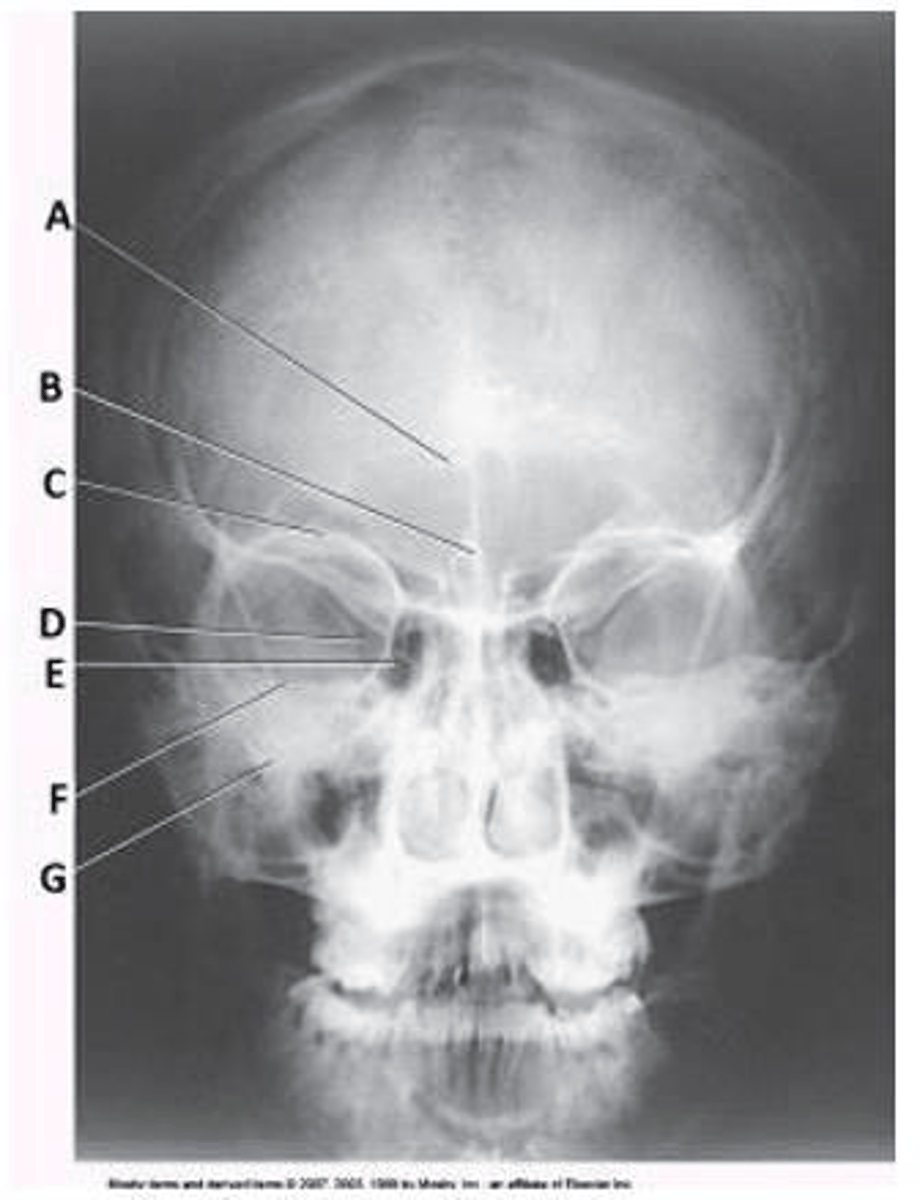
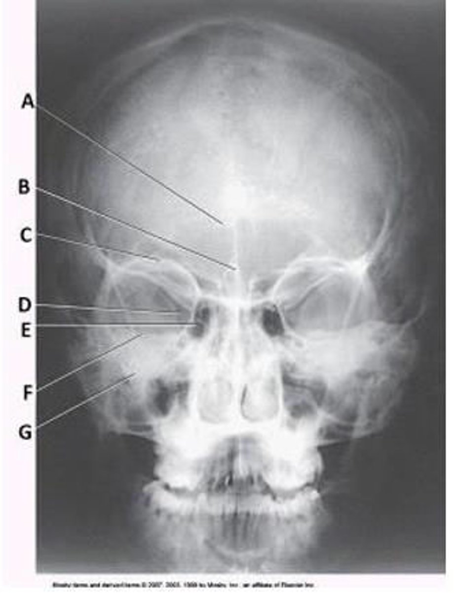
What bone is labeled as letter B in the diagram below of the orbit?
a. Frontal
b. Zygoma
c. Maxilla
d. Sphenoid
a. Frontal

Letter G in the diagram below of the orbit labels the:
a. frontal.
b. palatine.
c. maxilla.
d. lacrimal.
d. lacrimal.
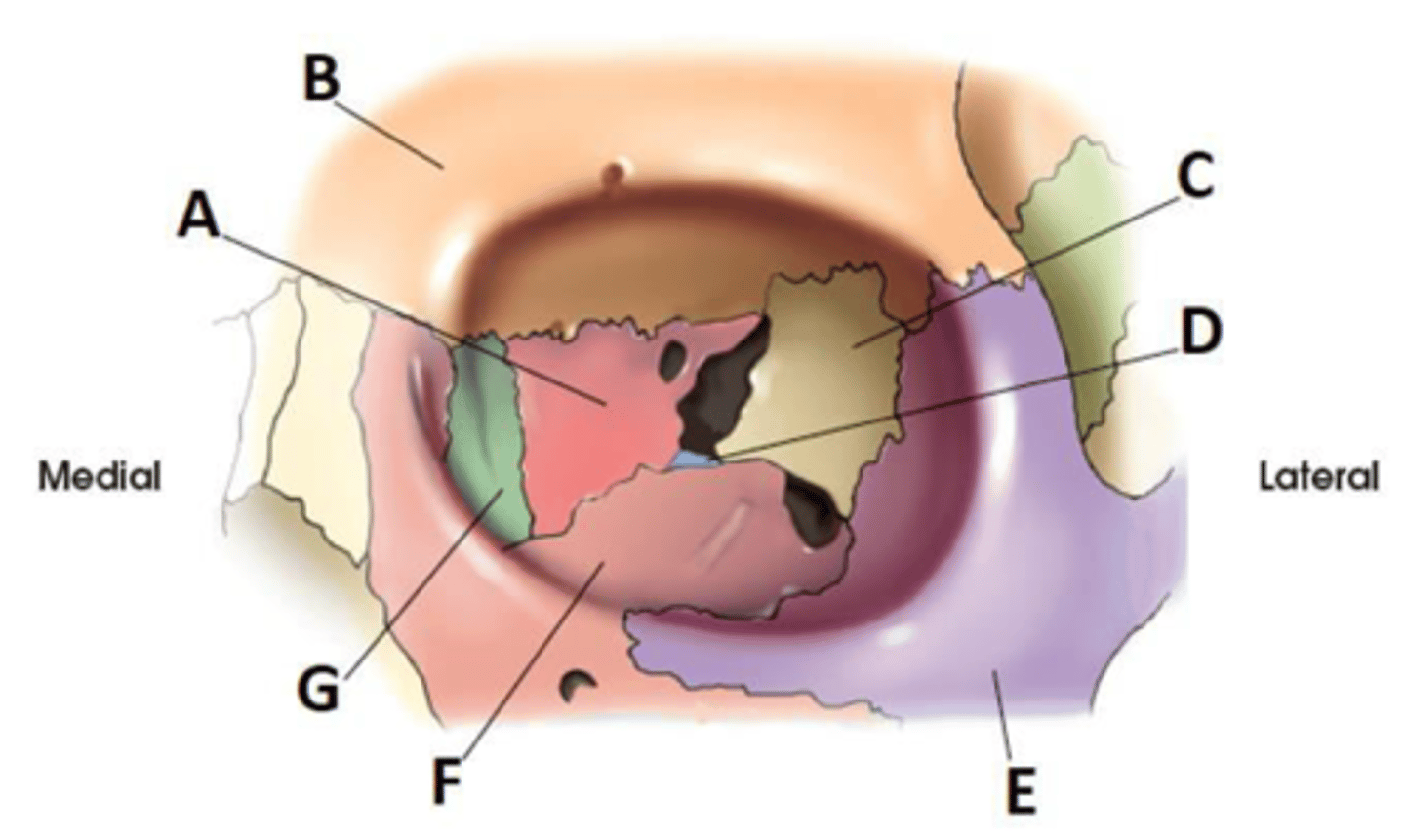
Letter E in the diagram below of the orbit labels the:
a. sphenoid.
b. zygoma.
c. maxilla.
d. frontal.
b. zygoma.

Letter A in the diagram below of the orbit labels the:
a. ethmoid.
b. sphenoid.
c. lacrimal.
d. maxilla.
a. ethmoid.

Letter C in the diagram below of the orbit labels the:
a. ethmoid.
b. sphenoid.
c. lacrimal.
d. maxilla.
b. sphenoid.

Letter D in the diagram below of the orbit labels the:
a. ethmoid.
b. sphenoid.
c. lacrimal.
d. palatine
d. palatine

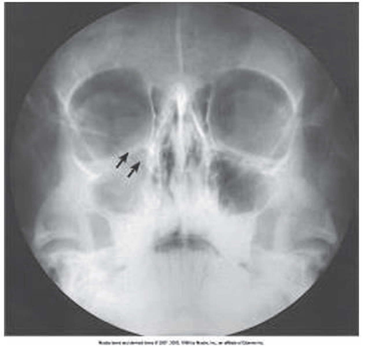
What is the projection (method) demonstrated in the image below used to evaluate trauma to the orbit?
a. Parietoacanthial (Waters)
b. PA axial (Caldwell)
c. AP axial (Towne)
d. PA axial (Haas)
a. Parietoacanthial (Waters)
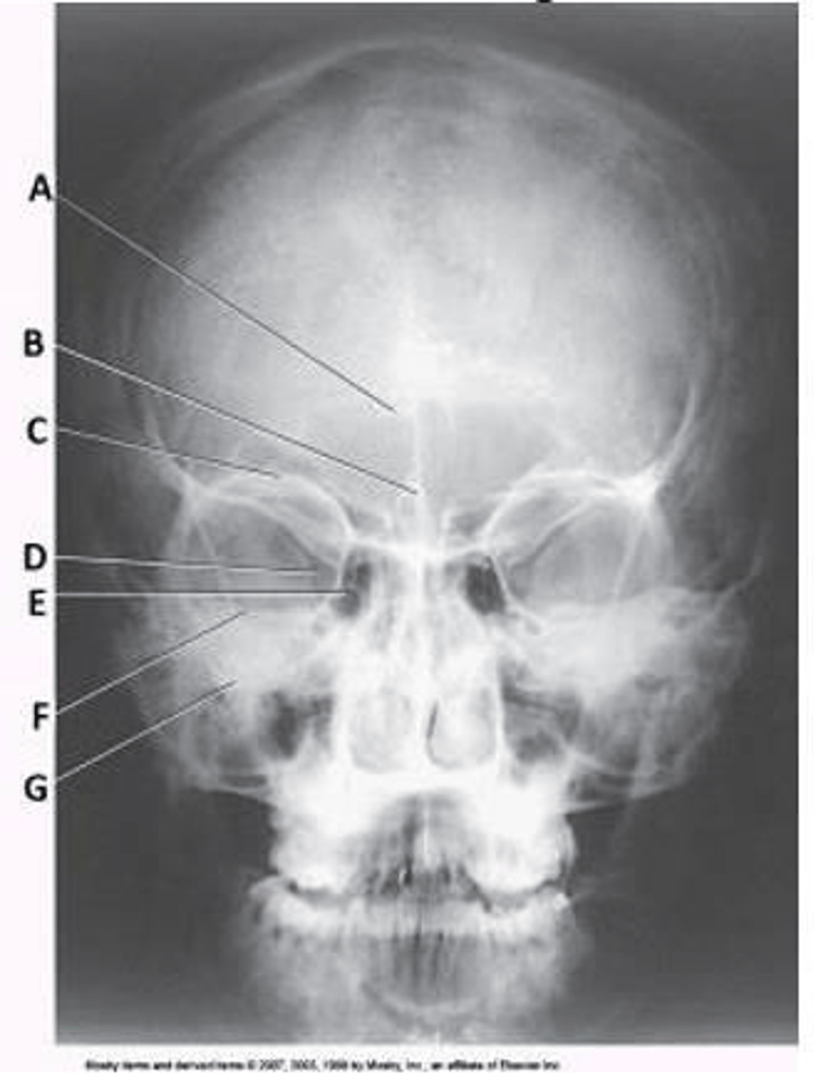
What projection (method) used to evaluate the cranium is demonstrated in the image below?
d. PA
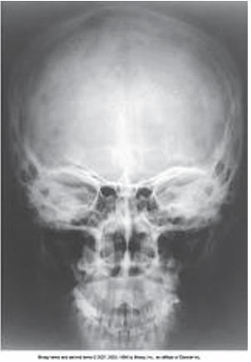
How was the central ray directed to obtain the image below, used to evaluate the cranium?
a. Perpendicular
b. 15 degrees caudad
c. 15 degrees cephalad
d. 37 degrees caudad
a. Perpendicular
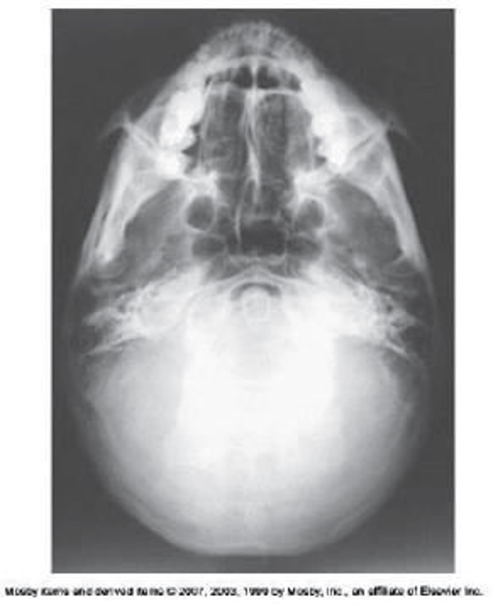
How was the central ray directed to obtain the image below, used to evaluate the cranium?
a. Perpendicular to IOML
b. 15 degrees caudad
c. 25 degrees cephalad
d. 37 degrees caudad
a. Perpendicular to IOML
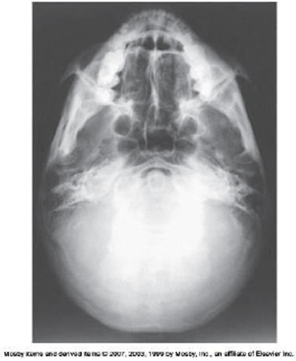
What projection (method) is demonstrated in the image below, used to evaluate the cranium?
a. PA axial (Caldwell)
b. AP axial (Towne)
c. PA axial (Haas)
d. SMV (Shüller)
d. SMV (Shüller)
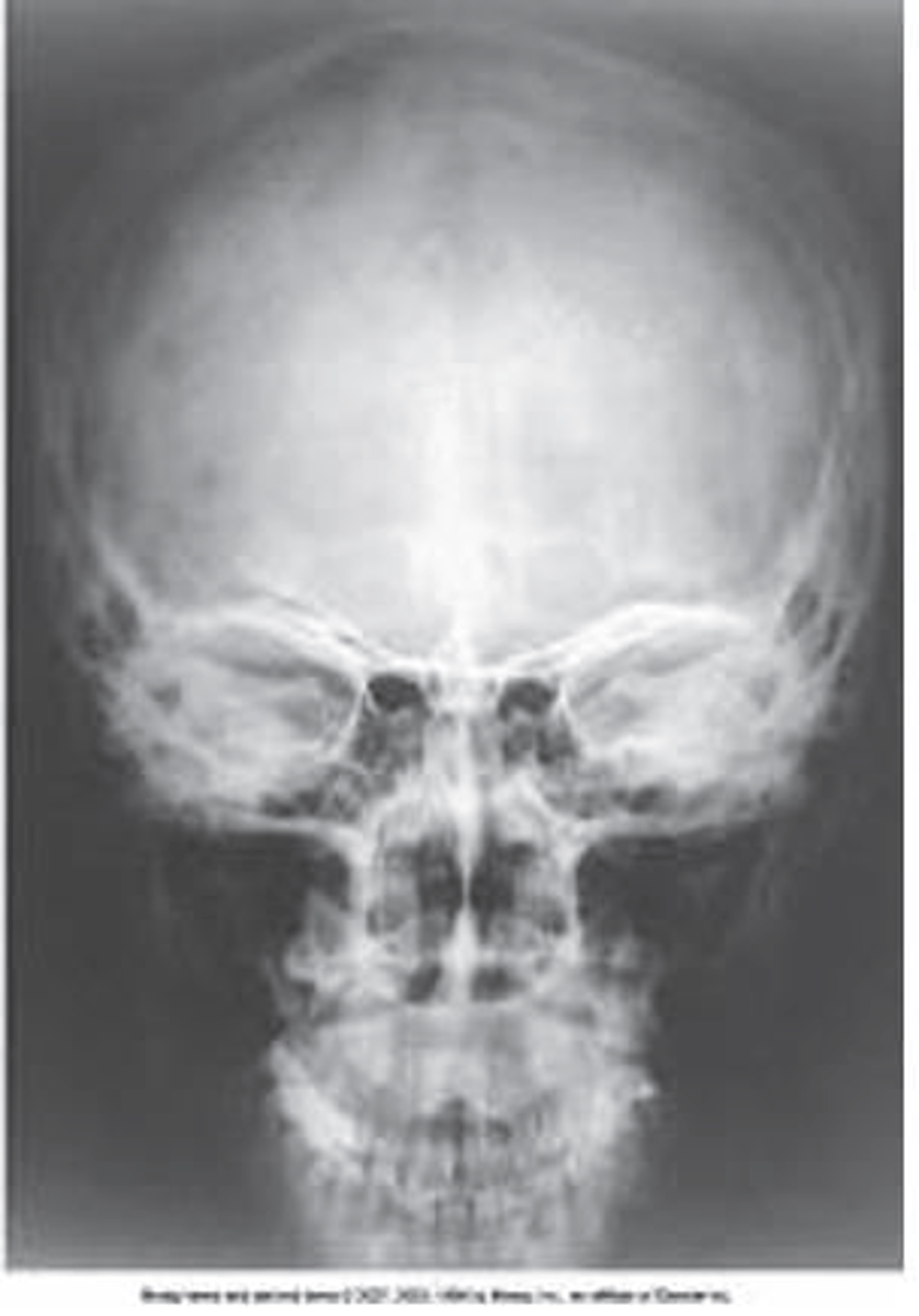
What projection (method) is demonstrated in the image below, used to evaluate the cranium?
a. PA axial (Caldwell)
b. AP axial (Towne)
c. PA axial (Haas)
d. SMV (Shüller)
a. PA axial (Caldwell)
What structure is labeled as letter B in the image below, used to evaluate the cranium?
a. Acanthion
b. Crista galli
c. Cribriform plate
d. Petrous ridge
b. Crista galli
Letter F in the image below used to evaluate the cranium labels the:
a. ethmoid sinuses.
b. crista galli.
c. petrous ridge.
d. inferior orbital margin
c. petrous ridge.
What projection (method) is demonstrated in the image below, used to evaluate the cranium?
a. PA
b. AP axial (Towne)
c. PA axial (Caldwell)
d. SMV (Schüller)
b. AP axial (Towne)

Letter A in the image below, used to evaluate the cranium, labels the:
a. dorsum sella.
b. foramen magnum.
c. petrous ridge.
d. parietal bone.
b. foramen magnum.

Letter B in the image below, used to evaluate the cranium, labels the:
a. dorsum sella.
b. foramen magnum.
c. petrous ridge.
d. parietal bone.
d. parietal bone.

Letter C in the image below, used to evaluate the cranium, labels the:
a. dorsum sella.
b. foramen magnum.
c. petrous ridge.
d. parietal bone.
c. petrous ridge.
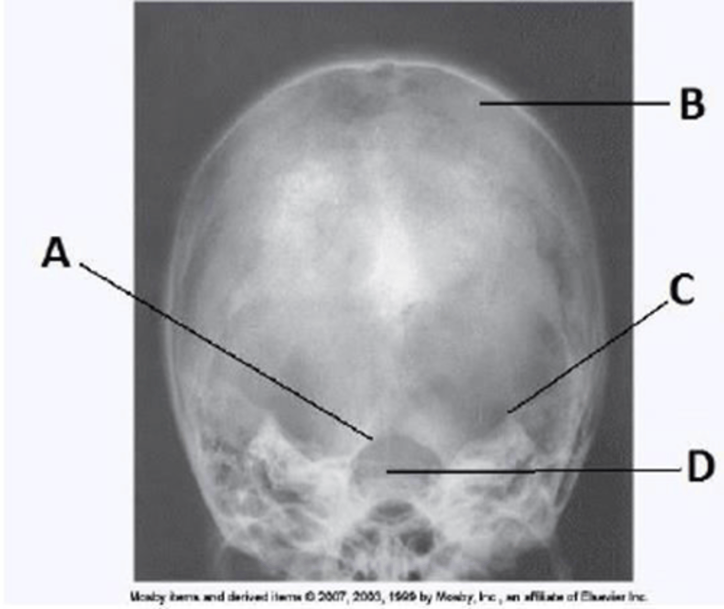
Letter D in the image below, used to evaluate the cranium, labels the:
a. dorsum sella.
b. foramen magnum.
c. petrous ridge.
d. parietal bone.
a. dorsum sella.

For a lateral projection of the facial bones, the image receptor is centered to the:
a. glabella.
b. nasion.
c. acanthion.
d. zygomatic bone.
d. zygomatic bone.
Which of the following is placed perpendicular to the front edge of the IR for a lateral projection of the facial bones?
a. Mentomeatal line
b. Acanthiomeatal line
c. Orbitomeatal line
d. Infraorbitomeatal line
d. Infraorbitomeatal line
For a lateral projection of the facial bones, the central ray will enter:
a. at the TMJ.
b. at the EAM.
c. at the outer canthus.
d. halfway between the outer canthus and the EAM.
d. halfway between the outer canthus and the EAM.
The lateral projection of the facial bones clearly demonstrates:
a. the maxillae.
b. the petrous ridge.
c. the petromastoid portion.
d. all facial bones in their entirety.
d. all facial bones in their entirety.
The parietoacanthial projection of the facial bones is commonly called the _____ method.
a. Towne
b. Waters
c. Caldwell
d. Rhese
b. Waters
For the Waters method for the facial bones, the orbitomeatal line is placed at what angle to the IR?
a. 30 degrees
b. 35 degrees
c. 37 degrees
d. 55 degrees
c. 37 degrees
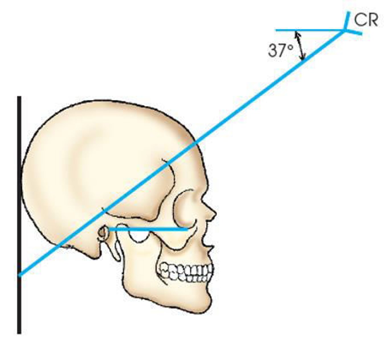
Which of the following is(are) true regarding positioning for the Waters method for the facial
bones?
1. The orbitomeatal line forms a 37-degree angle with the plane of the IR.
2. The infraorbitomeatal line forms a 37-degree angle with the plane of the IR.
3. The midsagittal plane is perpendicular to the IR plane.
a. 1 and 2
b. 1 and 3
c. 2 and 3
d. 1, 2, and 3
b. 1 and 3