CSB520 - Cell adaptation & death
1/9
There's no tags or description
Looks like no tags are added yet.
Name | Mastery | Learn | Test | Matching | Spaced | Call with Kai |
|---|
No analytics yet
Send a link to your students to track their progress
10 Terms
Examples of cell types that are considered connective
Connective tissue cells
Muscle – myocytes (3 types):
Smooth muscle - loose CT with smooth muscle cells - stable
Cardiac Myocytes - individual cells with single central nuclei, intercalated discs connect cells end to end allowing coordinated contraction - permanent
Skeletal muscle fibres - striated or voluntary muscle - permanent but tissue able to repair
Endothelium - Endothelial cells (lining of blood vessels)
Cartilage – Chondrocytes
Bone – Osteoblasts, Osteocytes
Fibroblasts/myofibroblasts - (secrete collagen protein)
Fat – Adipocytes (large cells whose cytoplasm is full of fat which is often lost in sections. Nucleus against edge)
What is meant by labile, stable & permanent cells, give some examples.
Labile (continuously dividing)
•Epithelial e.g. Skin, GIT, reproductive, urinary tracts , lining of exocrine ducts
•Haemopoietic stem cells
Stable (quiescent)
•Epithelial e.g. Liver, kidney, lung, pancreas
•Smooth muscle cells, fibroblasts, endothelial cells
Permanent (non-dividing)
•Cardiac & skeletal myocytes, CNS neurons
Examples of cell types that are considered epithelial
Simple Squamous Epithelium - Single layer of cells whose cytoplasm often appears thinner than their nuclei. Allows of gases, ions and small molecules e.g. alveoli
Stratified Squamous Epithelium - (Labile) Basal cuboidal-like cells that mature as they migrate up towards the surface. At some sites, a keratinised upper layer reduces absorption but increases strength. Line surfaces exposed to abrasion, friction, physical stress:
Skin, Oral cavity, Pharynx, Oesophagus, Anal canal, Outer cervix (ectocervix), Vagina
Simple Cuboidal Epithelium - (Labile/Stable) cells are as tall as they are wide with a central nuclei. Lines protected surfaces including: Ducts of exocrine glands, Collecting tubules of the kidney, Outer surface of the ovary
Simple Columnar Epithelium - cells line surfaces involved in secretion & absorption. Sometimes microvilli are present to increase the surface area of the absorptive membrane or cilia to aid movement across the surface. Mucous secreting cells appear to have a fragile looking or poorly stained cytoplasm due to the numbers of mucous-filled vesicles within their cytoplasm
Pseudostratified Epithelium - (Stable) Specialised epithelium located in the upper respiratory tract and sections of the male reproductive system. As the name suggests, although this epithelium appears to include multiple layers of cells, there is only a single layer however, the nuclei are located at different levels of the cytoplasm giving the illusion that there are many cells
Transitional Epithelium - Restricted to urinary system, (renal calyces, ureters, urinary bladder & parts of the urethra). Allow stretch & retraction and are lined by stratified cells that when relaxed appear stratified columnar but when stretched look like stratified cuboidal/squamous.
Stratified Cuboidal/Columnar Epithelium - A fairly rare type of epithelium
Some large ducts are lined by columnar on top of cuboidal cells with cilia when located within areas of the soft palate and epiglottis. Stratified cuboidal epithelium lines the mammary glands, parts of the cochlea, germ cells of the seminiferous tubules and granulosa cells of the ovarian follicles
Examples of cell types that are considered “other”
Mesothelial cells or Mesothelium
Melanocytes
Germ Cells
Lymphoid Tissue
Bone Marrow
Haemopoietic Cells
Central Nervous System Tissue
RBC
Neutrophils
Lymphocytes
Macrophages
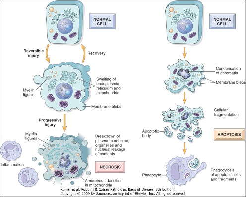
Describe the main differences between apoptosis & necrosis.
Apoptosis occurs in physiology & Pathology
Necrosis only in pathology
Apoptosis is active
Necrosis is passive
Apoptosis is single cell death
Necrotic cell can kill its neighbouring cells
Apoptosis does not stimulate inflammation
Necrosis stimulates inflammation
What is an infarct and how does it occur?
Neighbouring cells induced to undergo necrosis
Due to stimulation of acute inflammation
Loss of function of tissue
scarring
calcification
death
Infarct is an area of necrotic tissue
Tissue Patterns of Necrosis
Coagulative: Hypoxic death (not brain)
•Protein digestion preserved cell & tissue framework
•Necrotic tissue: Heterolysis/autolysis (lysosomal)
Liquefactive: Localised bacterial infections, brain
•Auto/heterolysis > protein denaturation
•Necrotic area is soft & fluid filled
Caseous: TB lesions
•Soft friable, ‘cheesy’ material
Fat: Adipose tissue
•Lipase activation: releases FAs from trigs→complex with Ca2+ → soaps
What determines whether a stimulus causes atrophy or infarction?
Atrophy involves apoptosis (decrease in cell number) & autophagy (decrease in cell size)
Infarct is ischaemic & haemorrhagic
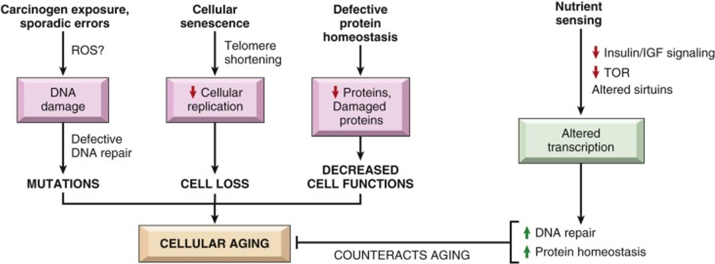
What effect does ageing have on cells & organs?
Define types of cellular adaptation & understand when these processes are not possible/reversible
Atrophy - reduced size of tissue/organ due to loss of tissue. reduction in size due to individual cells undergoing a combination of autophagy & apoptosis or just apoptosis in old cells
Hyperplasia - increased number of cells
Metaplasia - the change from one normal/well-differentiated cell type to another normal/well-differentiated cell type
Hypertrophy - increased size of cells
All are reversible - but irreversible when there is repeated/sustained stress in cells (e.g trauma, smoking) allows cell survival but there is also a loss in function