MBB 3
 Call Kai
 Learn
Learn Practice Test
 Spaced Repetition
 Match
Match Flashcards
 Knowt Play
1/175

There's no tags or description
Looks like no tags are added yet.
Name | Mastery | Learn | Test | Matching | Spaced | Call with Kai | Chat |
|---|
No analytics yet
Send a link to your students to track their progress
176 Terms
DNA to Protein
DNA Synthesis - Replication
RNA Synthesis - Transcription
Protein Synthesis - Translation
Amino Acids
DNA primary structure
nucleotide
phosphate
sugar
base (purine or pyramidine)
Pentose Sugar - RNA
Ribose
Pentose Sugar - DNA
deoxyribose
Difference of structure in DNA and RNA
RNA the 2’carbon has a hydroxyl group while in DNA the 2’ carbon has a hydrogen group
What is relevant in RNA splicing
2’-oh
Pentose in each nucleotide is attached to the base via?. The phosphate is attached to the ? carbon is called the ?
Pentose in each nucleotide is attached to the base via 1’carbon. The phosphate is attached to the 5’ carbon is called the 5’phosphate
What 2 parts of nucleotides are linked to crease the repeating sugar phosphate backbone
2’Hydroxyl and 5’PO4
Nitrogenous heterocyclic ring structures
Purines and Pyrimidines
highly conjugated aromatic rings
Why are bases called bases
because some of their ring nitrogen can be protonated
Purines
Adenine
Guanine
Pyrimidines
Cytosine
Uracil
Thymine
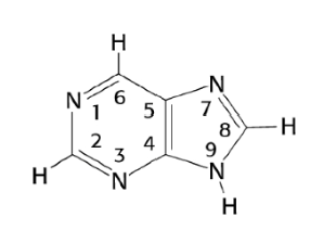
Where does pentose attach on Purines
On C9
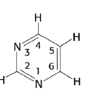
Where does pentose attach on Pyrimidines
C1
Pentose forms a covalent bond with the bases via a B-glycosidic linkage
a B-glycosidic linkage
Why not form covalent bonds with a-linkages
the base would lie below the plane where the H is
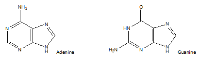
How to distinguish A from G
Guanine has a double bonded oxygen sticking up
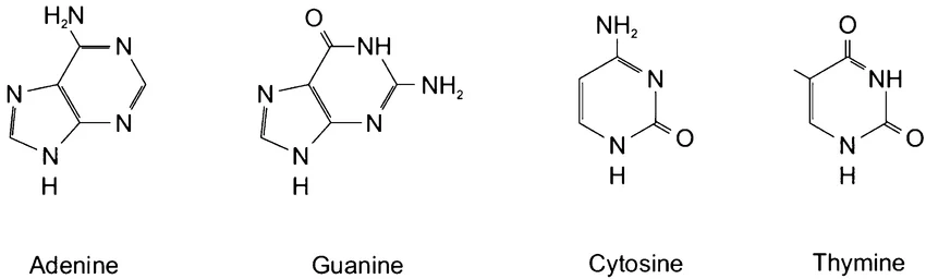
How to distinguish T from C
Thymine has 2 double bonded O groups while Cytosine has 1
What is the difference between deoxyribonucleotides and ribonucleotides
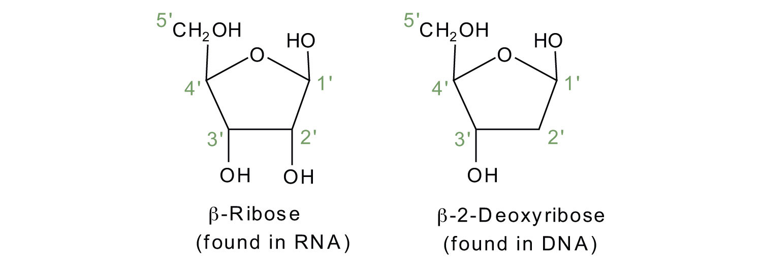
What is the difference between U and T
uracil does not have a methyl group (CH3)
Nucleotide Sequence
5’-3’
pACGTA is an example of how one would write a sequence
What tells you is its a 3’ or 5’ end
5’ has a free phosphate group
3’ has a free OH group
If it only has a few residues (nucleotides) it is a
an oligonucleotide
The phosphate group link the pentoses via
phosphodiester bonds
The strands are said to be
polar
The phosphate group is an acid or base what is the impact
Acid. At the physiological pH the phosphate group of every nucleotide within the strand is deprotanated so carries a net charge of -1
Why does the phosphate group carry a net charge of -1
This occurs because each internal phosphate is linked in a phosphodiester bond involving two of its oxygen atoms, leaving one remaining acidic hydroxyl group that is fully deprotonated at cellular pH levels
Each linked internal phosphate carries a single negative charge (-1) due to the ionized oxygen
This repeating negative charge gives the entire sugar-phosphate backbone of DNA and RNA its strong overall negative character
Why the name nucleic acids
because of the acidic phosphate groups
Two DNA strands interact via
hydrogen bonds
A has 3 bonds with T/U
G has 2 bonds with C
Who proposed base pairing specificity and based on what
Watson-Crick base pairing based on x-ray diffraction data
The specific pairing of bases permits ?
the duplication of genetic info because each strand is a template for its complimentary strand
In addition to H-bonding between base pairs, the
double helix is held together by
base stacking
Base Stacking
between the hydrophobic bases minimizes their contact with water and stabilizes the double helix
base-stacking is a form of van der Waals forces
bases are slightly offset so they are not directlynon top of one another
the bases lie in a plane almost perpendicular tomthe axis of the helix
the offset pairing of the two strands (i.e., bases not in the center of the double strand) forms
a major groove and a minor groove on the surface of the duplex
grooves lie on opposite faces of the double helix and twist around the helix axis
so that if you see a major groove facing you, the minor groove is on its back side
The base pairs are more exposed to solvent on the _______ groove side than on the ______ groove side. Why?
The base pairs are more exposed to solvent on the major than on the minor groove side.
Major is more spacious so water and protein easier access to base edges
Minor is narrow so limits direct access
The forms of the double helix
B-DNA
A-DNA
Z-DNA
B-DNA
most common
right handed helix
10.5 pairs per complete turn with wide major and narrow minor
A-DNA
occurs in dehydrating conditions - standards for double stranded RNA or hybrids
right handed but shorter and wider than B
10.7 pair per turn with bases tilted away from central
Z-DNA
high energy conformation forming under high salt or supercoiling stress
left handed helix with a zigzag backbone
narrower and elongated 12 base pairs per turn
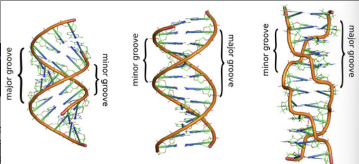
Label the DNA
A
B
Z
Nucleotide bases absorb UV light at? and can determine?
260nm
DNA concentration
What allows UV absorption in nucleotides
purines and pyrimidines are highly conjugated - resonance among rings give most of the bonds a partial double-bond character, allows UV absorption
Beers Law
the amount of light absorbed by a solution is directly proportional to its concentration and the distance the light travels through it
Beers law formula
A = ε ⋅ l ⋅ c [
A: The absorbance of the solution (which is unitless).
ε (epsilon): The molar absorptivity (or molar extinction coefficient). It measures how well the chemical species absorbs a specific wavelength of light (units are usually L ⋅ mol⁻¹ ⋅ cm⁻¹).
l: The path length, which is the distance the light travels through the solution (usually in centimeters).
c: The concentration of the absorbing substance in the solution (usually in molarity, mol/L).
A260 can also be used to distinguish between single-stranded and double-stranded DNA
double-stranded DNA (dsDNA) helix can be disrupted by heating
The melting point is determined by nucleotide sequence
Once cooled the strands re-anneal - come back due to base complementarity
The stacked bases absorb less UV that unstacked so absorbance is quenched when strands come together
DNA melting (denaturation) and re-annealing (renaturation) can be followed by studying the ?
hyperchromic shift
hyperchromic shift
the large increase in ultraviolet (UV) light absorption by nucleic acids when double-stranded DNA or RNA unwinds into single strands. It happens because unstacked bases in single strands absorb more light than stacked bases in a double helix
absorption intensity e is increased
shift due to change in structure
UV absorption at 260 nm spikes by about 37%
Why DNA Denaturation Causes the hyperchromic shift
Since the nitrogenous bases are stacked tightly on top of each other it limits their ability to absorb UV light
therefore when heat or chemicals denature the strands the bases get fully exposed to light
this allows the bases to absorb more UV light causing the shift
Tm
the temperature at which
half the DNA is in a ds form, half is in a ss
form.
Melting occurs at a specific Tm depending on
nucleotide sequence
length
concentration of salt in solution
The stacking energy is more negative (more stable) for ? pairs, so Tm is higher. The ? regions melt first.
The stacking energy is more negative (more stable) for GC pairs, so Tm is higher. The AT regions melt first.
Tm is also proportional to [salt] and sequence length
high salt concentrations and longer sequences stabilize the duplex and increase the Tm
More sodium or magnesium ions neutralize the negative charges of the DNA strands, which reduces the pushing force between them and helps them stick together tighter.
Longer DNA strands have more hydrogen bonds and base-stacking forces holding the two strands together, which requires more heat to pull them apart.
way of compacting DNA
DNA superhelicity
How do cells contain/package/handle their DNA?
Bacterial DNA can be compacted by a process called supercoiling into a nucleoid.
DNA is condensed, organized and segregated with the help of topoisomerase enzymes, nucleoid associated proteins and the Structural Maintenance of Chromosome (SMC) complex.
A closed double-stranded molecule of DNA can be compacted by
supercoiling
Supercoiling
can only exist in a DNA molecule where both strands of DNA are closed circles or otherwise fixed at one end. If one strands breaks the DNA rapidly loses its supercoiling
DNA molecules in different coiled forms that have the same nucleotide sequence are called
topoisomers
Supercoiling
The topology (shape) of dsDNA can be defined in terms of
Linking number
Linking Number
the number of times one strand
would have to be passed
through the other strand in
order for the strands to be
completely separated from each
other
Linking number formula
Lk=Tw+Wr
Tw
Twist
For dsDNA, twist is the number of full turns of the helix
one turn of a B form helix is 10.5 bps, Tw of a
segment of this relaxed DNA which is 105 bp long
will be:
Tw=105/10.5 = 10
Wr
writhe
measure of the degree of supercoiling
The number of times double helix crosses itself
if ds helix writhes in the left- handed direction
Wr is assigned a positive value
Overwinding (+) makes it more difficult to separate the strands of the double helix
If helix writhes in the right-handed direction
Wr is assigned a negative value
Underwinding (-) makes it easier to separate the strands of the double helix
Wr for relaxed dsDNA
0
Lk
a topological property of the circular DNA - it cannot be changed unless one or both of the strands of the duplex is broken
What moves faster in centrifugation or gel electrolysis supercoiled or relaxed DNA and why
A supercoiled DNA molecule is
more compact than a relaxed
DNA molecule of the same length:
it moves faster than relaxed DNA
when subjected to centrifugation or
gel electrophoresis.
What kind of structure is supercoiled DNA
tertiary structure
DNA gel electrophoresis
The migration also depends on their degree of supercoiling
Since DNA has a net - charge it will migrate towards the anode (+) in agarose gel.
Smaller DNA move faster through than less supercoiled on same size
More supercoiled fragments move faster than less supercoiled of the same size
The DNA bands can be visualized under UV light by staining with
ethidium bromide
ethidium bromide
a dye that binds the DNA by intercalating between the bases
DNA gel electrophoresis
Separating DNA fragments based on their sizes (larger
or smaller) and shapes (supercoiled or relaxed)
Supercoiled the fastest and furthest
Linear depends on true size in base pairs
Relaxed migrates the slowest and shortest distance
Topoisomerases change the linking number of DNA by catalyzing a three-step process:
the cleavage of one or both strands of double-stranded DNA
the passage of a segment of DNA through this break
resealing of the DNA breaks
Topoisomerases have a key
tyrosine residue in their active site that covalently attaches to a phosphate in the sugar-phosphate backbone that is transiently broken
Type I topoisomerase
Type I topoisomerase is an enzyme that relaxes DNA strands by cutting a single strand, passing another strand through the break, and resealing it.
No ATP
Changes DNA linking number by 1
Type 1 relaxes supercoiled DNA by letting one strand rotate around the other
Thermodynamically favorable process driven by release of energy of supercoiling
relax DNA by removing negative
supercoils (increasing Lk)
Type II topoisomerase
cuts both strands of the DNA double helix
needs ATP
DNA linkages by 2
untangle, unknot, and separate linked rings of DNA (decatenation) in addition to relaxing supercoils.
Type I mechanism
Cleavage:
Uses a tyrosine residue to attack and break one phosphodiester bond on a single DNA strand.
Passage:
The intact strand passes through the nick, or the cut strand rotates around the uncut strand.
Religation:
The enzyme seals the broken strand back together without consuming external energy
Type II Mechanism
Binding:
Grabs onto one DNA segment (the G-segment) and binds ATP.
Cleavage:
Breaks both strands of the G-segment, forming a transient protein-gate.
Passage:
Passes a second, intact DNA duplex (the T-segment) completely through the gate.
Religation:
Reseals both strands of the G-segment using energy from ATP hydrolysis.
Topoisomerases are the molecular targets of
antibacterial and anti-cancer drugs
The topological state of cellular DNA is intimately connected with its function
Without topoisomerases, cells cannot replicate or package their DNA, or express their genes, so they die
The quinolones inhibit bacterial DNA gyrase, a type II topoisomerase.
Quinolones act by blocking the last step of the topoisomerase reaction, the resealing of the DNA strand breaks. This action inhibits the unwinding of the bacterial chromosomal DNA during and after the replication, thus preventing bacterial cell division.
Three hypothesized mechanisms for DNA replication
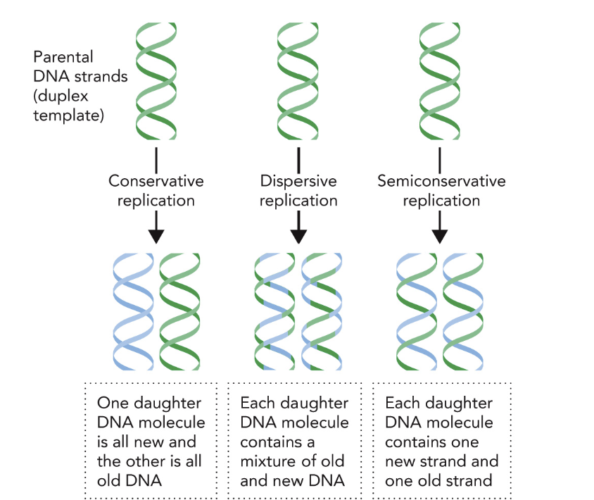
The Meselson–Stahl experiment
provided biochemical evidence
that DNA replication is
semiconservative.
How was this experiment done?
semi-conservative replication using nitrogen isotopes and density gradient centrifugation. coli were first grown in heavy nitrogen (¹⁵N) medium. They were then shifted to light nitrogen (¹⁴N) medium. DNA density was analyzed using cesium chloride gradient centrifugation.
The resulting DNA bands proved that each new double helix consists of one original parent strand and one newly synthesized strand
What are DNA building blocks?
nucleotides
How many phosphates do dNTPs have?
3
alpha
beta
gamma
starting from the one closest to sugar
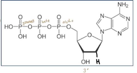
Primer to provide what
OH on 3’
DNA polymerase is an
enzyme
Required cofactors for DNA replication
Mg2+ to facilitate nucleophilic attack
The raw materials for DNA synthesis/replication are:
a DNA templatedeoxynucleoside triphosphates (dNTPs)
a DNA or RNA ‘primer’ to provide the first 3’-OH
Mg2+ ions (cofactor for thepolymerase)
DNA polymerase - a large protein complex that includes the enzyme that catalyzes the addition of dNTPs on the 3’ OH end of the primer
The DNA polymerase reaction adds a
deoxynucleotide to the 3' end of thegrowing DNA chain.
The Mg2+ ions
stabilize the negative charges on the
deoxynucleotide and assist in deprotonation of the 3'-OH by a base
The 3'-oxygen of the growing DNA strand serves
as the nucleophile in this reaction, displacing the pyrophosphate from the
deoxynucleoside triphosphate (dNTP) in the active site. The product is a DNA
strand that has been extended by one nucleotide in the 3' direction.
The shape of the DNA polymerase active site facilitates
formation of the correct Watson–Crick base pairs: correct bases pairs fit well into the DNA polymerase active site, mismatches do not
high fidelity
base selection
steric clashes
What is an endonuclease?
an enzyme that cleaves the internal phosphodiester bonds within a polynucleotide chain (DNA or RNA)
What is an exonuclease?
type of enzyme that degrades nucleic acids (DNA or RNA) by removing nucleotides one by one from the ends of the strand, rather than cutting them in the middle
What is a 5’-->3’ exonuclease?
A 5’-->3’ exonuclease is an enzyme that removes nucleotides one by one from the 5' end of a DNA or RNA strand, moving toward the 3' end
What is a 3’à5’ exonuclease?
A 3’→5’ exonuclease is an enzyme that removes DNA or RNA building blocks (nucleotides) one by one from the 3' end of a nucleic acid chain
DNA synthesis occurs at
replication forks simultaneously for both parent strands, always in the 5’→3’ direction.
DNA is partially unwound at “replication forks.” Both strands are used as templates for the
synthesis of new strands in a 5’→ 3’ direction.
The leading strand is synthesized continuously
the lagging strand is, by necessity, is synthesized in short pieces termed Okazaki fragments (100s to 1000s of nucleotides).
Both new strands are synthesized in a coordinated fashion by a single multimeric DNA polymerase III complex (DNA pol III).