Lab 3: Restriction Digests, Agarose Gel Electrophoresis, & Gel Purification
 Call Kai
 Learn
Learn Practice Test
 Spaced Repetition
 Match
Match Flashcards
 Knowt Play
1/76

There's no tags or description
Looks like no tags are added yet.
Name | Mastery | Learn | Test | Matching | Spaced | Call with Kai | Chat |
|---|
No analytics yet
Send a link to your students to track their progress
77 Terms
Lab objectives
- Understand how restriction enzymes function & what they can be used for
- Explain the process of agarose gel electrophoresis
- Explain the process of gel purification
What were we doing in lab 3?
Digesting the pET24a MaSp1 vector to release the kanamycin resistance genes, so the product (AmpR gene) can be inserted into the expression vector
- Release kanamycin resistant gene
- Insert AmpR gene
What can you do once the recombinent cloning vector is built?
Use it for expression studies
How can we recombine two different pieces of DNA, from two distinct sources (PCR-amplified AmpR gene and the digested pET24a MaSp1 4x vector)?
Restriction enzymes
- We want to do this because we want to create new DNA molecule with new function that neither source had by itself
Restriction enzyme / Restriction endonuclease (RE)
An enzyme that binds to dsDNA at a specific sequence of nucleotides and makes a double-stranded cut in the phosphodiester backbone, at or near the sequence, without damaging the nucleotide bases
What do restriction enzymes contain that allows them to have their own unique DNA sequence that it binds to and cuts
Sequence specificity
The position of "cuts" within a DNA molecule can be predicted (as long as you know the DNA sequence)
What is so significant about restriction enzymes?
They enable DNA molecules to be cut at defined positions
What are the "defined positions" that restriction enzymes cut at?
Restriction site / Recognition sequence for that enzyme
Which restriction enzyme cuts at the recognition site, 5'-ATCGAT-3'
ClaI
Which restriction enzyme cuts at the recognition site, 5'-GGATCC-3'
BamHI
Which restriction enzyme cuts at the recognition site, 5'-GTGCAG-3'
PstI
What is the pattern that restriction sites often follow?
They are palindromic
- They have the same 5' --> 3' sequence on both DNA strands
How long are recognition sequences usually?
4 to 12 nucleotides in length
- Occur naturally in most genomes
- Can be manipulated by scientists for use in the laboratory
What do many plasmids contain that can be used to cut open the vector to allow it to accept a piece of foreign DNA?
A multiple cloning site filled with different restriction sites
What are the two different types of vector cutters?
Sticky-end cutters and Blunt-end cutters
In this lab, what type of cutter did we use in the PsiI (PsiI-v2), where its restriction site is 5'-TTATAA-3'?
Blunt-end cutter
- The PCR product was designed to have a blunt end at one side, and a sticky end on the other (compatible with ClaI)
If you want to clone a gene into a plasmid, what is the first step?
Cut your plasmid with the restriction enzyme, and then cut the DNA for your gene with the SAME restriction enzyme
Why do we want to use the same restriction enzyme when cutting our plasmid and DNA for our gene?
Restriction enzymes can cut the two DNA strands (plasmid and gene) of the restriction site at different positions, usually two to four nucleotides apart, so that the resulting fragments have short, single-stranded overhangs at each end
- So that the plasmid and gene have matching DNA ends so they can attach to each other; you want to use restriction enzymes that create compatible ends on the plasmid and insert, often using the same enzyme on both DNA pieces
What are the short, single-stranded overhangs at the end of DNA fragments called?
Sticky-ends
Base-pairing between them can stick the DNA molecule back together again
Example of ClaI recognizing and cutting its restriction site in dsDNA
When ClaI cuts its restriction site, it produces the following complementary sticky ends

In this exercise, we are performing a DOUBLE DIGESTION. What is that?
We're using two different restriction enzymes on the same DNA sample in one reaction
What is the purpose of doing the double digestion?
We're doing it on pET24a MaSp1 4x prokaryotic expression plasmid to remove the kanamycin resistant gene, preparing it to receive the ampicillin resistant gene from PCR
What are the two different enzymes that we are using for the double digestion?
ClaI (sticky-end cutter) and PsiI (blunt-end cutter)
Imagine using a pair of scissors to cut the circular plasmid at both locations (ClaI and PsiI). How many pieces of DNA would it yield?
Two pieces!
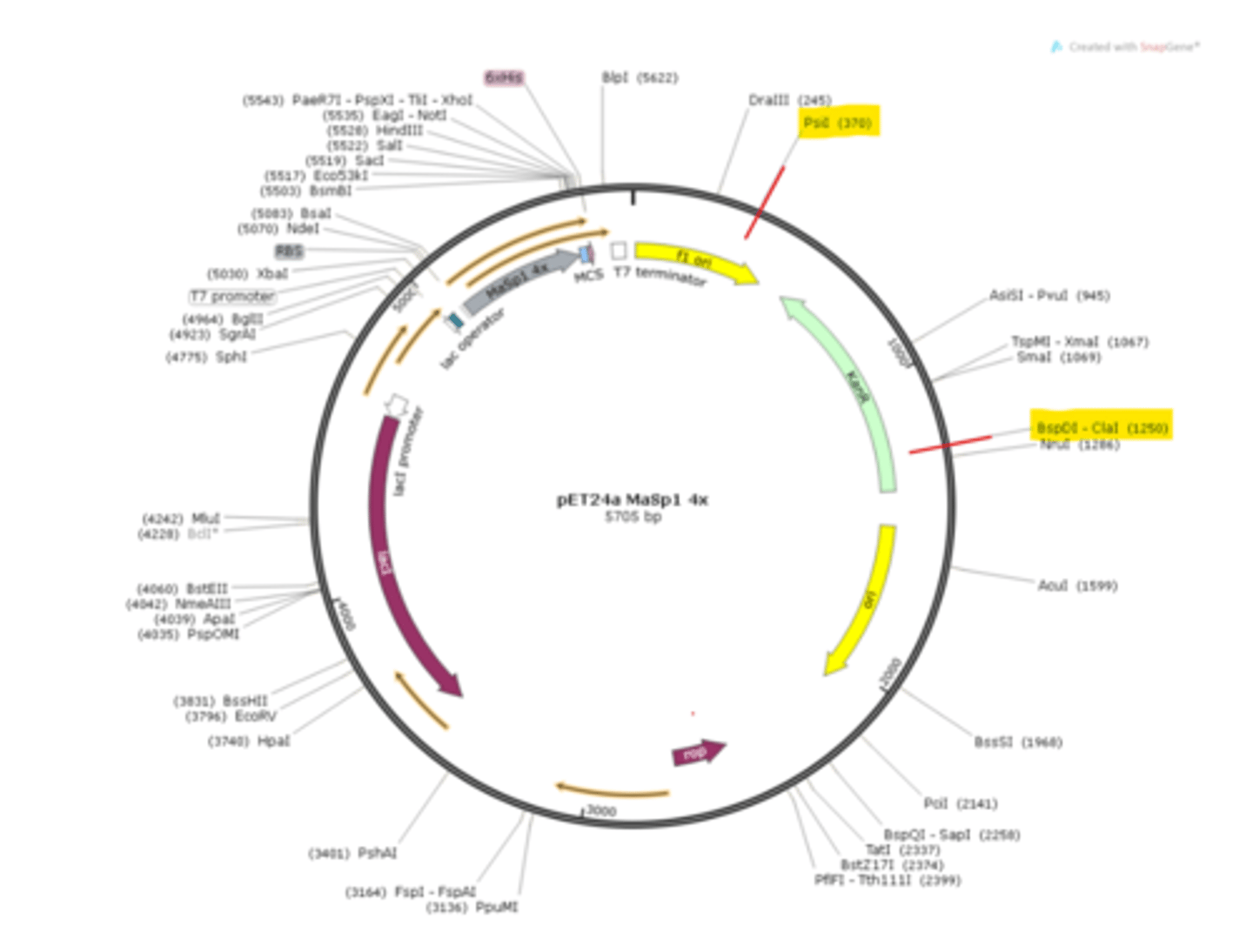
What are the sizes (in base pairs) of the two fragments cut by ClaI and PsiI?
The two fragments are 880bp and 4825bp
Calculation:
- PsiI cuts at 370bp
- ClaI cuts at 1250bp
- Distance from 370bp to 1250bp is 880bp
- Entire plasmid is 5705bp (given in the middle of image), so remaining segment is 4825bp
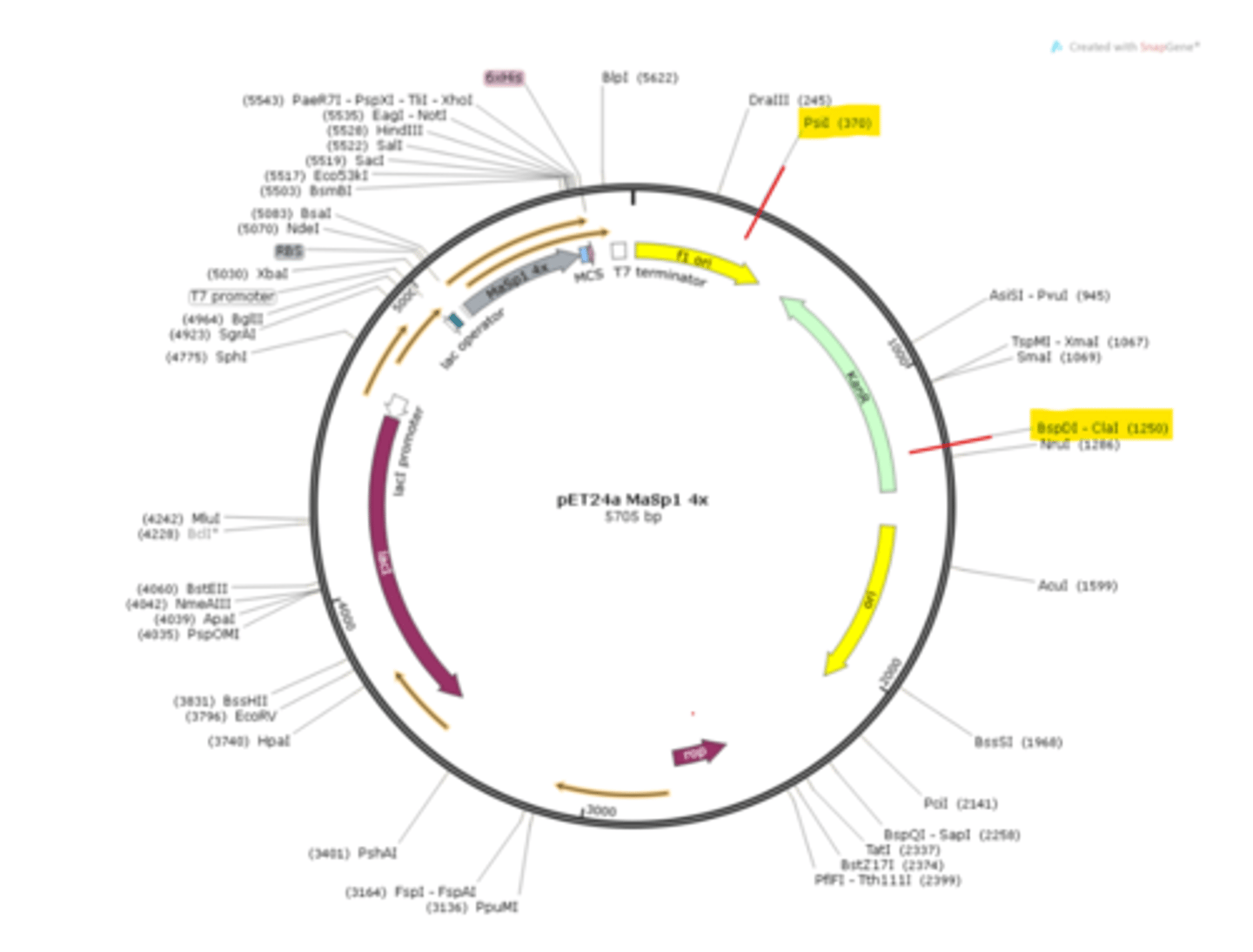
What is the restriction site ClaI subject to when propagated in most E. coli strains, and what does this do?
Subject to methylation, which prevents digestion by ClaI
- ClaI will not cut plasmid if plasmid DNA is methylated!
How do you make the restriction site suitable for ClaI digestion?
The plasmid must be copied/replicated in a dam-/dcm- strain (free of Dam and Dcm methylases)
- This is a special strain of E. coli that doesn't methylate DNA using Dam or Dcm
Sticky-end cutters are often used in cloning, but we will use both sticky-end and blunt-end cutters. What does blunt-end cutting do?
When using a blunt-end cutter, the other molecule that is being ligated (joined together) must also contain blunt ends— called blunt-end ligation
Our PCR will have one end that is blunt and the other that is sticky. Which of the two is less efficient and why?
Blunt-end ligations are less efficient relative to sticky-end ligations
- This is because blunt ends do not naturally stick to each other first. There are no exposed bases that can base-pair, and the two DNA pieces have to randomly collide and line up perfectly for ligase to join them (which happens less likely)
- With a sticky end, the cut leaves short-single stranded overhang where a matching DNA end has complementary bases and the two pieces can temporarily base-pair through hydrogen bonds
How are blunt-ends and sticky-ends stuck together?
Via hydrogen bonds
How do you covalently attach blunt-ends and sticky-ends together?
Using an enzyme
- Note that the ends need to be hydrogen-bonded to each other first before they can be attached
Scientists have engineered plasmids to contain restriction sites in them, but how did we get restriction sites on the ends of our gene?
We added them to our primers that we used in the PCR
How do you know if the PCR reaction worked or not, especially if there is a clear liquid in the tube when you start and when you finish?
Agarose gel electrophoresis to help visualize DNA
Electrophoresis
A standard technique used for separating molecules based on their size and electrical charge
Which is the most common type of electrophoresis used to separate DNA molecules?
Agarose gel electrophoresis
Which is the most common type of electrophoresis used to separate proteins?
SDS-PAGE (SDS-polyacrylamide gel electrophoresis)
Agarose
A polysaccharide purified from agar, which is itself isolated from seaweed
- Makes up the porous gel of agarose gel electrophoresis
What is agarose made up of?
Repeating units of a disaccharide composed of galactopyranose and cyclic form of galactose (both rings) that has an ether chemical group in the sugars
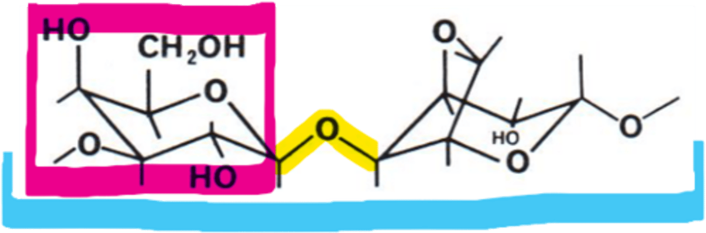
How is agarose dissolved, and what is its melted form used for?
Dissolved by heating it in a buffer solution and then melted agarose is poured into a gel caster
What happens when you let the melted agarose cool?
Agarose solidifies at room temperature, making a matrix that resembles stiff, jello-like substance in the shape of the gel caster
What do we do during the gel polymerization step to hold the DNA samples being studied?
We will use a plastic "gel comb" to make small indentations called "wells" in our gel, and these will hold the DNA samples
What happens to the gel when it's placed into a "gel box"?
It will be submerged in a running buffer that allows the generation of an electrical current
- The electrical current will flow through the gel to separate the DNA fragments that we have loaded into the wells
How are DNA fragments separated by the electrical current?
Because DNA backbone phosphate groups carry a NEGATIVE CHARGE in the slightly basic pH of the buffer used, the DNA fragments migrate towards the positive (+) pole during electrophoresis in the apparatus
Molecular sieve
A molecular sieve is a material with lots of tiny holes or pores that lets smaller molecules move through more easily than larger molecules
How do agarose matrices serve as a molecular sieve?
As the DNA molecules migrate towards the positive pole, they move through the gel pores and, with time, separate based on their size
- Smaller fragments move through pores faster & travel longer distances relative to larger molecules
If you make gels with different amounts of agarose (ex. 1% agarose vs. 3% agarose), what occurs?
It can change the size of the pores
- More agarose = Smaller pores
How do we know the size of the DNA we are looking at in our gels?
DNA fragments of known lengths (called markers) are placed in separate, distinct wells and used to compare with the migration distance of unknown fragments (your PCR reactions)
- With the distance traveled by the marker fragments, one can determine the approx size of the unknown fragments (think of it as a DNA ruler)
What will become of your DNA fragments after electrophoresis?
They should be separated according to size; however, they will be invisible and cannot be seen unless they are subject to staining
What is the most common method of DNA fragment detection?
Staining the DNA fragments
What chemical is used to stain DNA fragments?
Ethidium bromide, which slides (intercalates) in between the base pairs of the double helix
What are the two ways ethidium bromide can be applied to stain fragments?
1) Gel submerged in an ethidium bromide solution after completion of gel electrophoresis
2) Ethidium bromide can be directly added to agarose before casting the gel (we did this)
Why did we use the second method of adding ethidium bromide directly to agarose before casting the gel?
The DNA fragments stain during electrophoresis, so this approach is faster
Why do you need to wear gloves when handling ethidium bromide?
It's a suspected carcinogen
Why is UV light required for the visualization of the DNA?
When exposed to UV light, the ethidium bromide molecules emit a bright orange fluorescent color, allowing the DNA fragments to be visualized within the gel
Is it possible to visualize DNA without UV light?
If DNA concentration is high enough, you might be able to see DNA directly in gel by eye
- Most of the time, you'll need UV light illumination
What is the difference between ethidium bromide staining and loading dye?
Ethidium bromide: Stains the DNA itself so you can see DNA bands under the UV light. You add it to the agarose gel.
Loading dye: Helps you load and track your sample, but does NOT show the DNA bands. The dye helps indicate how far the DNA has run within the gel. Mixed with the DNA sample before putting it into the wells
When is the loading dye added to the DNA fragments?
Prior to electrophoresis
What does the loading dye contain?
Compound EDTA: Helps to inactivate DNA-destroying enzymes (DNAases) that might be present
Glycerol: To sink the sample to the bottom of the well
One or more tracking dyes: Bromophenol blue or xylene cyanol
Bromophenol blue molecules
Known to migrate with ~500 bp fragments of DNA at the percentage of agarose we're using
What is the major use of agarose gel electrophoresis?
Analysis or purification of DNA fragments
- You're using this technique in the lab to see if your PCR reactions were successful
Gel purification
The process of getting a specific DNA fragment out of an agarose gel and into a clean tube so you can use the DNA in the next step, such as ligation or cloning
What are the three things that you will do when carrying out agarose gel electrophoresis
1) Determine if your PCR was successful
2) Check the size of your PCR fragment to see if it is the right size
3) Separate it away from any background bands that might also have been accidentally amplified
What are you doing following electrophoresis?
Extracting the clean DNA from the agarose gel so that we can "clone" it
What happens after you excise the DNA from the entire gel?
The gel slice containing your DNA fragment can be placed in a special solution containing a chaotropic salt
What does chaotropic salt do?
It is used to break down the agarose gel and release the DNA fragments
How do we capture the DNA fragments released by the chaotropic salt solution?
Using a silica (SiO2) resin that binds the DNA fragments
What are examples of chaotropic salts and their mechanism?
Sodium iodide or guanidinium chloride
Helps to disrupt intermolecular interactions, such as hydrogen bonds or hydrophobic effects, and can remove water from hydrated molecules
How do the chaotropic salts affect agarose polymers?
They disrupt the agarose polymers and keep it from re-forming a gel at low temperatures (which would trap the DNA again)
What is the significance of the pH of the salt solution?
pH of the salt solution helps the DNA interact with the silica resin
What can you do with the DNA once you capture it?
DNA fragments can be washed free from salts, enzymes and impurities, and then eluted from the silicia resin for use in other procedures such as cloning or sequencing
Why are we using reagents provided by a kit from the company Qiagen?
They provide the researcher with all the reagents and easy-to-follow protocol, and are designed to be more foolproof than if you did it yourself
Procedure 1: Restriction Enzyme Digestion
1) Label a 1.5mL microfuge tube
2) Add the following reagents to the tube:
a) 36.0uL of undigested pET24a MaSp1 4x expression vector (from last lab)
b) 4uL 10x rCutSmart digest buffer (provides correct salt concentration and pH)
3) Mix thoroughly by pipetting up and down, but avoid bubbles
4) Add 1uL ClaI and 1uL PsiI-v2; this will give final reaction volume of 42uL (known as double-digest)
5) Incubate reactions at 37°C for 30mins. Use water bath for the incubation
Why do we use a water bath for the incubation in procedure 1 instead of an air incubator?
Air incubators are not recommended due to slow heat transfer to the reaction mixture
Why do different restriction enzymes require different kinds of buffers?
They've been isolated from many different species of bacteria, so different buffers provide them with ideal conditions for digestion
- The one we're using works with a variety of enzymes
EcoRI Buffer Conditions
EcoRI: Comes from E. coli
- Likes buffers with 50mM NaCl, a 7.9 pH, and 100ug/mL BSA
What is BSA?
Bovine Serum Albumin
- A blood protein that is used to protect enzymes from heat, degradation, and interference by detergents
- Can keep your enzymes from sticking to plastic of microfuge tube
Kpn1 Buffer Conditions
- Likes no salt, 7.0 pH