CCRN-EKG
 Call Kai
 Learn
Learn Practice Test
 Spaced Repetition
 Match
Match Flashcards
 Knowt Play
1/24

There's no tags or description
Looks like no tags are added yet.
Name | Mastery | Learn | Test | Matching | Spaced | Call with Kai | Chat |
|---|
No analytics yet
Send a link to your students to track their progress
25 Terms
Lateral wall (LCA, left circ)
I, aVL and V5, V6
- occlusion of Left circumflex artery
II, III, and aVF
Inferior wall.
RCA. 2nd most dangerous.
Right sided EKG to look for reciprocal changes.
Avoid nitrates and vasodilators and give fluid if confirmed. This is volume dependent.
R sided EKG. Changes in V3R-V6R
V1
Ventricular septum
V2, V3, V4
Anterior wall.
V1-V4
Anteroseptal (LAD).
Most dangerous
BBB
PA catheter is contraindicated with LBBB d/t risk of injury to RBB leading to complete heart block
V1+V6.
WilliaM and MorroW.
Left BBB V1 looks like a W, V6 looks like an M
Right BBB V1 looks like M, V6 looks like W.
Heart auscultation
S1- Tricuspid/mitral valve
S2-Aortic/pulmonic
S3
Lub-dub-da
Sounds like "Sloshing in"
Ventricular gallop.
FVO
S4
Da-Lub-Dub
Sounds like "A-stiff wall"
Blood being forced into a stiff ventricle
Bell of stethoscope over apex
Murmurs
Sten0sis- Valve 0pen
regurg or insufficiency-valve closing issue
Systolic Lub, murmur, dub
Diastolic Lub, dub, murmur.
Heart pressures
RA/CVP-2-8mmHg
RV-15-30/2-8
PA-15-30/8-15
Mean PAP 9-18, (>25=P. HTN)
PAOP- 6-12
PAOP ranges
Normal 6-12
18-20 onset of P congestion
20-25 moderate
26-30 severe
above 30-P edema
Cardiac Output
Measured with thermodilution. Think of thermoster
Normal 4-8L/min
Cardiac index is weight based CO.
CI normal is-2.5-4.3 L/min/m2
PA wave form
A,C,V
A-left atrial contraction
C-closure of mitral valve. Not often seen
V-LV systole, if V wave is higher than a wave=mitral regurg.
Mixed venous oxygen saturation
Serves as an early trouble indicator and can drive changes to care plan
Obtained from PA catheter. Normal values 60-80%
SvO2=increased supply and decreased demand.
Mixed venous oxygen saturation (<60%)
Insufficient supply vs demand
Increased metabolic requirements
-seizures, restlessness, shivering, hyperthermia.
Oxyhemoglobin Dissociation curve (Left)
Hbg more sticky for O2. SvO2>80%
Alkolosis
Hypothermia
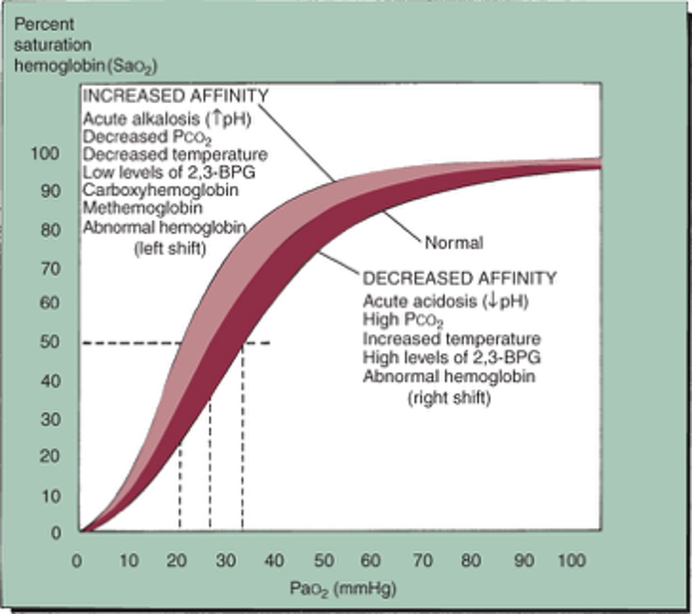
Oxyhemoglobin Dissociation curve (Right)
Less affinity for O2, SvO2<60%
Acidosis
hyperthermia
Allen's test
If the hand flushes within 5-15 seconds then the ulnar artery is patent an there is good blood supply.
Flo-trac
Helps recognize fluid status and connects to arterial line
Measures CO and stroke volume variation. SVV normal <13%.
>13% the patient is dry and needs more fluid.
Alternative methods to determining fluid status
Passive leg raise
Clear sight system-SV, Stroke volume variation, CO, SVR, Continuous BP.
Look at Stroke volume index after 250 cc bolus or passive leg raise.
Stroke volume changes with Passive leg raise
Stroke volume increase >10% give fluids
<10% FVO. Myocardium is distended. Consider diuretic
IABP
Bridge to surgery
Augments diastolic BP
Increase Coronary perfusion
Improves CO up to 15%
Must have a-line and EKG input for timing.
IABP cont
Inflates during diastole. Dicrotic notch=aortic valve closing.
Blood forced inferiorly and increases perfusion to distal organs.
Deflates during systole reduces afterload.
decreased O2 demand
IABP complications
Renal perfusion- monitor UOP
Balloon rupture-rapidly diffused gas
Contraindicated in Aortic valve insufficiency, Aortic aneurysms, suspected aortic dissection