Chapter 21: First Trimester Obstetrics (0-12 weeks)
1/28
There's no tags or description
Looks like no tags are added yet.
Name | Mastery | Learn | Test | Matching | Spaced | Call with Kai | Chat |
|---|
No analytics yet
Send a link to your students to track their progress
29 Terms
First Trimester
based on 28-day menstrual cycle, defined as 12 weeks after the first day of your last period; the term gestational age (GA) is synonymous with menstrual age and is used to date the age of pregnancy; includes ovarian, embryonic, and fetal phases of development
Ovarian Phase: First 2 Weeks
First 2 weeks: ovarian follicle matures;
Ovulation: occurs around day 14 but depends on FSH to promote follicular maturity and rise in LH to cause ovulation
Fertilization: occurs at ampulla of fallopian tube; complete when egg and sperm fuse to form zygote
Zygote divides into cluster of cells, a morula: morula exits tube and enters endometrial cavity; endometrial fluid penetrates morula, creating blastocyst
Blastocyte has inner cell mass, embryoblast: embryoblast develops into embryo and outer shell (trophoblast) forms placenta; by day 28, blastocyst is fully embedded into endometrium
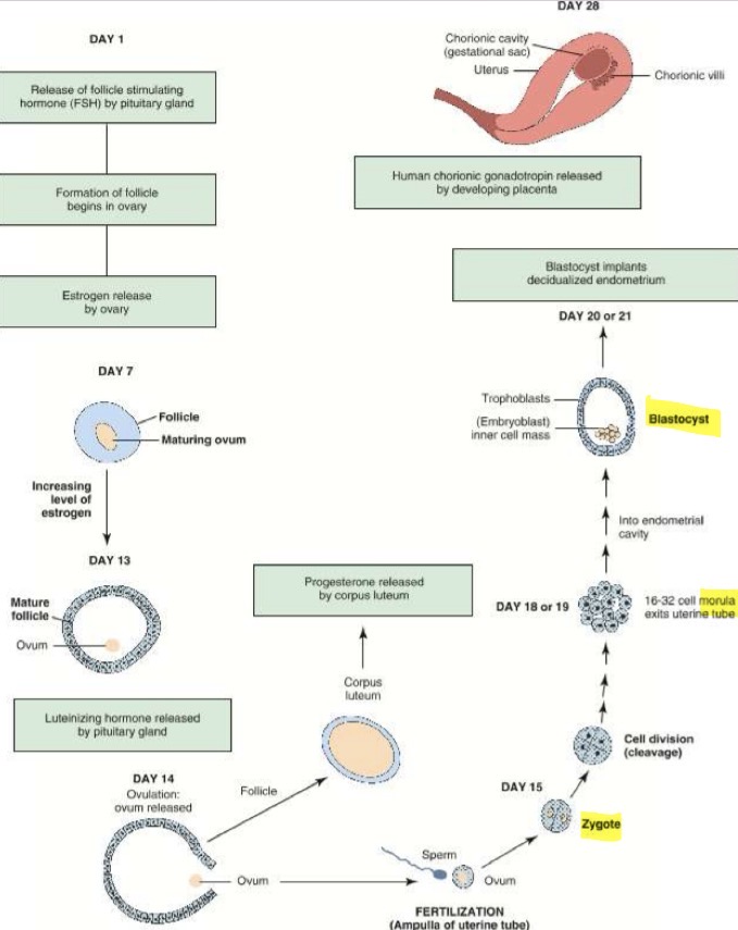
Post-ovulation
Ruptured follicle transforms into corpus luteum, which produced progesterone and small amount of estrogen and prepares uterus for pregnancy; during pregnancy corpus luteum may enlarge; endometrium becomes thick
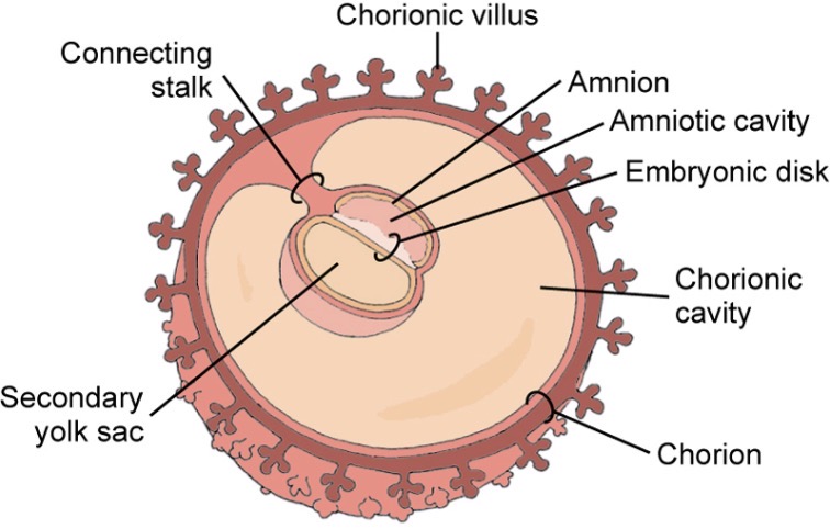
Ovarian Phase: Weeks 4-5
Second yolk sac forms between the amnion and chorion; an embryonic disk lies between the secondary yolk sac and amnion (called “double bleb sign”)
Initiation of placental development, develops alimentary canal (GI tract), neuroplate develops into neurotube (beginning of brain/spinal cord development), fetal lungs beginning development
Embryonic Phase: Week 6
week 6-10 are critical phase for human development; primitive heart begins to beat; primitive brain consists of:
Prosencephalon (forebrain): develops into cerebrum, lateral ventricle, and thalamus
Mesencephalon (midbrain): becomes adult midbrain and forms aqueduct of sylvius
Rhombencephalon (hindbrain): develops into adult pons, medulla, and cerebellum
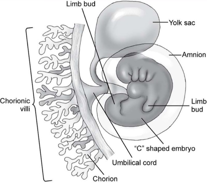
Embryonic Phase: Week 7-8
Formation of anterior and lateral abdominal walls; midgut forms from root of yolk sac; yolk sac fuses with vitelline duct to become umbilical cord; c-shaped embryo with limb buds
Week 8: mineralization of skeleton begins; embryo becomes c-shaped (gets larger) with limb buds; small bowels herniates (comes out of embryo) at base of umbilical cord
Week 10
Functional fetal kidney appears; by end of 10 weeks, major organ system are established; embryo demonstrates human feature
Fetal Phase: weeks 11-12
Growth is rapid; organ development continues; fetal intestinal activity begins in 11th week of development; fetal swallowing (of amniotic fluid) usually starts in week 12; skull and femur are adequately mineralized by 11.5 to 12 weeks; fetal head is disproportionately large compared with body and constitutes one half of length
Placenta Development
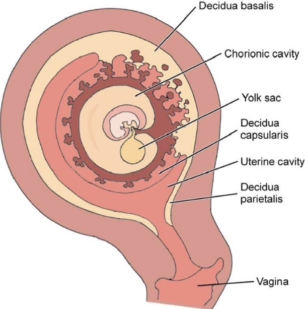
Decidualized endometrium differentiates into 3 distinct areas:
Decidua Basalis: portion of thick decidua at implantation site, maternal portion, deepest layer of placenta
Decidua Capsularis: Thin portion of endometrium, overlies section of gestational sac facing uterine cavity
Decidua Parietalis: (decidua vera), remaining endometrium or peripheral portion, unoccupied by implanted ovum
Decidua capsularis and parietalis are shed at parturition (birth)
Placenta serves as endocrine gland: produces human chorionic gonadotropin-hCG- communicates to the body that gestation is present ; produces estrogen and progesterone throughout pregnancy
Placenta is Divided into:
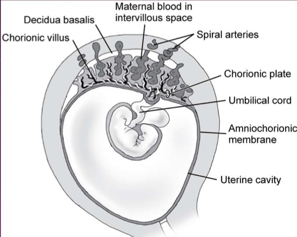
Chorionic Plate: Portion toward inside of sac that touches amniotic membrane
Base Plate/Basal layer: Portion on outside that touches uterus
Placental Substance: Placental material between basal layer and chorionic plate
Chronic Villi
Finger like projections of trophoblast; invade decidua basalis, forming embryo-maternal circulation; some villi breakdown chorion frondosum (surrounded by maternal tissue called lacunar network rich in blood vessels; after contact with villi, broken down to form small pools of blood or lacunae)
Development of Fetal Membrane: Amnion
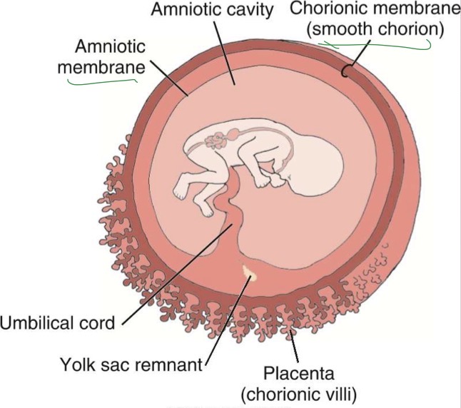
Innermost membrane of developing embryo; encloses amniotic cavity; remains attached to embryo at cord insertion site; covers umbilical cord
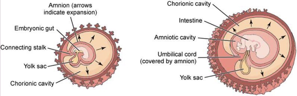
Development of Fetal Membrane: Chorion
Outermost tissue of developing embryo; vascular structure encloses chorionic cavity (chorionic cavity encloses amnion, yolk sac, and embryo); after 16 weeks, fuses with amnion
Development of Amniotic Fluid
Structures Responsible for Production of amniotic fluid: chorion frondosum, chorionic and amniotic membranes, skin, respiratory, and urinary tract
Structures involved in reduction of amniotic fluid: gastrointestinal system and amniotic-chorionic interface
Functions of Amniotic Fluid
permits symmetric growth of embryo/fetus
prevents adhesion from forming in fetal membranes
cushions the embryo/fetus and acts as shock absorber
helps to maintain proper temp of embryo
allows normal development of respiratory, gastrointestinal, and musculoskeletal systems
helps prevent infection
possibly serves as source of nutrients for developing embryo
Amniotic Fluid Volume (AFV)
Depends on balance between amniotic fluid production and removal or absorption; reflects the state of gestational well-being, many abnormalities are associated with marked increases (polyhydramnios) or decreases (oligohydramnios) in the AFV
Measurement Guidelines in First Trimester for Dating Pregnancy
gestational sac (no yolk sac, embryo or heartbeat) at 5 weeks
Gestational sac with yolk sac (no embryo or heartbeat) at 5.5 weeks
gestational sac with yolk sac (living embryo too small to measure) at 6 weeks
crown rump length (CRL) measurement of embryo from 6 weeks + days to 12 weeks
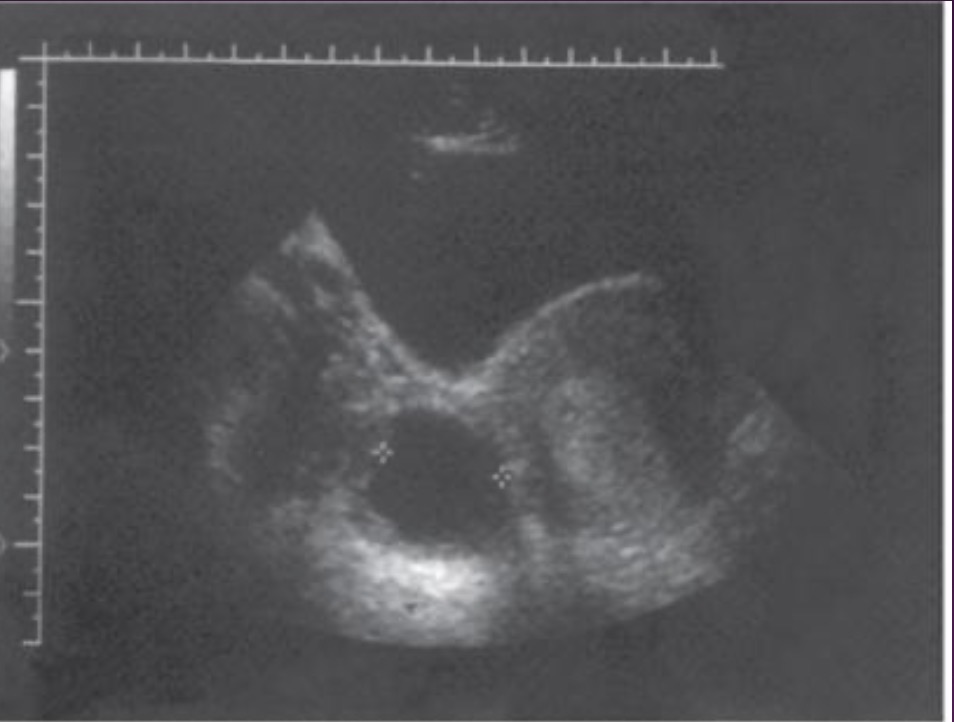
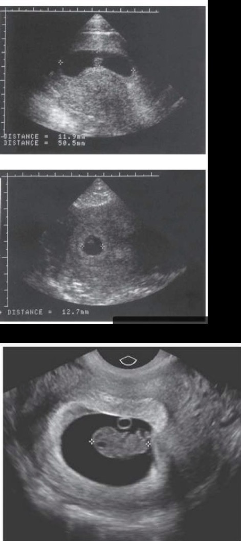
Gestational Sac
Seen as early as 3-5 weeks; small round/oval anechoic (fluid filled); enclosed by thick echogenic walls;
Double sac sign: fluid filled gestational sac and uterine cavity identified together and rules out pseudo sac
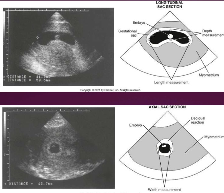
Mean Sac Diameter (MSD)
Sum of length, depth, and width of gestational sac (excluding wall) divided by 3; GA (in days) is calculated by adding 30 to MSD (in millimeters); machine usually calculates this on its own
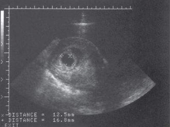
Yolk Sac
Small round cystic anechoic structure; within gestational sac; bright thin echogenic walls; normally measures less than 6mm; image shows normal size and if larger, tend to not make it
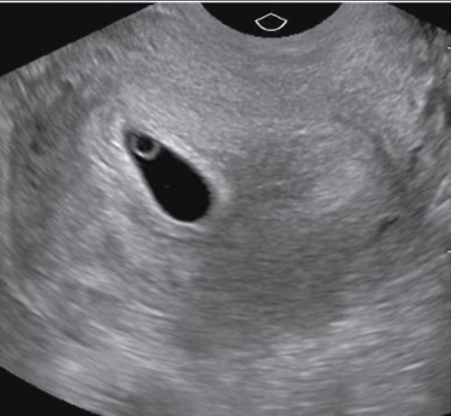
Embryo
Varies depending on age of embryo, with TV can see around 5.5 weeks; echogenic; cardiac activity by 6 weeks
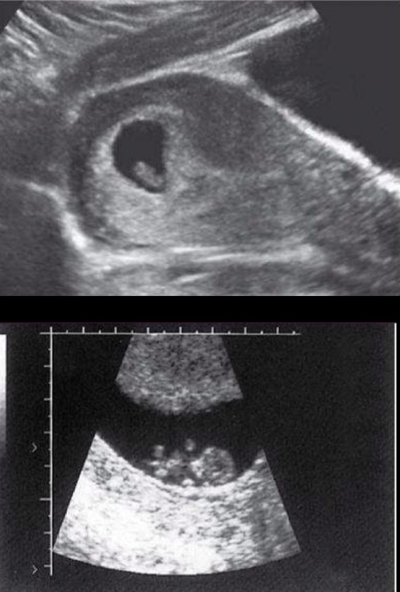
Crown Rump Length
Measured from top of head (crown) to middle of buttocks (rump); anechoic area is rhombencephalon; provides most accurate date for pregnancy
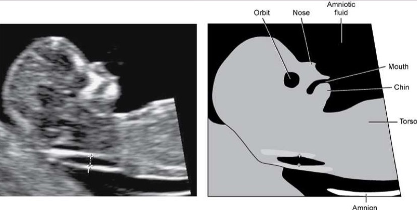
Nuchal Translucency
Used in combination with maternal serum screen (blood test) and maternal age to determine fetal risk; fluid that normally collects at back of fetus’s neck between 11.3-13.6 weeks of gestation; head should not be flexed or extended; spinal column should face bottom of imaging screen; calipers must be placed on inner borders of widest portion of NT (if NT is too large, could indicate to chromosomal abnormalities)
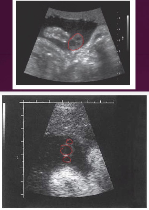
Umbilical Cord in First Trimester
Well visualized at 8 weeks; grows at rate similar to embryo; composed of 2 umbilical arteries and 1 umbilical vein; develops multiple spiral turns
Placenta in First Trimester
Homogeneous to heterogeneous; begins as thick echogenic ring around gestational sac; primarily medium-level echoes
Uterine Synechia
Membranes formed from scarring or adhesions secondary to surgery or infection; extend from uterus with amnion and chorion growing around them; synechiae do not attach to embryo/fetus; not common
Myometrial Contractions
Frequently seen on sonograms and are distinguishable by their inward bulge without disturbing uterine contour and their temporary nature
Sonographic Applications
Indicated for gestational age, vaginal spotting/bleeding, large/small for date of pregnancy, pelvic pain, fetal growth, substance abuse/prescription drugs during early pregnancy, trauma, or history of miscarriage or multiple gestations/fertility drug treatment
Lab Values
Human Chorionic Gonadotropin (hCG): hCG of 2000 is level at which sac will be seen in most normal early pregnancies; should be doubling every 48-72 hours
Alpha-fetoprotein (AFP): found in maternal blood and amniotic fluid; elevated levels indicate fetal abnormalities or defects
Triple Marker Screening (AFP,uE3, hCG): abnormal levels of AFP; unconjugated estriol (uE3); hCG
Associated Tests
Pregnancy test, Beta-hCG test, maternal/paternal blood typing, chorionic villus sampling (CVS), MRI, or CT as last resort