HAN 200 ,, Special Senses
 Call Kai
 Learn
Learn Practice Test
 Spaced Repetition
 Match
Match Flashcards
 Knowt Play
1/87

There's no tags or description
Looks like no tags are added yet.
Name | Mastery | Learn | Test | Matching | Spaced | Call with Kai | Chat |
|---|
No analytics yet
Send a link to your students to track their progress
88 Terms
Eyebrows
Shade from the sun
Eyelids (Palpebrae)
Protection/glands
Conjunctivia
Transparent membrane
Lacrimal Apparatus
Tears
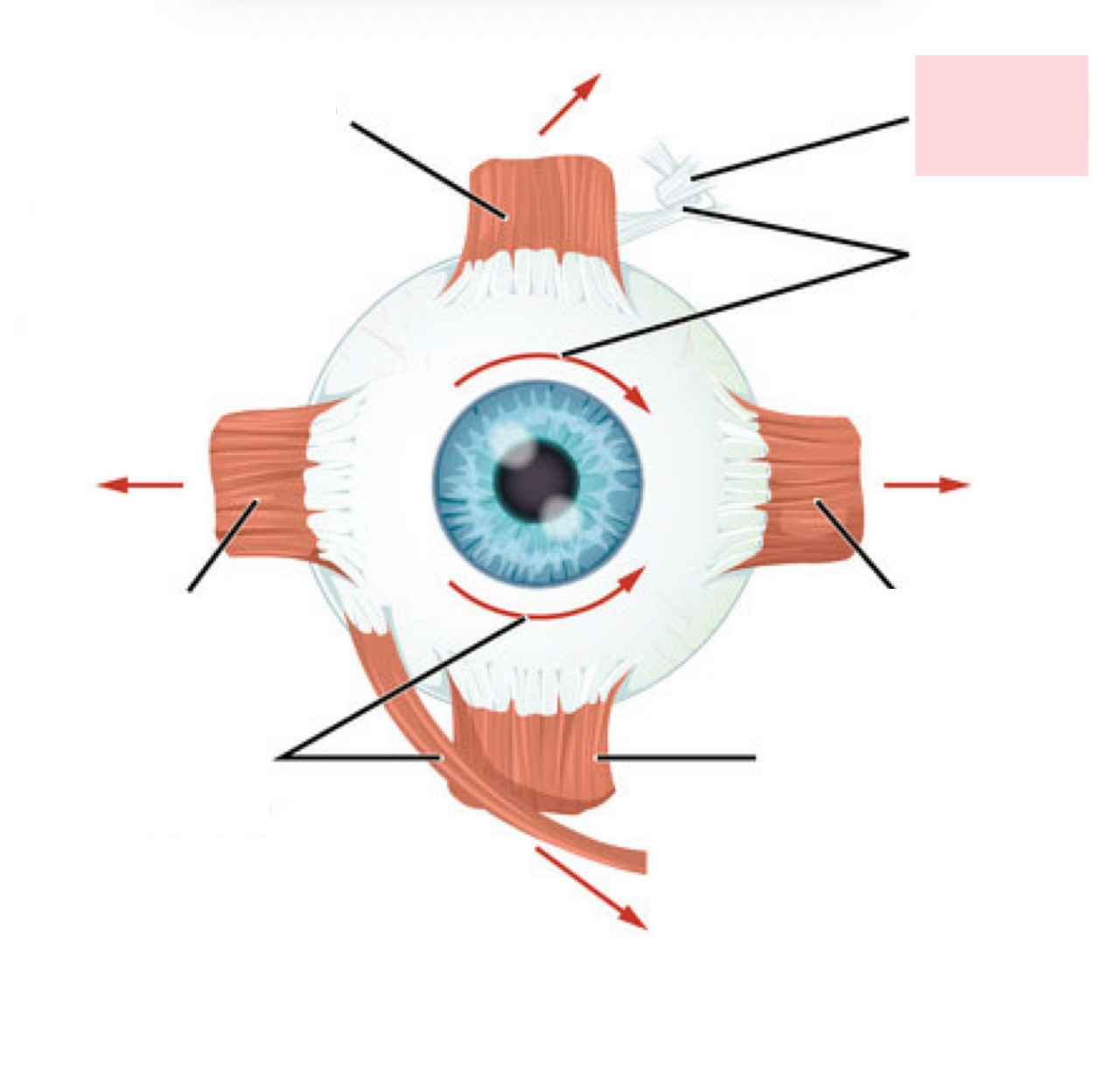
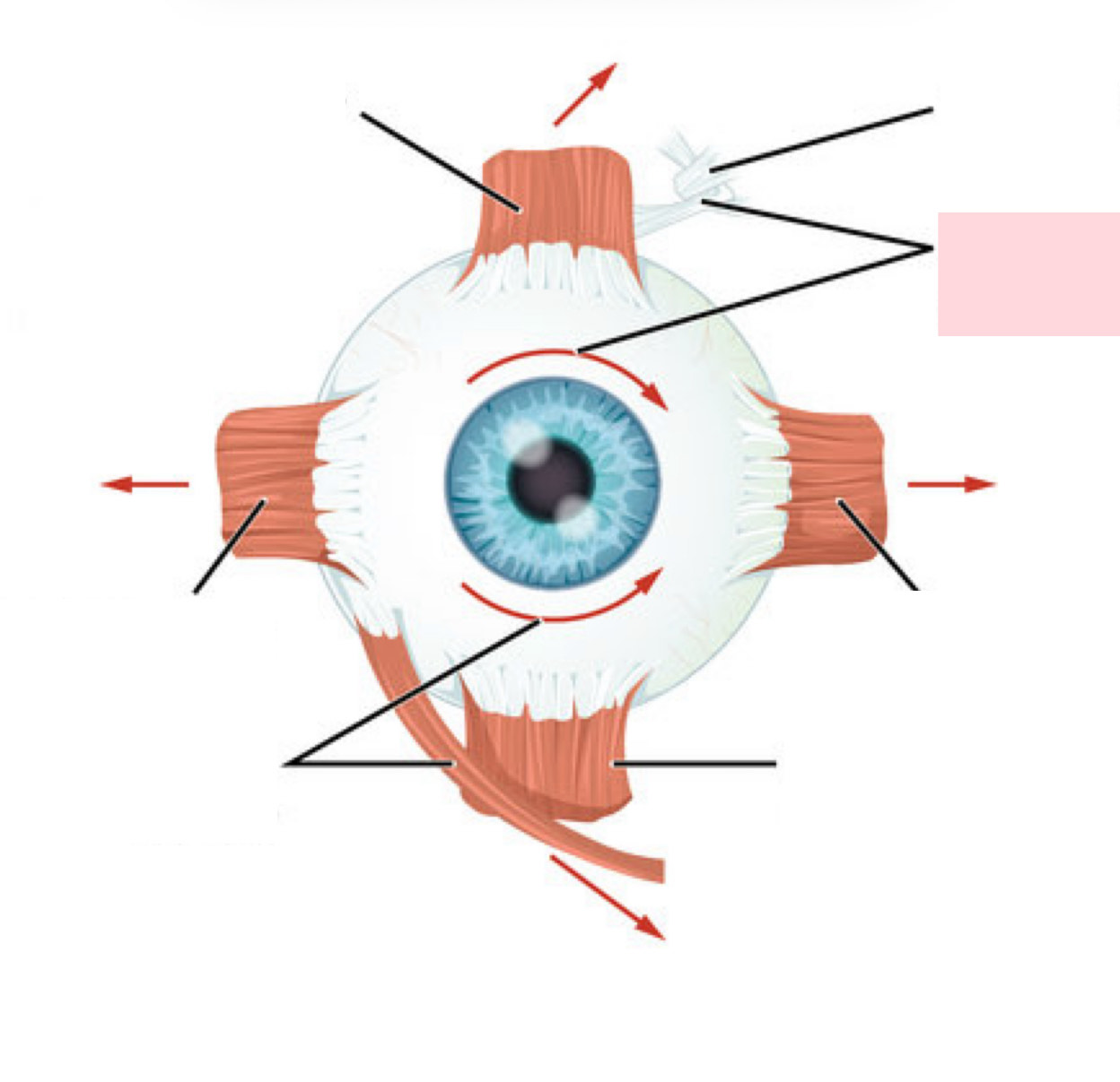
Extrinsic Eye Muscles
CN 3, 4, and 6
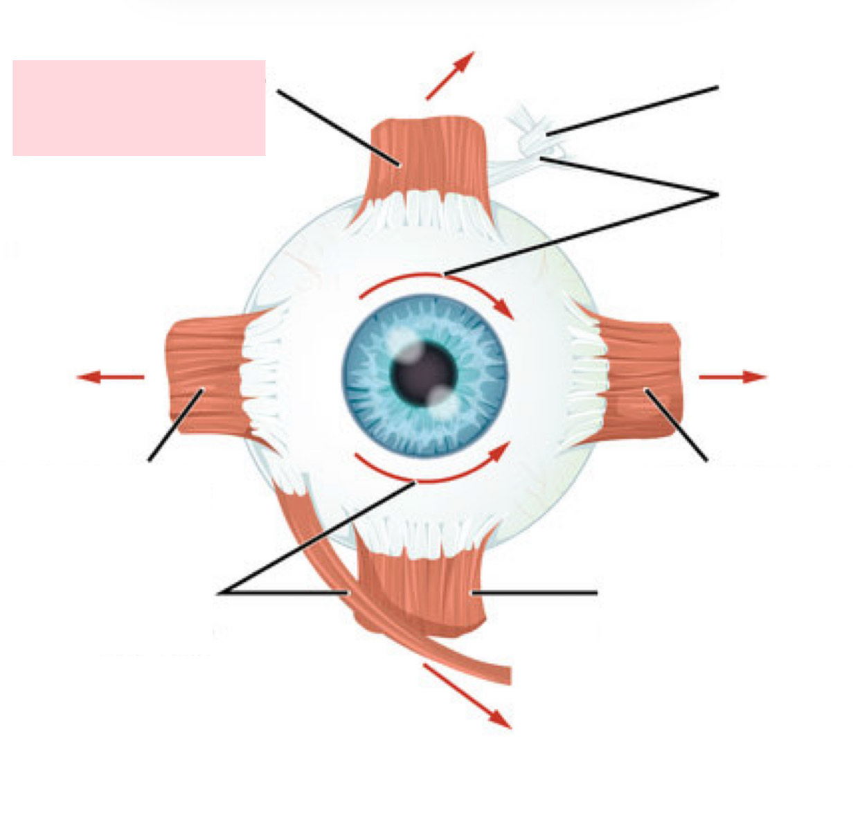
Label the Diagram
Superior Rectus
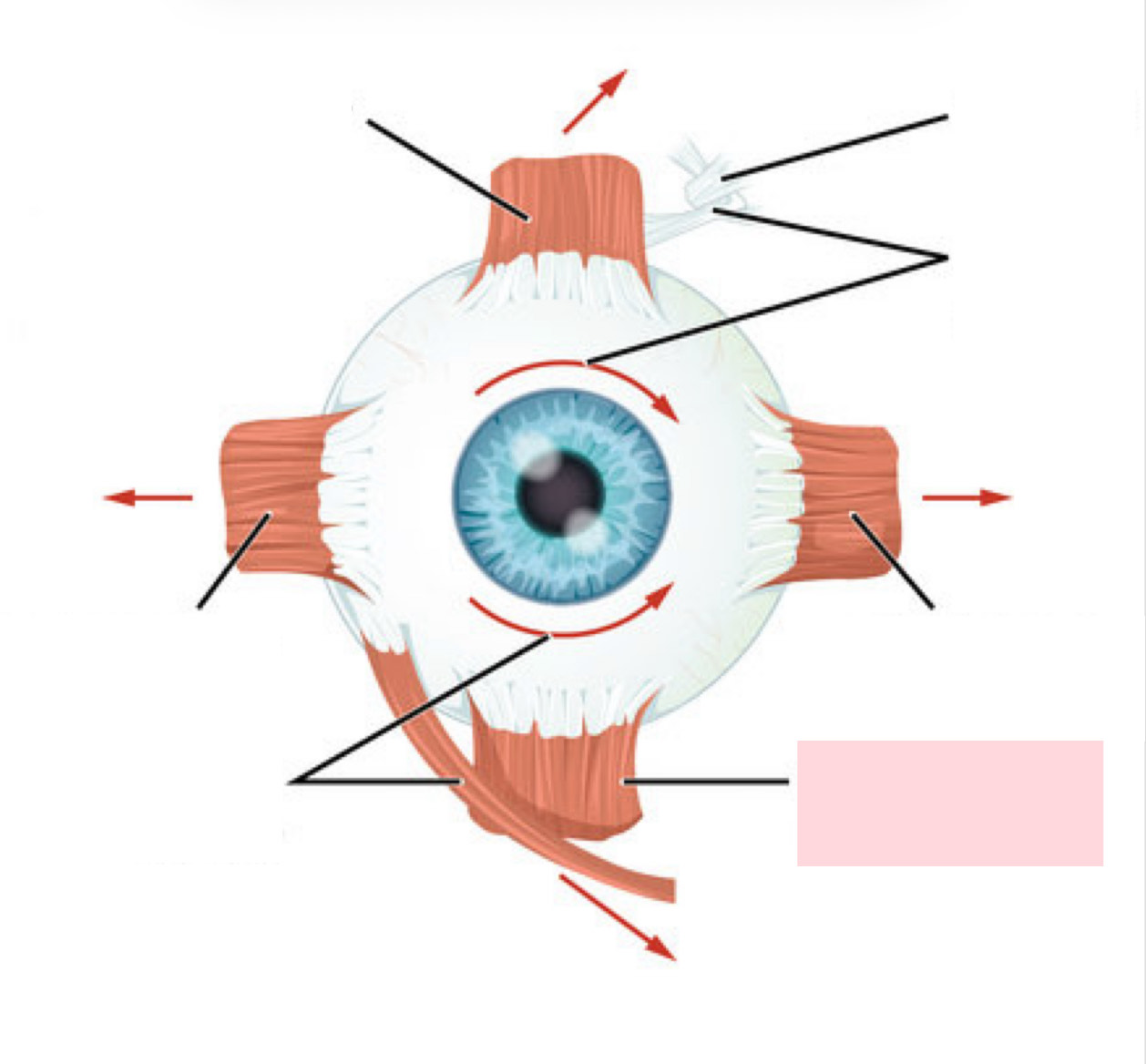
Label the Diagram
Inferior Rectus
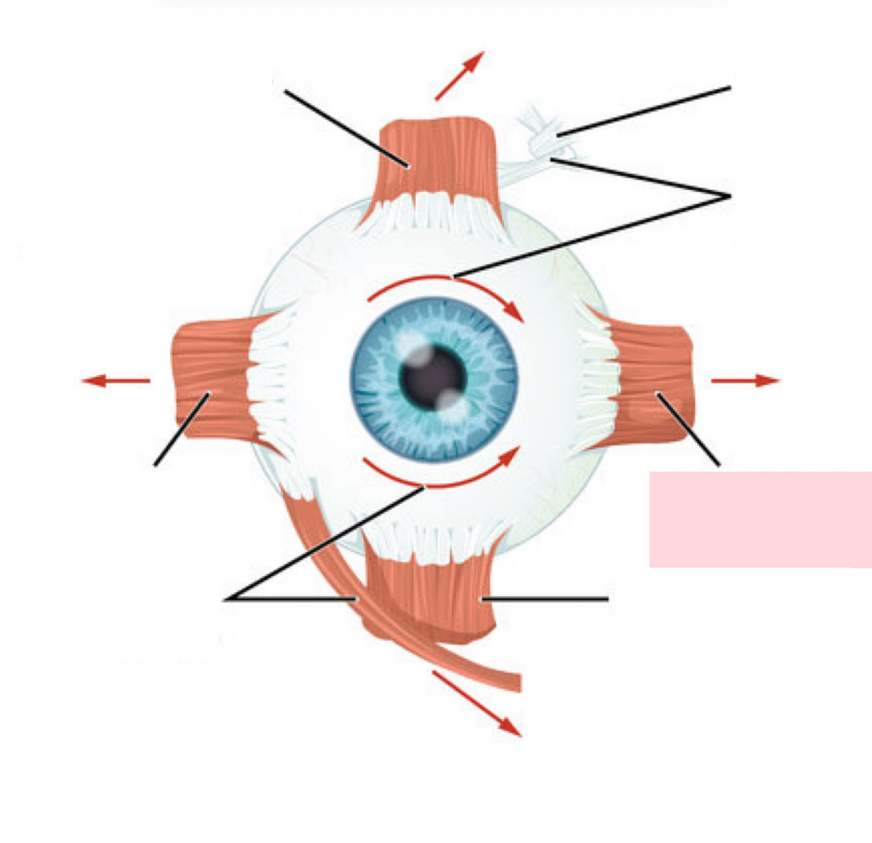
Label the Diagram
Medial Rectus
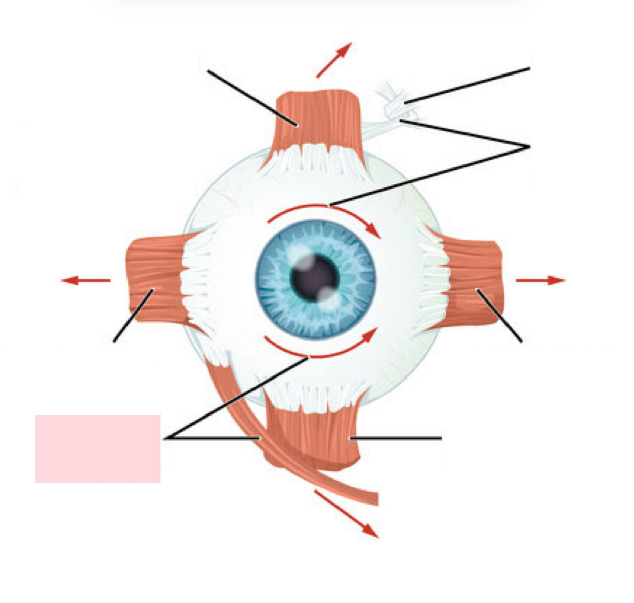
Label the Diagram
Inferior Oblique
Label the Diagram
Trochlear
Label the Diagram
Superior Oblique
Moves Eyes Laterally
CN VI (Abducens)
Moves Eyes Medially
CN III (Oculomotor)
Elevates Eyes and Turns it Medially
CN III (Oculomotor)
Depresses Eyes and Turns it Medially
CN III (Oculomotor)
Elevates Eyes and Turns it Laterally
CN III (Oculomotor)
Depresses Eyes and Turns it Laterally
CN IV (Trochlear)
Fibrous Wall of the Eyeball
Sclera and cornea (avascular, outermost CT)
Vascular Wall of the Eyeball
Iris, ciliary body, choroid (middle pigmented layer)
Sensory Wall of the Eyeball
Retina (delicate two-layered membrane)
Internal Cavity is FIlled With ____.
Humors
Label the Diagram
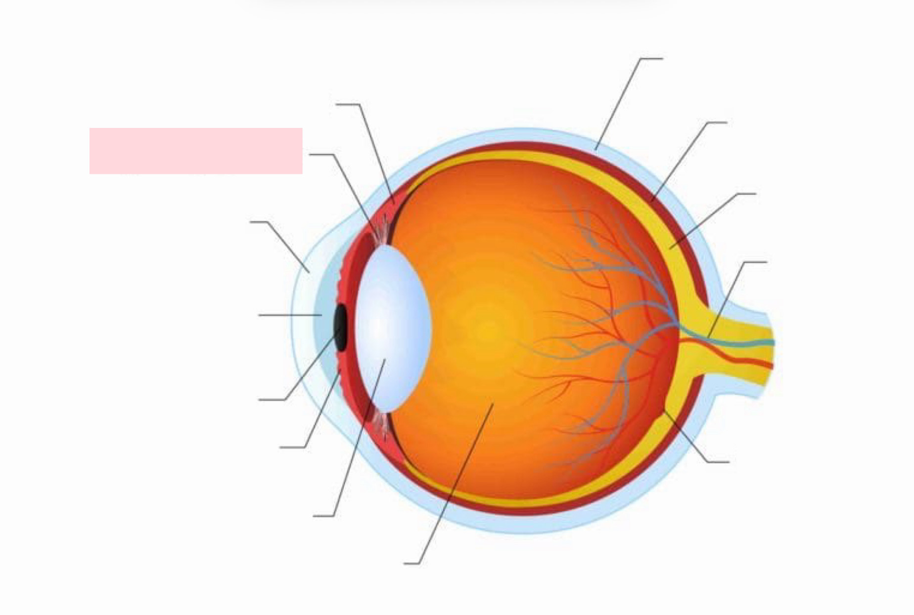
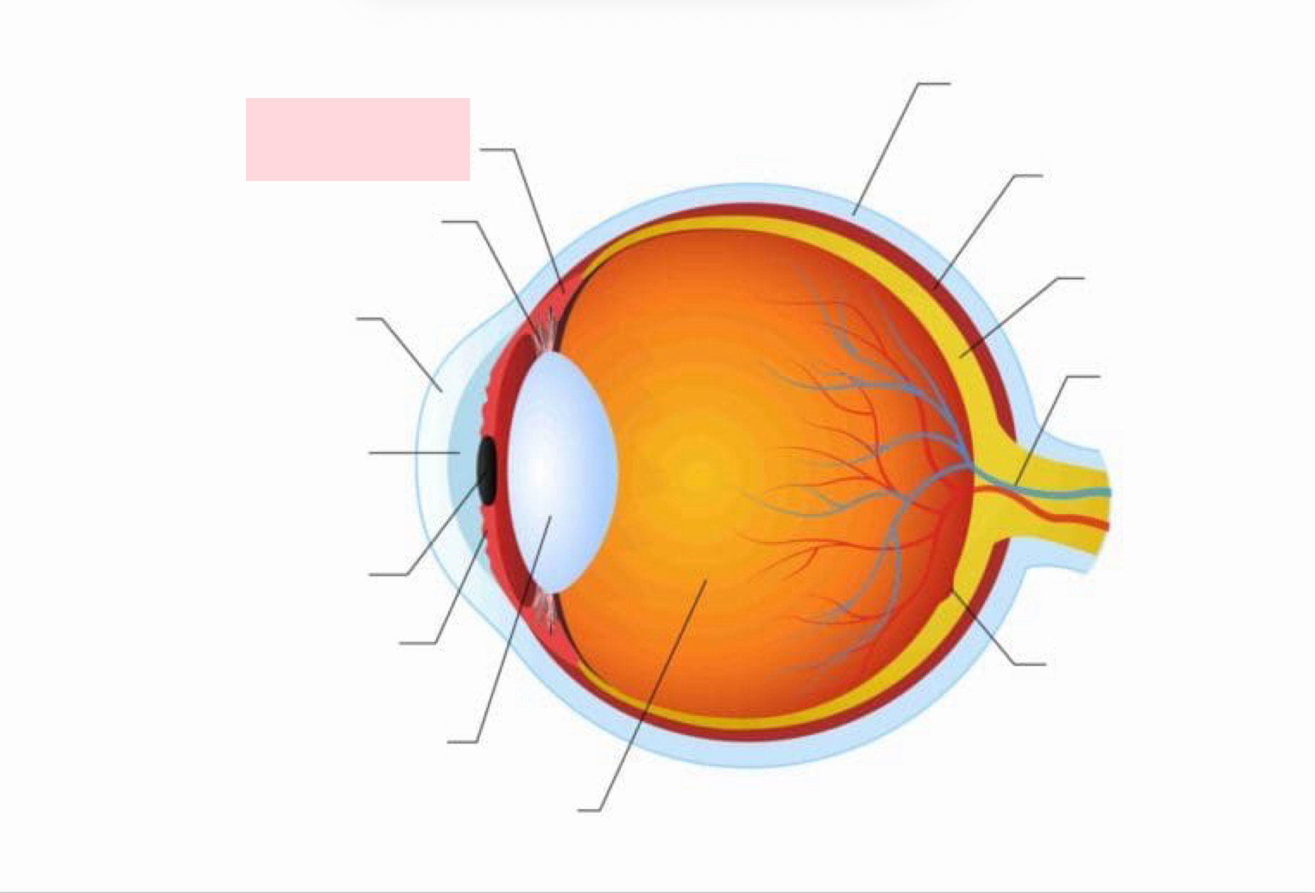
Suspensory Ligament
Label the Diagram
Ciliary Body
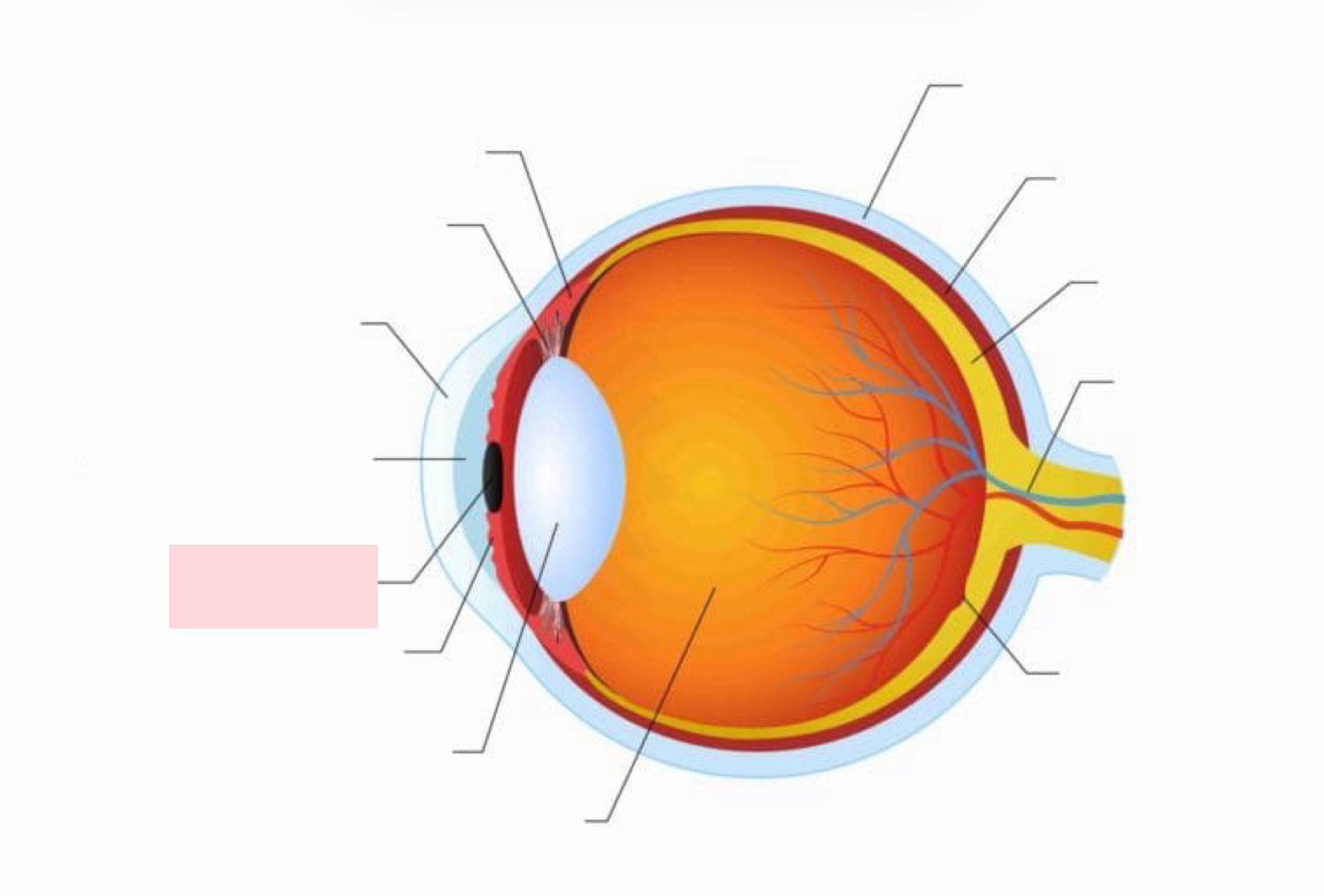
Label the Diagram
Pupil
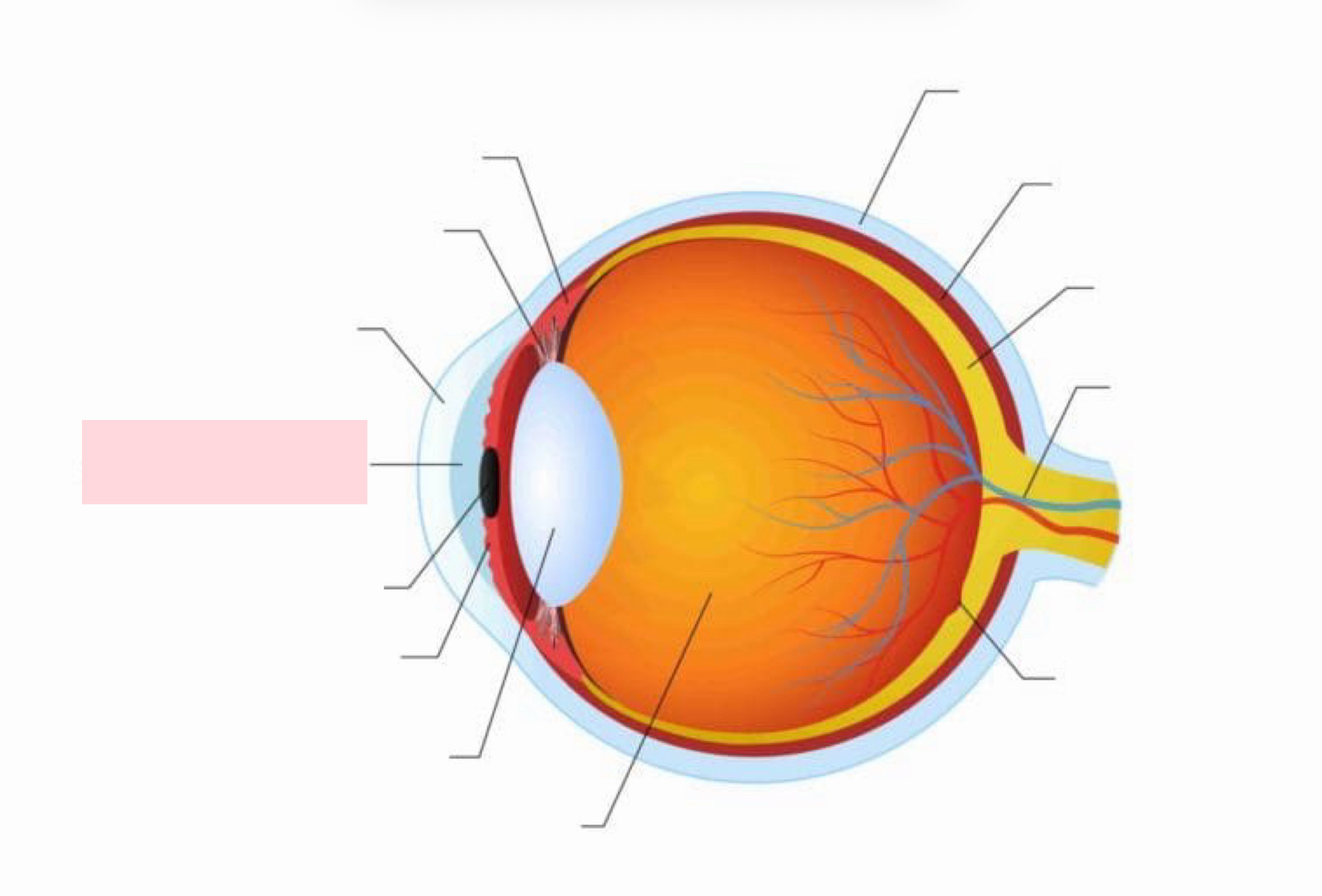
Label the Diagram
Anterior Chamber
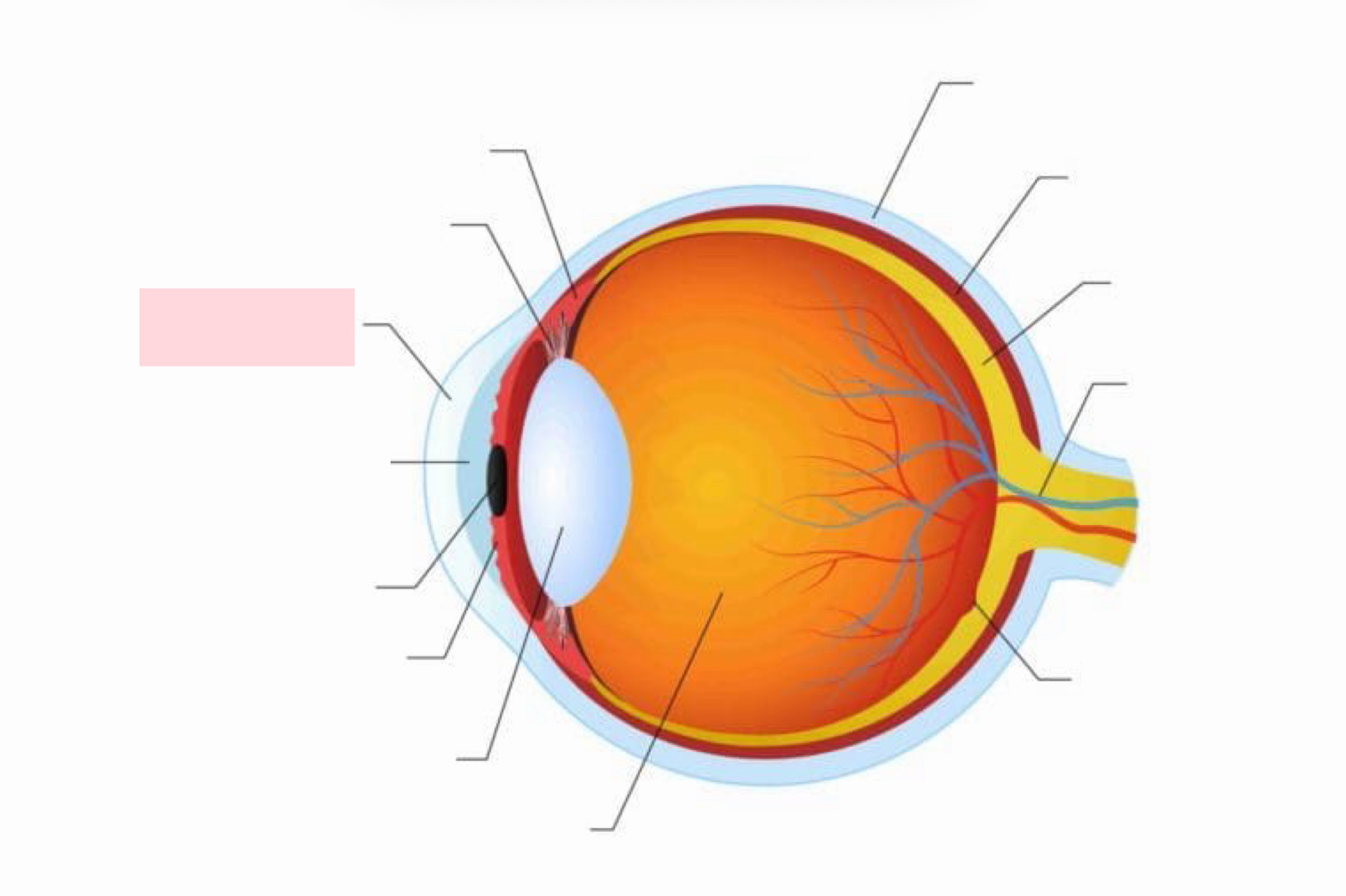
Label the Diagram
Cornea
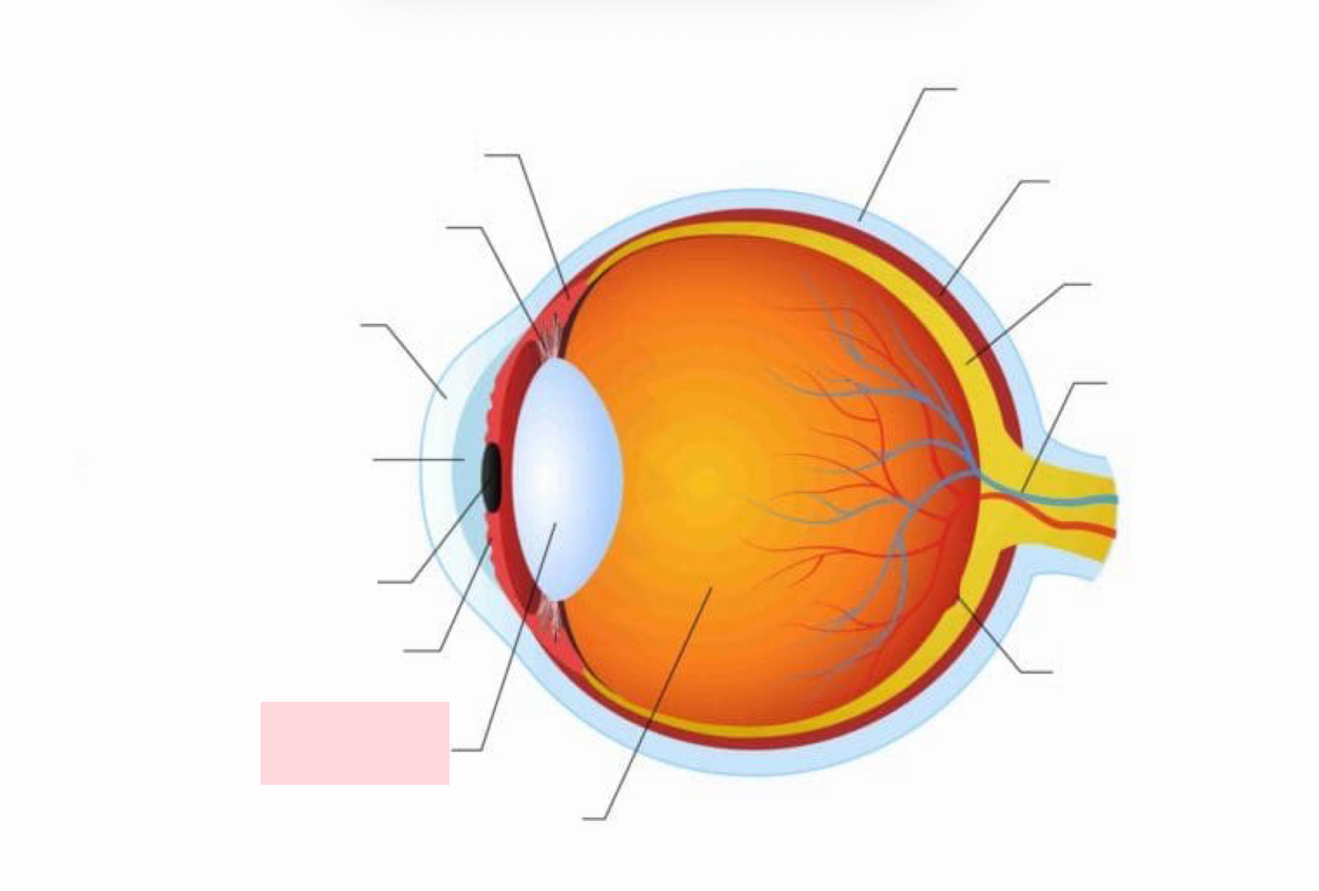
Label the Diagram
Lens
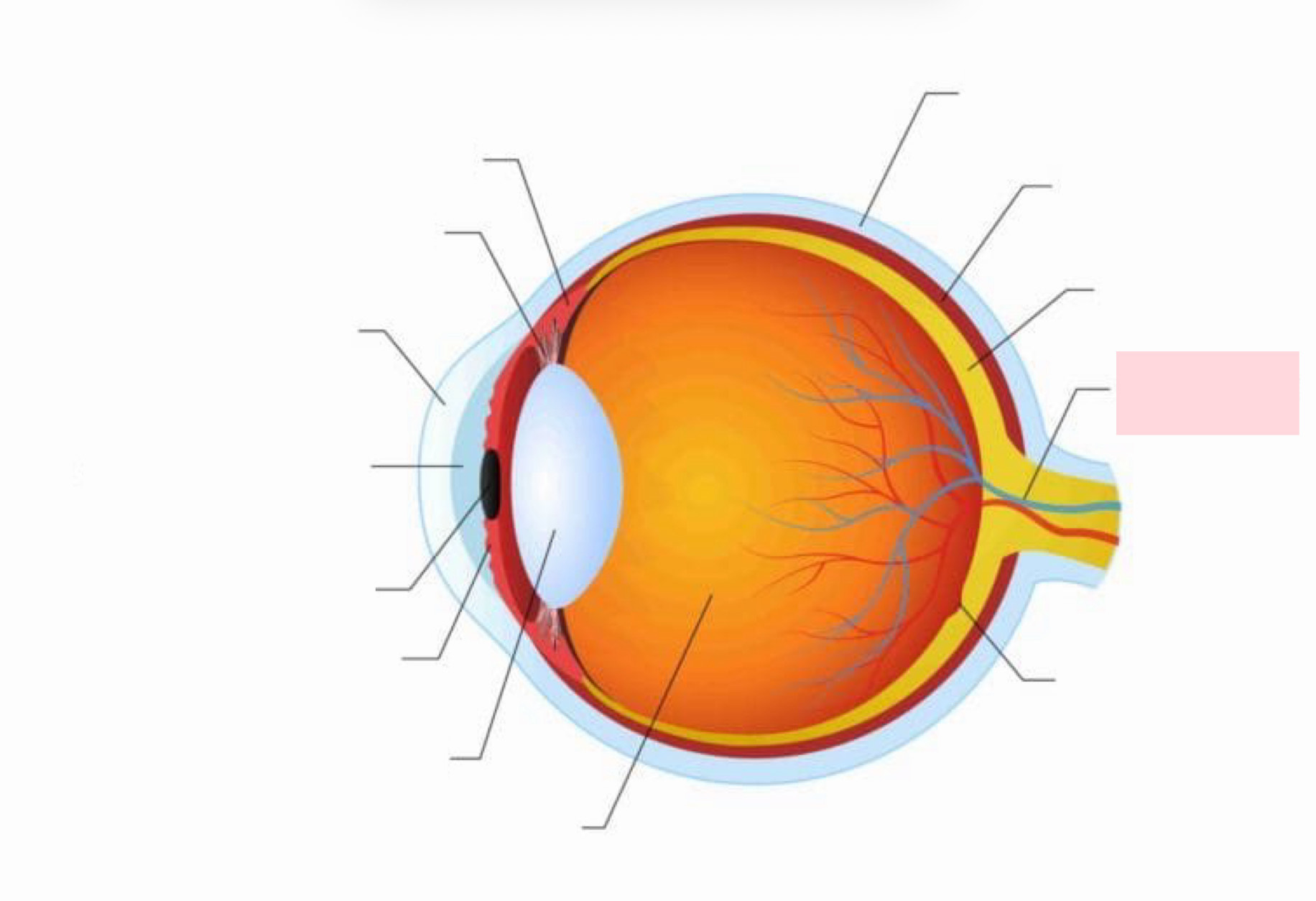
Label the Diagram
Optic Nerve
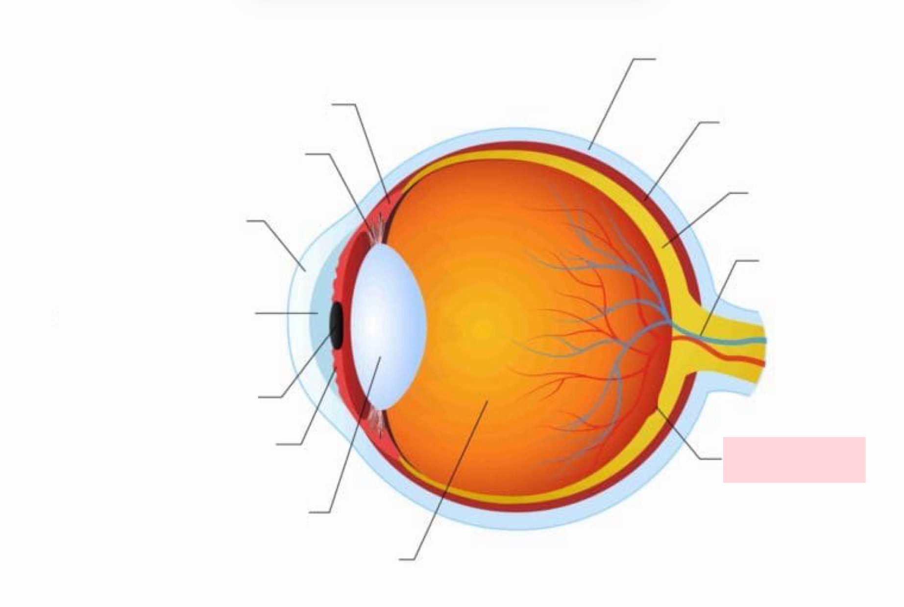
Label the Diagram
Macula
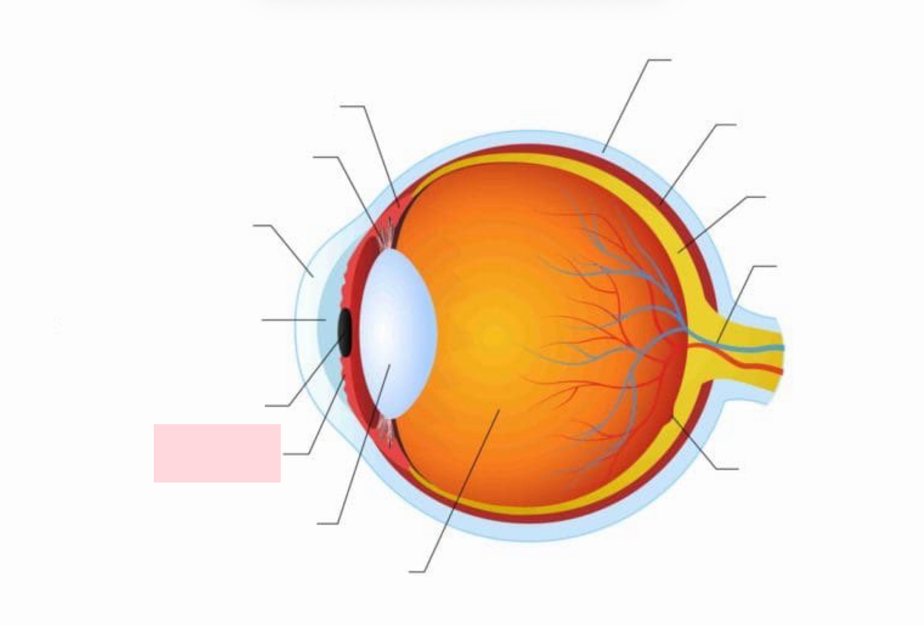
Label the Diagram
Iris
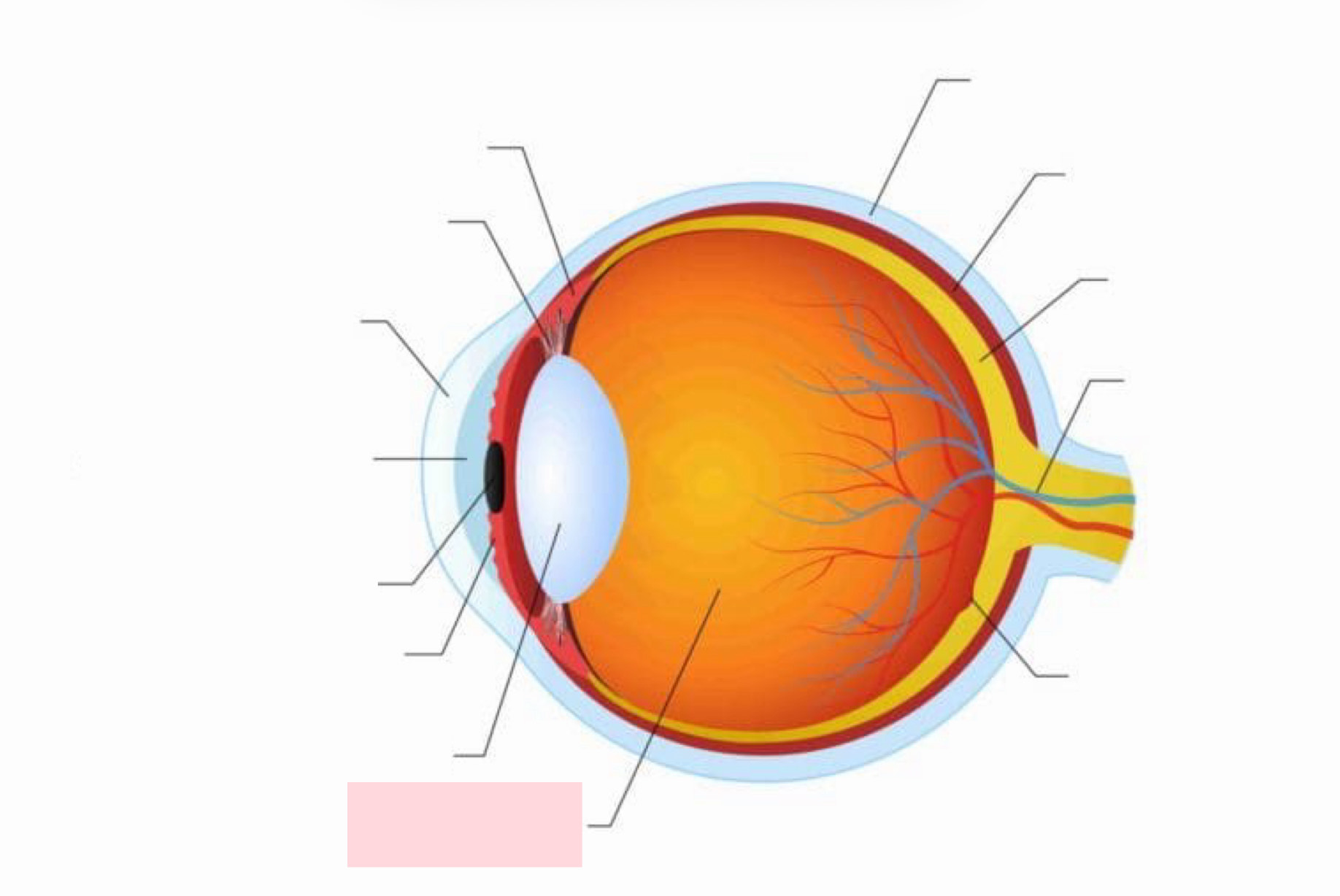
Label the Diagram
Vitreous Body
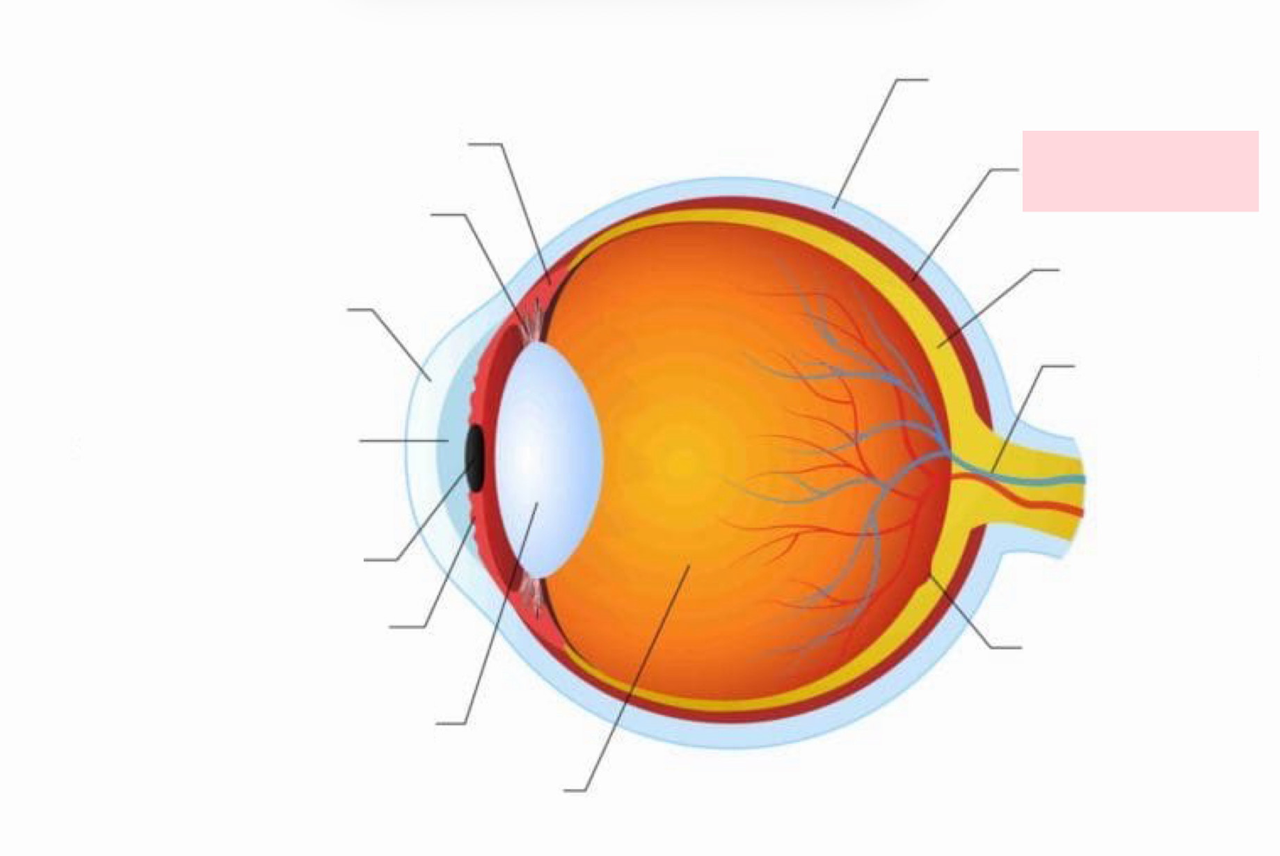
Label the Diagram
Choroid
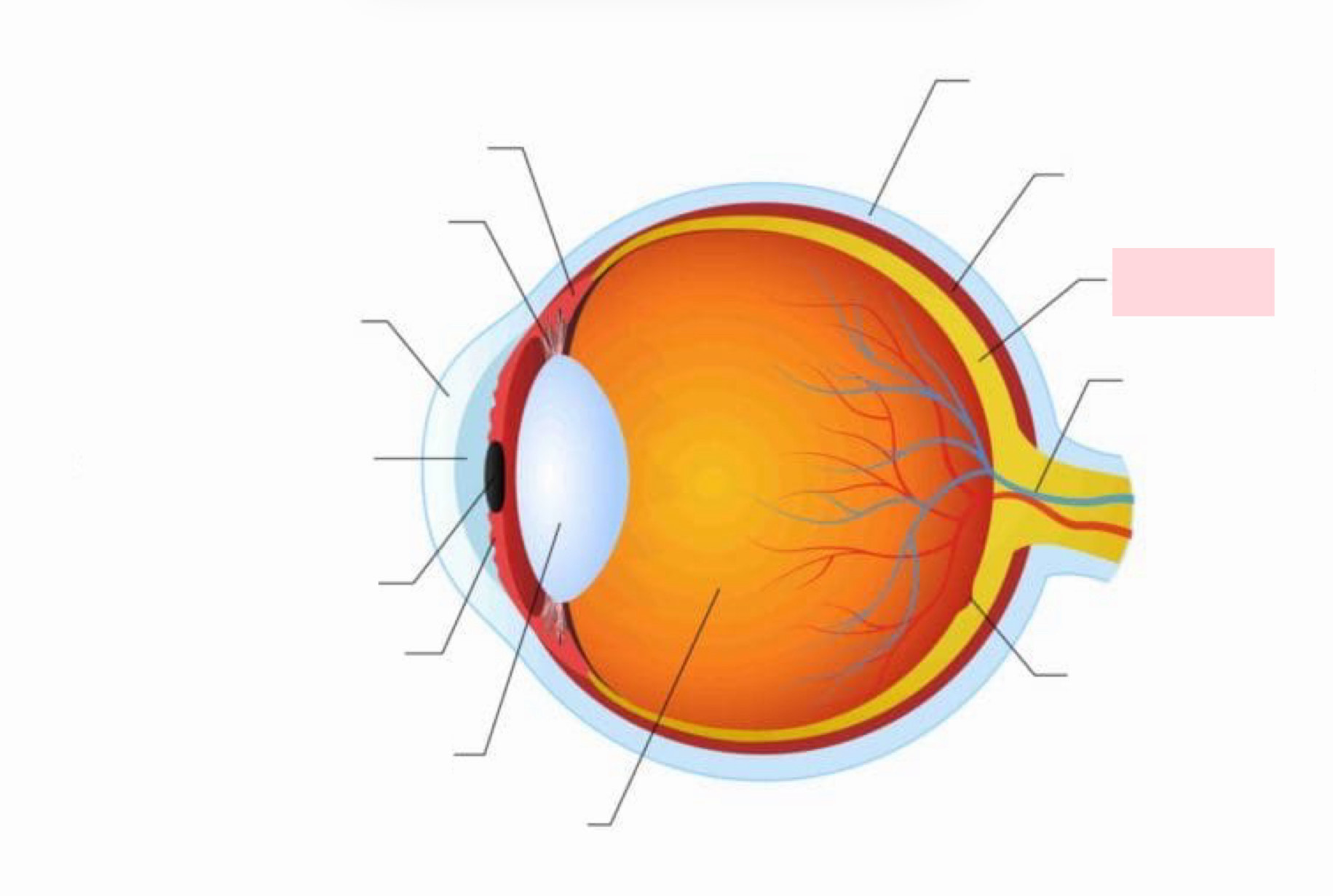
Label the Diagram
Retina
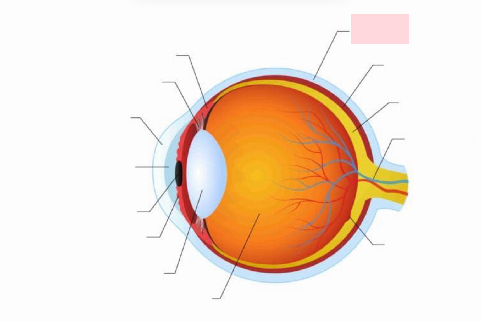
Label the Diagram
Sclera
Sclera (Whites of the Eye)
Protects and shapes the eyeball; continuous with the dura mater of brain
Cornea
Bends light as it enters the eye; numerous pain receptors contribute to blinking and tearing reflexes
Choroid Region
Supplies blood to all the layers of the body
Ciliary Body
Smooth muscle bundles control lens shape
Iris
The colored part of the eye
Pupil
Central opening that regulates the amount of light entering the eye
Pigmented Outer Layer of the Retina
Absorbs light and prevents its scattering
Neural Inner Layer of the Retina
Photoreceptor to transduce light energy
Photoreceptors Pathway
Rods/Cones → Bipolar Cells → Ganglion cells where action potential is generated
Blind Spot
The optic disc has no photo receptor cells and the brain fills in the area (visual filling)
Rods
Operates in dim lights; provides indistinct, fuzzy, non color peripheral vision
Cones
Operate in bright lights; provide high acuity color vision; concentrated in the fovea centralis
Fundus of Eye
The only place where small vessels can be observed directly
Posterior Segment of the Internal Chamber
Contains vitreous humor that transmits light and holds the neural retina firmly against the pigmented layer
Anterior Segment of the Internal Chamber
Contains aqueous humor
Anterior Chamber of the Internal Chamber
Between the cornea and the iris
Posterior Chamber of the Internal Chamber
Between the iris and the lens
Glaucoma
Compression of the retina and optic nerve if drainage of humors is blocked; second leading cause of blindness in the US
Lens
Biconvex, transparent, flexible, elastic, and avascular; allows for precise focusing of light on the retina
Cataracts (Clouding of the Lens)
Occur as a consequence of aging, diabetes, heavy smoking, and frequent exposure to intense sunlight
Pathway of Light Entering the Eye
Cornea → aqueous humor → lens → vitreous humor → neural layer of retina → photoreceptors
Light is Refracted Three times
At the cornea, entering the lens, and leaving the lens
Focusing for Close VIsion
Parasympathetic activation
Light Adaptation
Occurs when moving from darkness into bright light; cones and neurons rapidly adapt
Dark Adaptation
Occurs when moving from bright light into darkness; cones stop functioning in low-intensity light
Visual Pathway
CN 2 leaves the eye → some fibers cross at the optic chiasma → most fibers continue to the thalamus → fibers connect to the primary visual cortex in the occipital lobes
Depth Perception
Both eyes view the same image from slightly different angles, resulting in cortical fusion
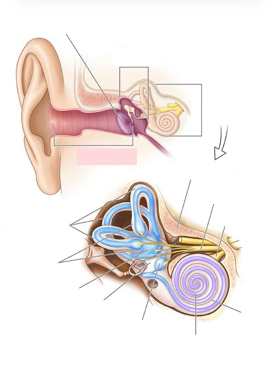
External and Middle Ear
Involved with hearing
Internal Ear
Hearing and equilibrium
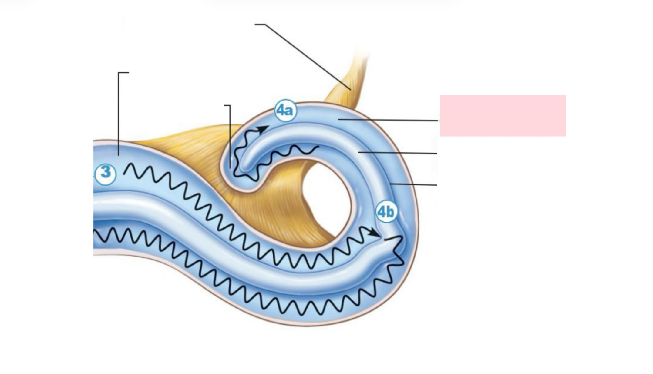
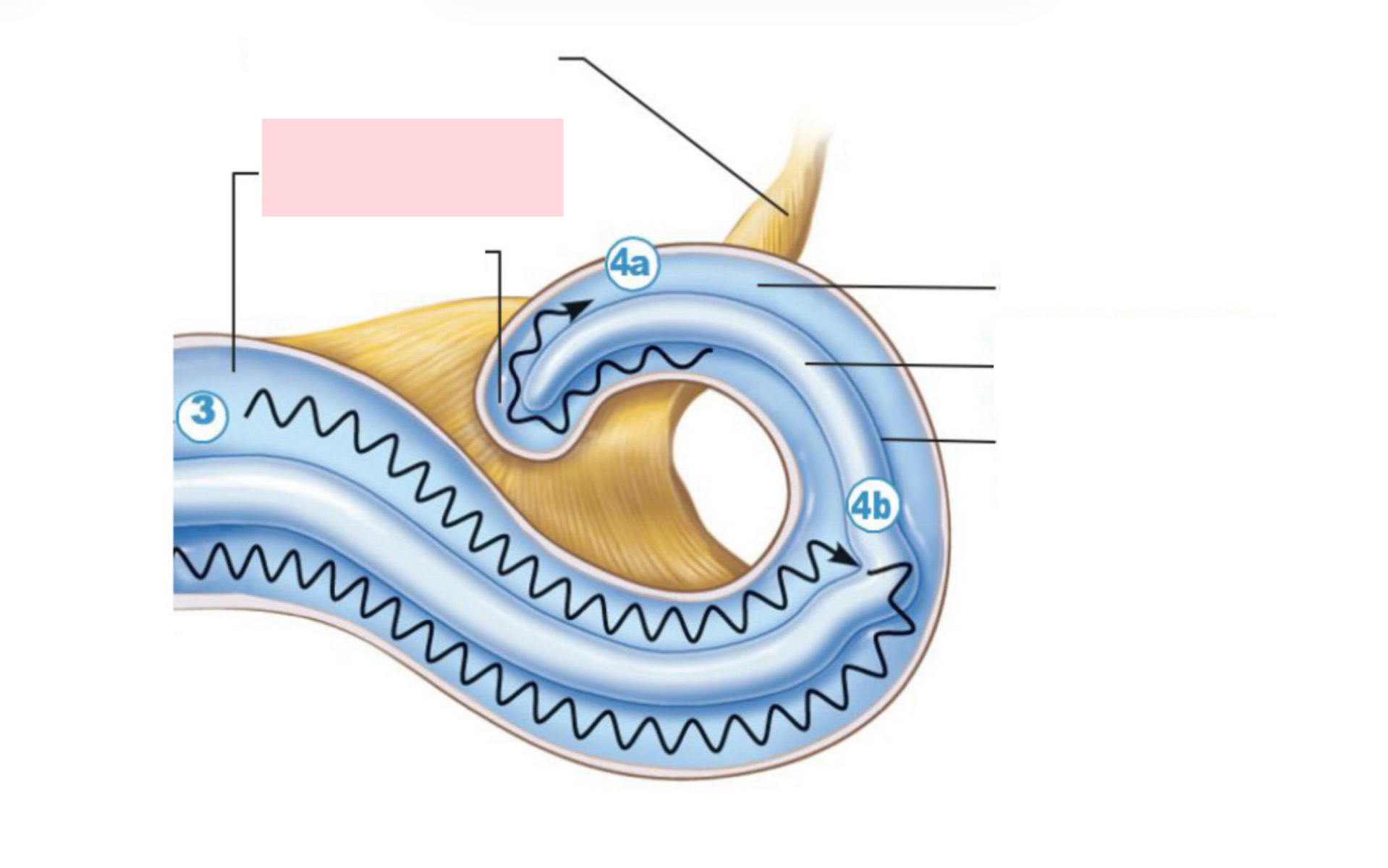
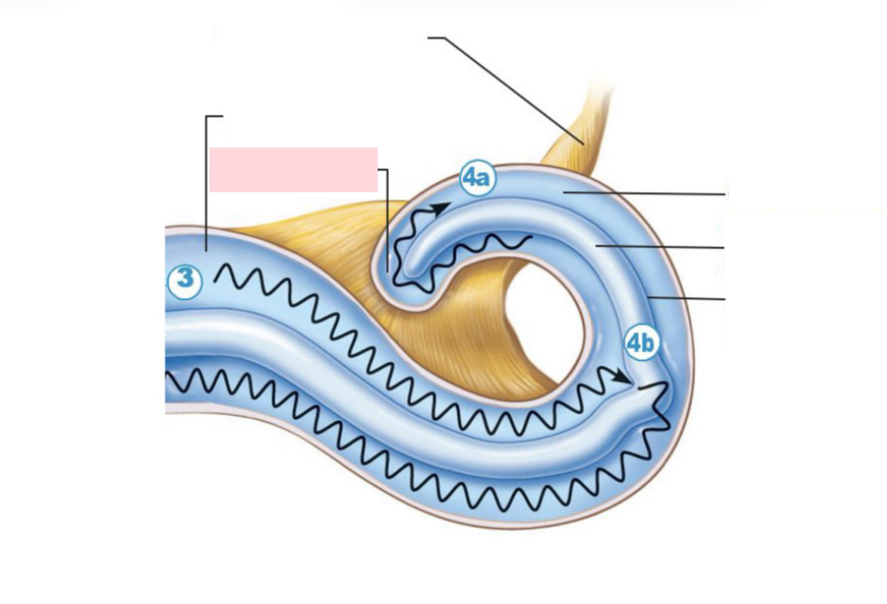
The Cochlea
Spiral bony chamber
Physiology of Hearing
Sound waves cause eardrums to vibrate → movement of auditory ossicles → fluid of inner ear moves → tiny hair cells are excited and stimulate local neurons of CN VIII
Auditory Processing
Impulses from specific hair cells are interpreted as specific pitches
Loudness
Detected by increasing numbers of action potentials that result when the hair cells experience large deflections
Three modes of input for balance and orientation
Vestibular receptors, visual receptors, and somatic receptors
Sensory Mismatch
Causes car sickness, seasickness, and dizziness on heights
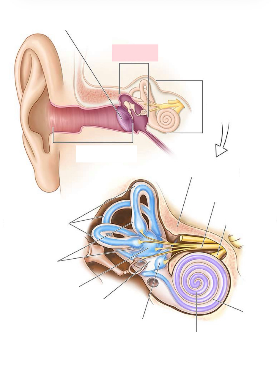
Label the Diagram
Middle ear
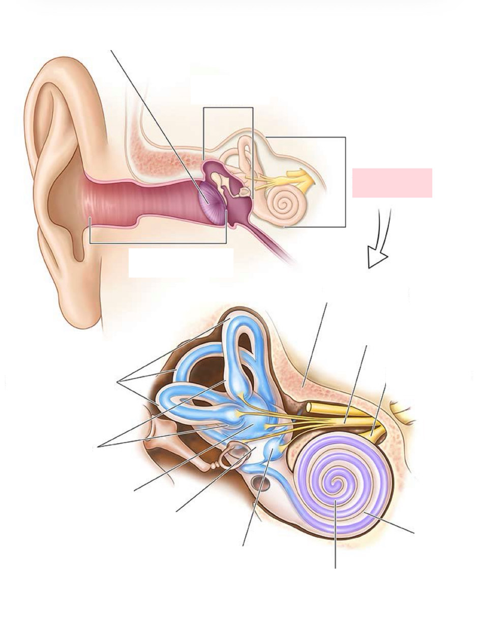
Label the Diagram
Inner ear
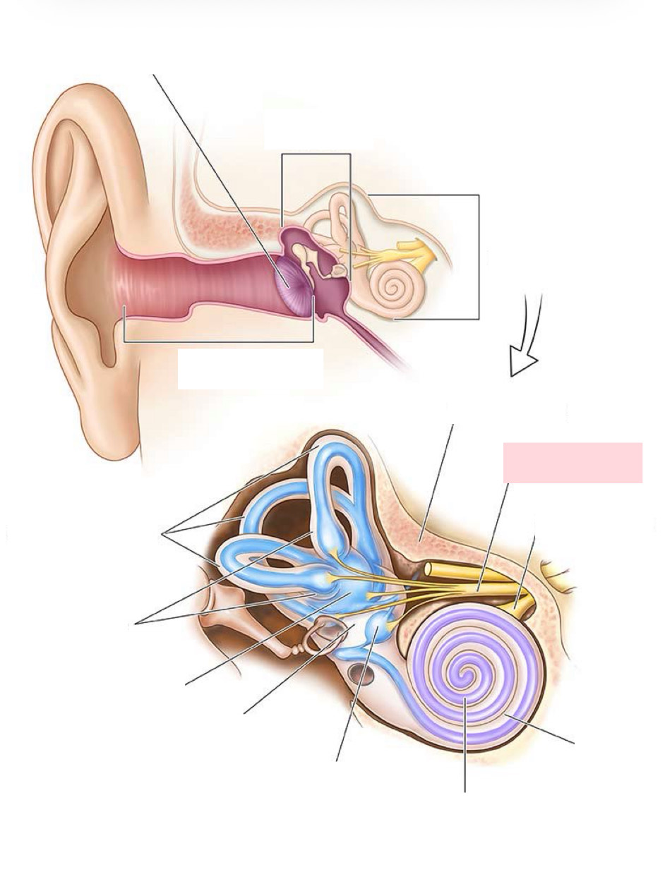
Label the Diagram
Vestibular nerve
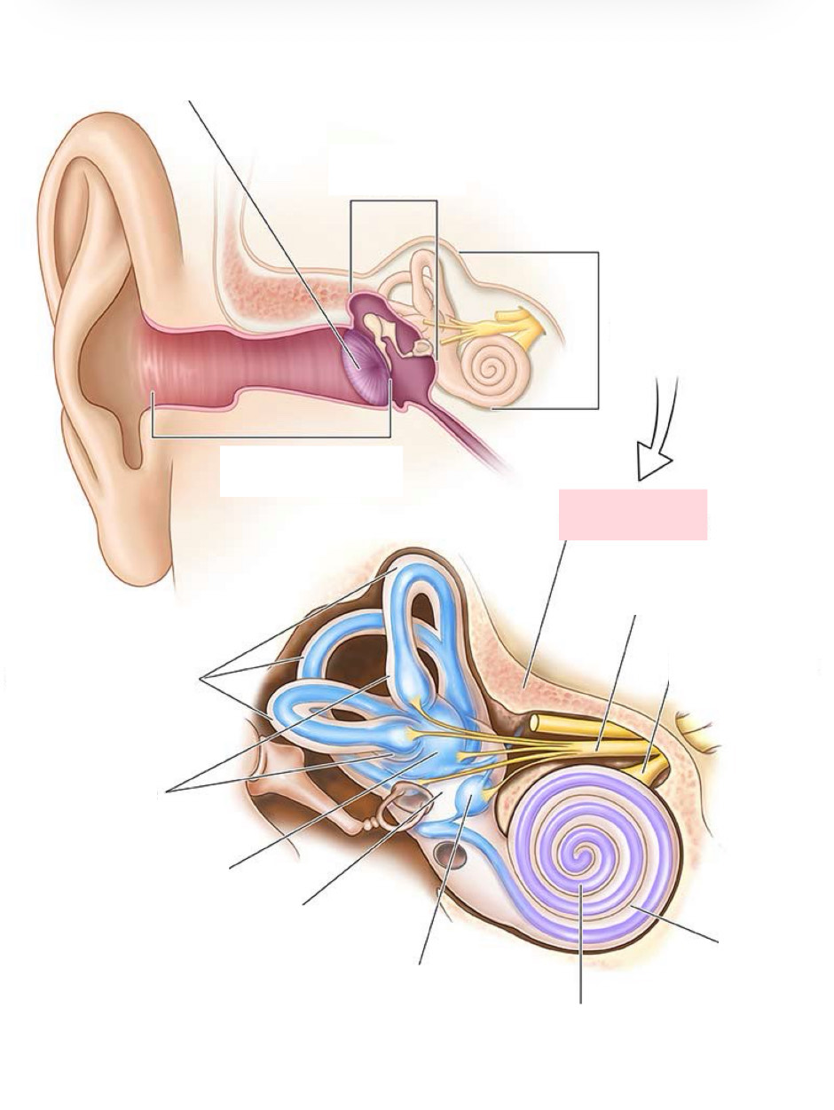
Label the Diagram
Temporal bone
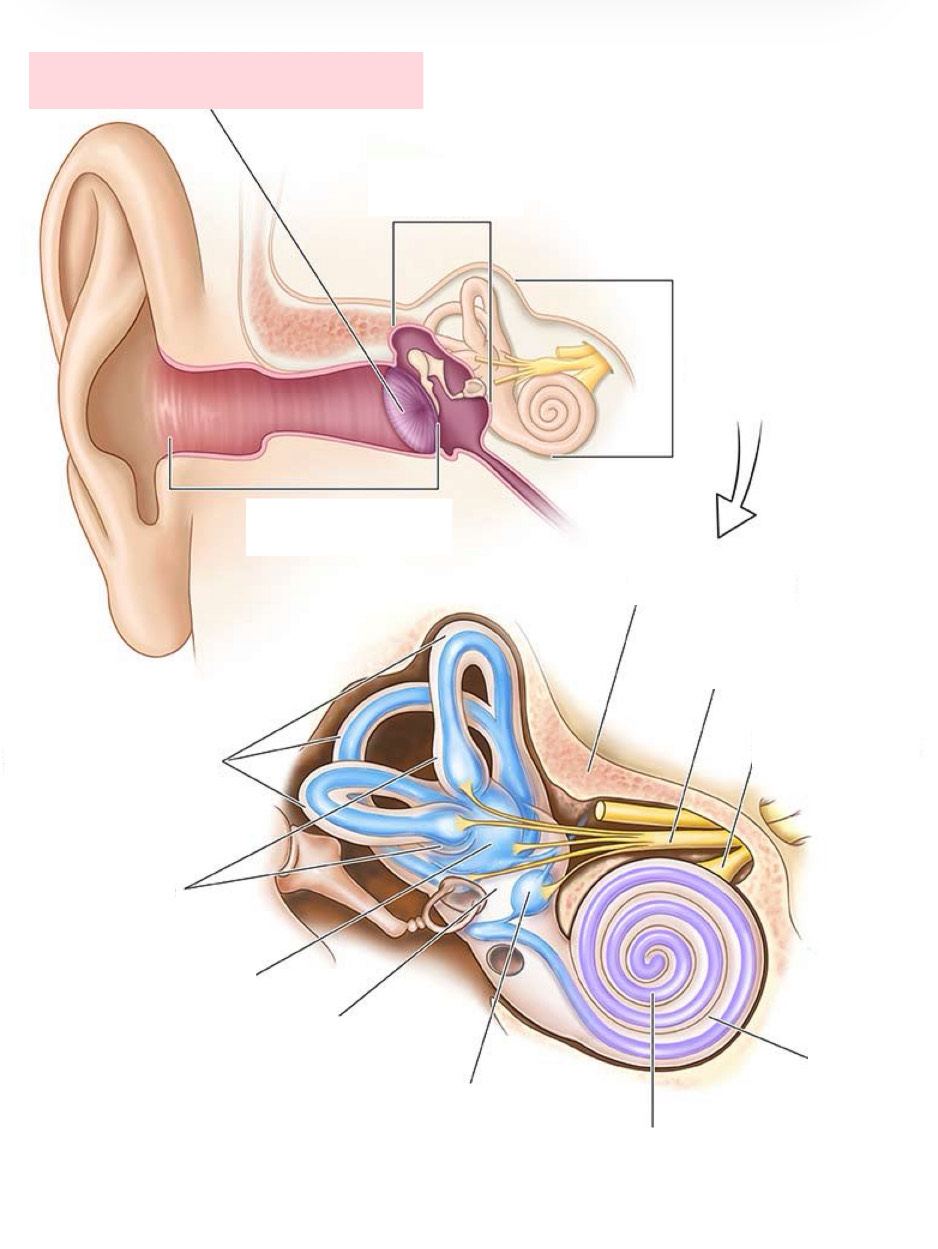
Label the Diagram
Tympanic membrane
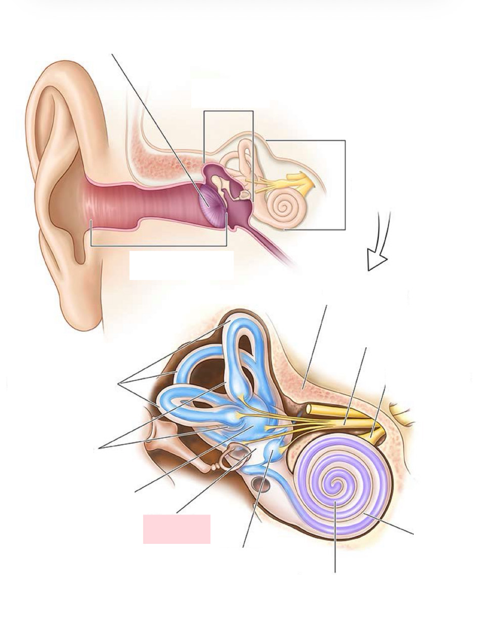
Label the Diagram
Vestibule
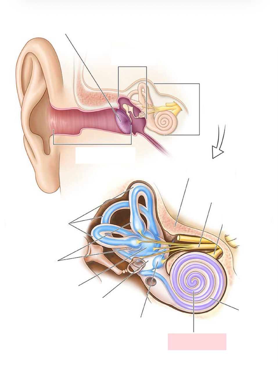
Label the Diagram
Cochlear duct
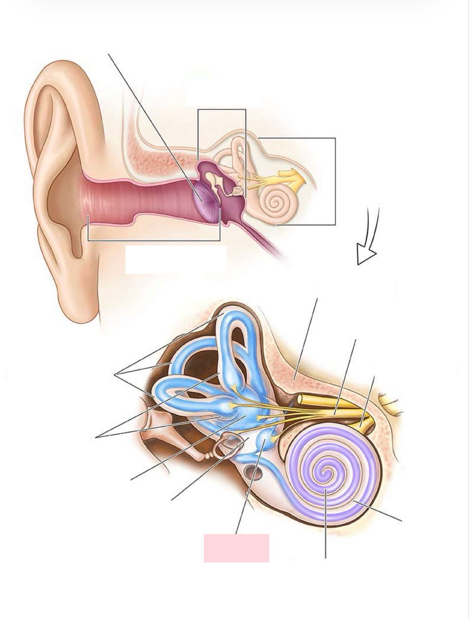
Label the Diagram
Saccule
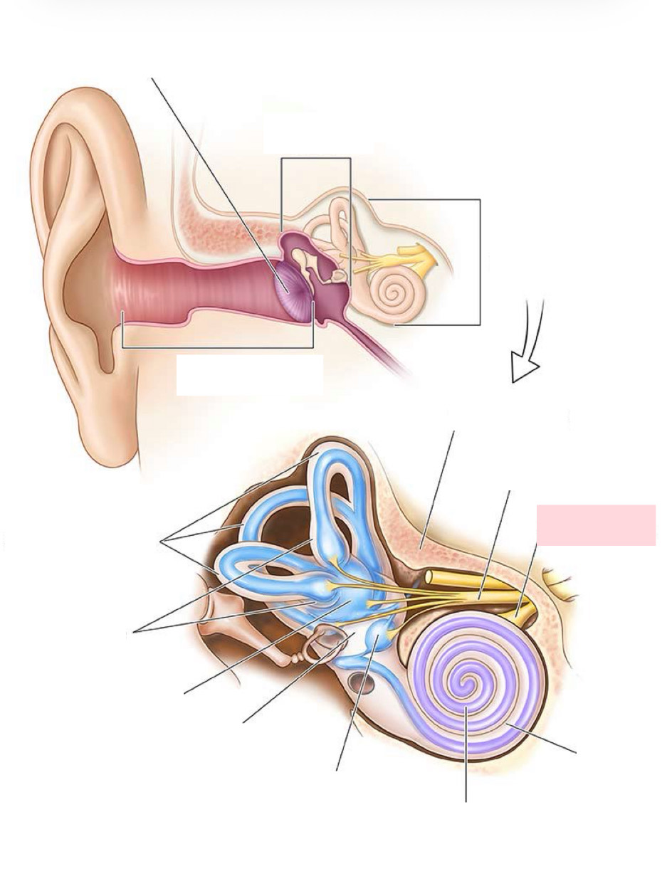
Label the Diagram
Cochlear nerve
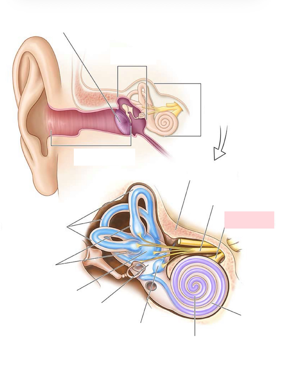
Label the Diagram
Cochlea nerve
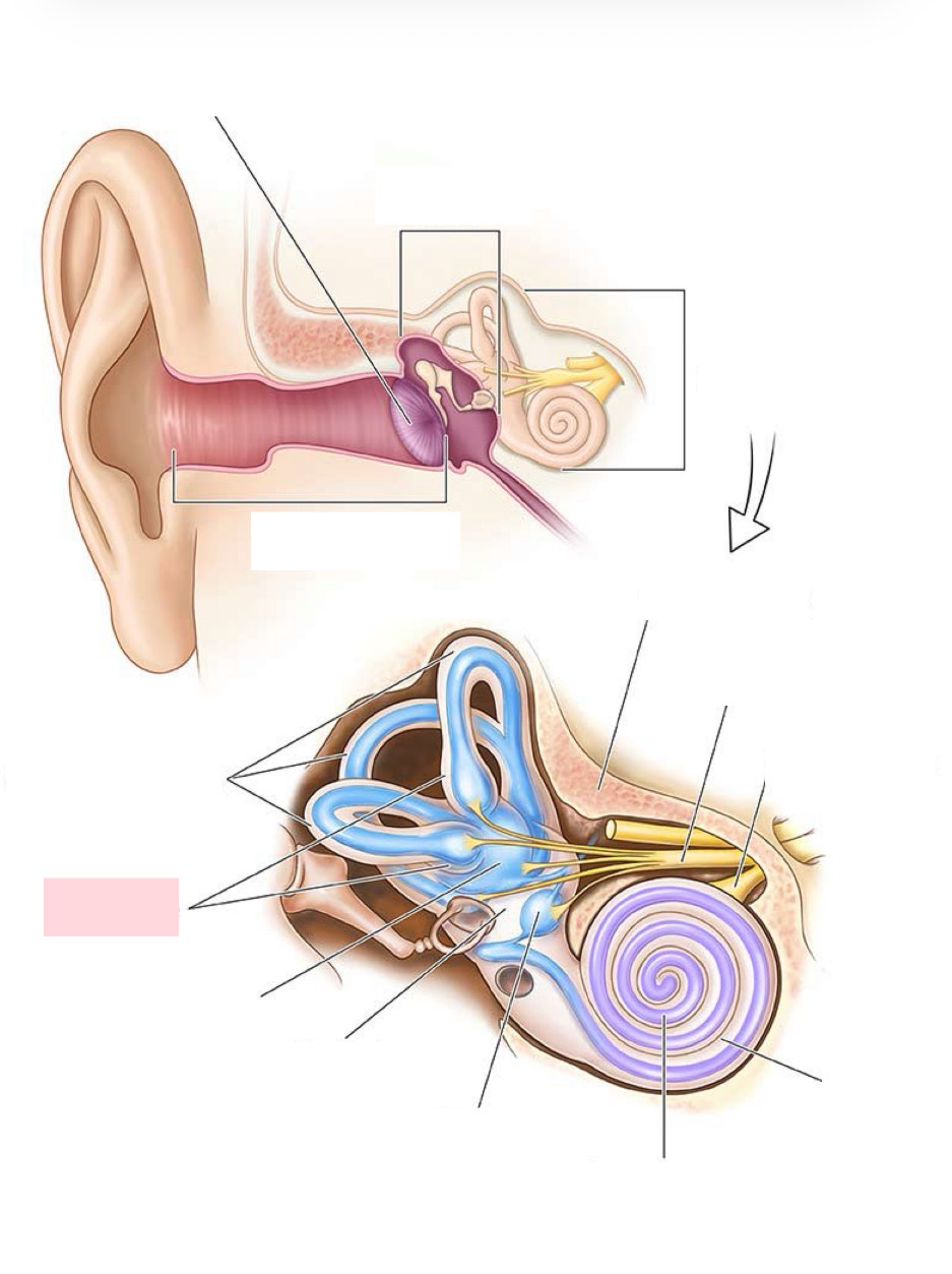
Label the Diagram
Ampullae
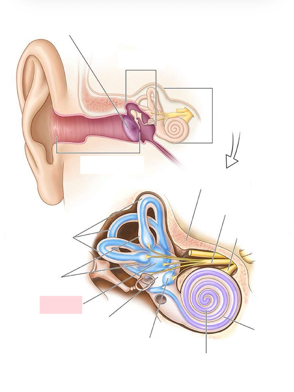
Label the Diagram
Utricle
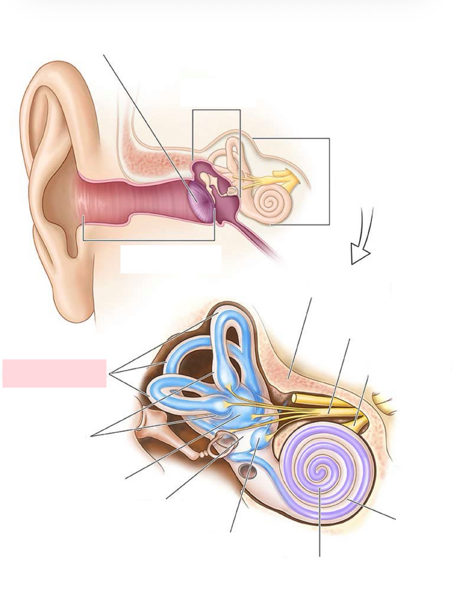
Label the Diagram
Semicircular canals
Label the Diagram
Outer ear
Label the Diagram
Scala Tympani
Label the Diagram
Scala vestibuli
Label the Diagram
Helicotrema
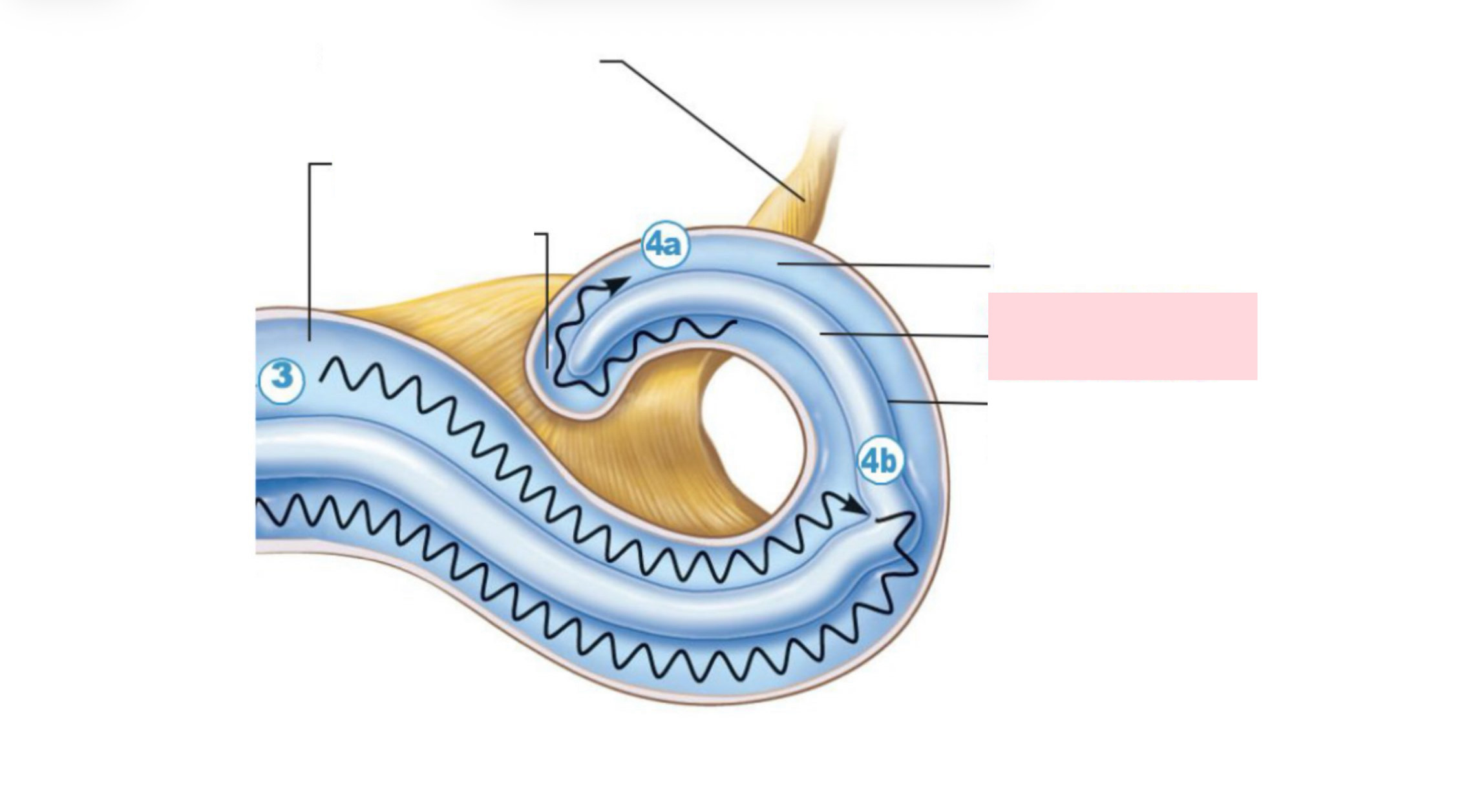
Label the Diagram
Scala media
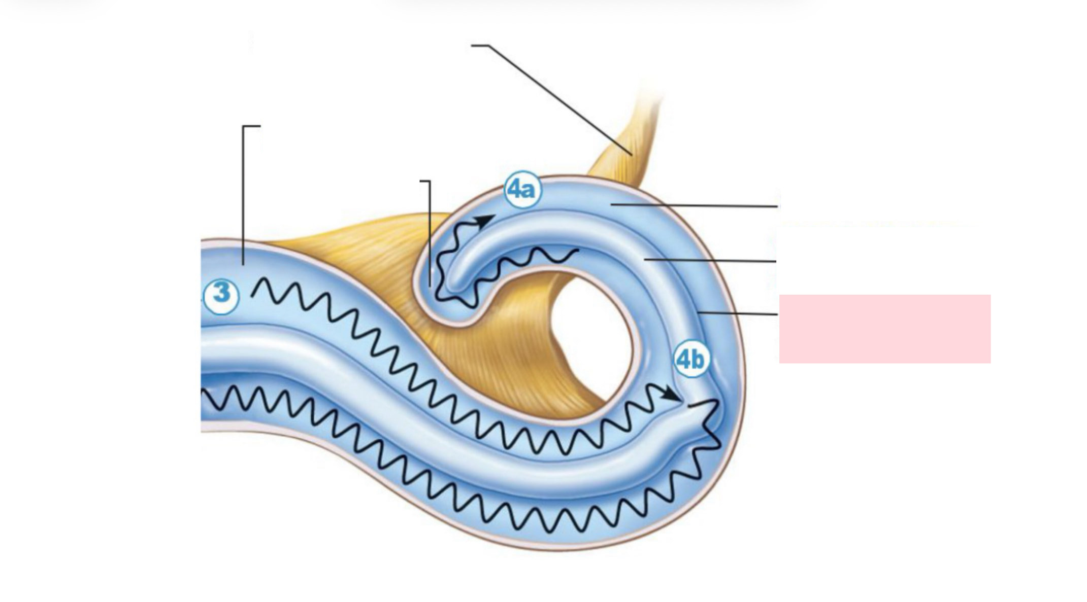
Label the Diagram
Basilar membrane