PMS III Week 6 (Intro to Trauma)
 Call Kai
 Learn
Learn Practice Test
 Spaced Repetition
 Match
Match Flashcards
 Knowt Play
1/70

There's no tags or description
Looks like no tags are added yet.
Name | Mastery | Learn | Test | Matching | Spaced | Call with Kai | Chat |
|---|
No analytics yet
Send a link to your students to track their progress
71 Terms
first line of defense
Physical & mechanical barriers
-Tightly bound epithelial cells fight off bacteria
-Skin, GI, GU, and respiratory tract
Biochemical barriers are substances meant to trap or destroy pathogens
-Mucus, perspiration, saliva, tears, earwax
If these barriers are invaded, the second line of defense is activated, and inflammation occurs
Second Line of Defense - Signs and Microcirculatory Changes
Inflammatory Signs: Redness, Heat, Swelling, Pain
Microcirculatory changes:
-Blood vessel dilation (increase blood flow)
-Increased vascular permeability
-Migration of WBC to site of injury (pus = dead WBC)
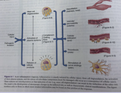
image of pathophysiology with cellular injury and pathogenic invasion
pain can arise directly from neuro injury but also inflammation
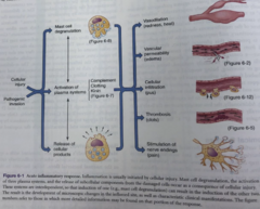
Stress Response to Injury
The body's stress response is initiated by the CNS and the endocrine system
Typically begins in the limbic system
Results in release of catecholamines from the adrenal gland and ultimately the release of cortisol
stress response lead to release of catecholamines which stimulate 2 classes of receptors - α & β
identify the role of α1, α2, β1, and β2
α1 - Increases smooth muscle contraction
α2 - GI & vessel muscle contraction, platelet aggregation, insulin secretion
β1 - Increases cardiac contraction (contractility and rate)
β2- Bronchial, vessel, GI/GU muscle relaxation, gluconeogenesis, releases insulin
Epinephrine works on?
Norephephrine works primarily on?
» Epinephrine works on α & β
» Norephephrine works primarily on α
epinephrine has a greater influence on?
greater influence on cardiac action
norepinephrine has a greater influence on?
primarily effects blood pressure through vessel smooth muscle contraction
cortisol
allows for gluconeogenesis and elevation of blood sugar
Systemic Reaction to Trauma/Stress
cardiovascualr
•Increased cardiac output
•Increased blood pressure
•Increased cardiac contractility
Systemic Reaction to Trauma/Stress
pulm
bronchodilation
Systemic Reaction to Trauma/Stress
GI
liver increases gluconeogenesis
Systemic Reaction to Trauma/Stress
MSK
decrease glucose uptake in skeletal muscle and adipose
Systemic Reaction to Trauma/Stress
integumentary
•Piloerection (goosebumps)
•Increase sweat gland action
Systemic Reaction to Trauma/Stress
immune
•Anti-inflammatory effects
•Decreased circulating WBCs
Systemic Reaction to Trauma/Stress
endocrine
•Decreased insulin excretion by pancreas
•Hyperglycemia
Systemic Reaction to Trauma/Stress
renal
•Increased water retention
•Decreased urinary output
Patients who arrive in the trauma bay have injuries that exceed their natural defenses and have triggered the stress response
Multisystem trauma patients presenting in shock are at risk of?
Multisystem trauma patients presenting in shock are at risk of total disruption of their homeostasis
SHOCK
3 risk factors to patients' mortality ?
-Coagulopathy (hypercoagulation bc platelets DON'T work --> can't clot)
-Hypothermia
-Acidosis (circulating blood is acidic, heart does not function properly)
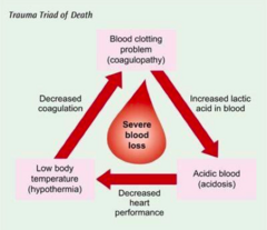
Coagulopathy
presentation? what do we do to help patient?
Severely injured trauma patients are at risk of HYEPRcoagulopathy bc clotting factors do not work when their body temperature is lowered
To prevent coagulopathy, we need to focus on rewarming the patient to avoid worsening hypothermia and stop blood loss
Hypothermia
presentation? how do we help patient?
May be present when the patient arrives
Can develop in ER if patient is left uncovered
Measures to rewarm patient should be taken:
-Temperature of trauma bay >80ºF
-Administer warmed fluids (around 102)
-Apply warm blankets
-Control hemorrhage rapidly
Acidosis
what occurs? can lead to?
Inadequately perfused cells are deprived of essential substrates for normal aerobic metabolism and energy production
Anaerobic metabolism occurs and lactic acid is formed resulting in metabolic acidosis
Prolonged acidosis can lead to end organ damage and multisystem organ failure
Blunt vs Penetrating Trauma
Blunt Trauma - A direct blow
Ex: falls, motor vehicle collisions (MVC), motorcycle crash (MCC)
Penetrating Trauma - enters into body cavity
Ex: GSW, stab wounds (SW), impalements
Sometimes combined!
Basics of Trauma (ABCDEs)
A - airway
B - breathing
C - circulation
D - disability
E - exposure
primary vs secondary vs tertiary surveys
Primary survey - the quick initial exam, ABCDE evaluated (and resolve ABCDE FIRST)
Secondary survey - the follow-up, more detailed exam (complete H&P done!!)
Tertiary survey - done approx. 24 hours after injury, detects additional injuries (can be done even if pt is intubated)
A- Airway
Inadequate delivery of air to the lungs prevents oxygenated blood from getting to the brain and vital structures
First area of assessment and frequent reassessment
Examine for patency and obstruction
physiologic signs of airway obstruction
hypoxia
-Patient agitated
-Low pulse oximetry
-Cyanosis (late finding)
physiologic signs of airway obstruction
hypercarbia
Not enough carbon dioxide being expelled
-Patient obtunded
-Monitor capnography / end tidal CO2
O2 can't get in, CO2 can't get out
physiologic signs of airway obstruction
abnormal sounds
-Stridor
-Gurgling
-Snoring
-Hoarseness
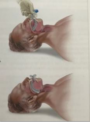
Managing Airway Obstruction
-Clear the airway
-Head tilt, jaw thrust
-Suction
-Administer oxygen
-Open and secure an airway (if excessive bleeding, for example)
-Insertion of artificial airway
-Prevent excessive movement of
cervical spine (cervical collar)
-Call anesthesia STAT
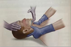
B - Breathing
Evaluation of breathing & ventilation
-Adequate gas exchange within alveoli
-Alterations may be due to chest trauma or destruction of alveoli
Examination includes observation of chest wall expansion, assessing breath sounds
Initial diagnostics may include checking ABG! esp if considering vent/intubation
alterations in ventilation: hypoxemia
not enough oxygen being absorbed in alveoli
alterations in ventilation: hypoxemia
pulse oximetry, ABG results?
Pulse Ox
-Goal above 95%
-COPD above 92%
ABG Results
-Normal PaO2 80-100
alterations in ventilation: hypercapnia
not enough CO2 being offloaded through alveoli
alterations in ventilation: hypercapnia
ABG results?
-Normal PaCO2 35-45
-Tachypnea
managing breathing abnormalities
-supplemental O2
-insertion or nasal or oropharyngeal airway
-definitive airway
-mechanical ventilation
-management of chest trauma
C - Circulation
Is the patient perfusing?
Includes monitoring of vital signs
-Blood pressure
-Pulse rate and
character
-Respiratory rate
-Pulse pressure (SBP-DBP)
circulation exam includes
-Auscultation heart
-Palpating central and distal pulses (may be decreased in the periphery)
-Capillary refill
Shock
classifications/types of shock
An abnormality in the circulatory system that results in inadequate organ perfusion and tissue oxygenation
Shock in trauma patients is classified as hemorrhagic or non-hemorrhagic
Types of shock: hypovolemic, cardiogenic, neurogenic, septic, anaphylactic
Shock associated with injury is most often ?
Shock associated with injury is most often hemorrhagic/hypovolemic in nature.
any patient who is cool to the touch and tachycardic should be considered in shock
________ may be the first sign of shock
tachycardia may be the first sign of shock
Hypovolemic Shock: what happens as a patient loses blood volume?
As a patient loses blood volume:
•The body attempts to preserve cardiac output
•HR increases... Tachycardia may be the first sign of shock
•The body attempts to maintain venous return
Shock is not simply a change in one vital sign but recognizing inadequate tissue perfusion! Relying solely on BP can delay recognition.
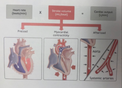
Amount of circulating blood volume determined by?
Amount of circulating blood volume determined by cardiac output
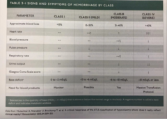
Class 1 Shock
Class 1 -
Class 2 Shock
Class 2 - 15-30% loss
-Tachycardia, tachypnea, decreased pulse pressure
-Most patients stabilize with IV fluids (NS or LR, euvolemic IVF)
Class 3 Shock
Class 3 - 31-40% loss
-Non perfusing tissues
-Tachycardia, tachypnea, decrease blood pressure
-Requires blood product AND 1 L bolus of NS or LR!
Class 4 Shock
Class 4 - >40% loss
-Immediate life threat
-Needs rapid transfusion and surgical interventions (more than 1 unit, not JUST RBCs... involves PRBC, FFP, platelets, cryoprecipitate, etc!)
Shock Classes - Comparing 1-4
D - Disability
•A rapid neurologic evaluation to determine a patient's LOC
•Also includes assessing pupillary size and reaction
•Lateralizing signs (elicit pain --> is only one side reacting? and how?)
•Spinal cord injury and level
GCS scoring
Eyes
4 Open spontaneously
3 Opens to verbal command
2 Opens to pain
1 Unresponsive
Verbal
5 Speaks spontaneously, makes sense
4 Speaks spontaneously, but is confused
3 Speaks spontaneously, does NOT make sense
2 Makes only sounds (goans, mumbles)
1 Unresponsive
Motor
6 Obeys commands
5 Localizes pain
4 Withdrawal from pain
3 Flexion to pain (turn inward)
2 Extension to pain (turn outward)
1 Unresponsive
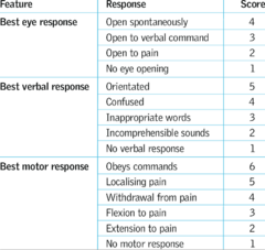
GCS LESS THAN ___ = INTUBATE
GCS LESS THAN 8 = INTUBATE
alterations in GCS can be caused by....
-shock (decreased cerebral perfusion)
-direct cerebral injury
-hypoglycemia
-alcohol
-drugs
the goal is to prevent secondary injury!! (anoxic brain injury)
E - Exposure
Facilitate a thorough examination
-Cut off all clothing
-Remove shoes and socks
-Log roll patients to see posterior side
-Be mindful of axilla and groin
Remember: These patients are at significant risk of hypothermia!
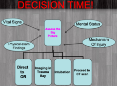
DECISION TIME! Treatment after assessing the big picture (VS, PE, Mental status, MOA)
- direct to OR
- imaging in trauma bay
- intubation
- proceed to CT scan
operative interventions for a trauma
- some pts will need to go directly to OR to manage injuries
- damage control surgery
- ongoing resuscitation
patients tend to get more sick before they get better
postoperative complications
Tailor towards your surgical specialty
Most common complications:
•Bleeding
•Pain
•Fever
•Urinary Retention
•Hyperglycemia
•Infection
Postoperative Bleeding
one of the most dangerous and obvious complications ... URGENT, see ASAP
Requires prompt examination
-Focus on dressings, drains, and compartments
-Drain management (to monitor volume)
Diagnostics for Postoperative Bleeding
- serial H&H and tracking trends
- possible CT with contrast (may be needed if there is no drain)... be specific with order too
Postoperative Pain
important to consider multimodal pain regimen such as?
-Oral and IV forms of medication
-Non-narcotic medications: NSAIDs, Toradol, etc.
-Adjuvant therapies
-Repositioning
-Adjustment of splints and wraps
-Augmenting pain medications
-Pain specialists
Fever - Most Common Causes (6 W's)
-Wind: atelectasis (MC IMMEDIATELY POST OP) and pneumonia (prevent atelectasis --> prevent PNA)
-Waves: EKG changes and MI
-Wonder drugs: drug or transfusion (MC) related
-Water: UTI
-Walking: venous thromboembolism
-Wound: infections (POD 5-7)
Postoperative Urinary Retention
Patient unable to void for 6-8 hours, suprapubic pain
if they haven't, do a straight cath
Urinary Retention Management
•Close monitoring for those who have had spinal epidural blocks
•Requires bladder scanning
•Threshold for placing catheters (straight cath- determines need to keep cath in)
•Repeat catherization may require placement of indwelling catheter (Foley)
when does the bladder start over-distending
>300 mL
at this point, have to leave catheter in bc muscle tone in the bladder is decreased (musculature is affected from stretching)
Postoperative Hyperglycemia
Stress and trauma (from surgical intervention) can cause persistent hyperglycemia
liver undergoing increased gluconeogenesis (this is expected after surgery)
Postoperative Hyperglycemia - Management
Management
-Restarting of home oral hypoglycemics
-Insulin sliding scales
Target range below 180
-Persistent hyperglycemia can lead to poor wound healing and increased risk of surgical site infections
Postoperative Hyperglycemia - Management with Insulin
2 blood sugars >200 significantly decreases wound healing
may need to give insulin temporarily while they heal
postoperative infections occurs
typically occurs 7-10 days after surgery
physical signs of postoperative infection
-Erythema surrounding wound
-Purulent drainage / persistent serous drainage
-Fluctuance surrounding or within wound
-Increased HR, decreased BP, fevers
**remember Tylenol can mask a fever**
labwork and imaging for postoperative infection
Labs - Increased WBC
Imaging - Ultrasound or CT scan
treatment for postoperative infection
culture wounds!
- IV or PO antibiotics depending on patient status and site of infection
- Potential drainage of abscess/fluid collections
Postoperative Reassessments
•Frequent rounding on postoperative patients can prevent complications
•Diligent postoperative checks
•Treatment of trauma patients requires continual reassessments
•Tertiary surveys
•Inform attending/surgeons of changes in clinical examinations