Practical 1 I BIOL 106 LA
 Call Kai
 Learn
Learn Practice Test
 Spaced Repetition
 Match
Match Flashcards
 Knowt Play
1/117

There's no tags or description
Looks like no tags are added yet.
Name | Mastery | Learn | Test | Matching | Spaced | Call with Kai | Chat |
|---|
No analytics yet
Send a link to your students to track their progress
118 Terms
divides body into front and back
Frontal Plane
divides body into right and left
Sagittal Plane
divides body into top and bottom
Transverse Plane
The standard body position used as an anatomical reference point
Anatomical Position
above/below
Superior / Inferior
closer/farther from middle of the body
Proximal / Distal
on the inner/outer side of
Medial / Lateral
Palmar / Dorsal: applies to the hand
Plantar / Dorsal: applies to the foot
Plantar / Palmar / Dorsal
in front/behind of
Anterior / Posterior
in front/behind of in animals
Ventral / Dorsal
above/below
Superficial / Deep
longer than they are wide
Ex.) femur, tibia, fibula, metacarpals, metatarsals…
Long Bones
at least one flat surface
Ex.) ribs, sternum, scapula, parietal bone, occipital bone
Flat Bones
equally as long as they are wide
Ex.) lunate, pisiform, calcaneus, talus, navicular, cuneiforms…
Short Bones
don’t fit into other categories of types of bones
Ex.) vertebrae, sacrum, coccyx
Irregular Bones
housed within tendons
Ex.) patella, hallux, pisiform (wrist)
Sesamoid Bones
Tendon: muscle to bone
Ligament: bone to bone
Tendon vs. Ligament

skull, spine, rib cage
Forms longitudinal axis of the body
Supports the head, neck, and trunk
Protects the brain, spinal cord, & internal organs
Axial Skeleton

bones of the limbs
enables movements of daily living
Appendicular Skeleton
Important Functions:
support
protection
anchorage
mineral storage
blood cell formation
fat storage
hormone production
Boney Functions
Weakest parts of the skeleton
Most likely to be stretched or
compressed - leads to injury
More movement = Less
stability
Factors of a synovial joint
Articular Cartilage
Joint Cavity
Articular Capsule
Synovial Fluid
Reinforcing Ligaments
Nerves & Blood Vessels
Synovial Joints
Bones joined by cartilage
Types
Synchondrosis: immobile
Symphysis: slightly movable
Cartilaginous Joints
Bones united by collagen fibers
Types
Suture - immobile
Syndesmosis - slightly moveable and immobile
Gomphosis - immobile
Fibrous Joints

Formed by the articulation of the surfaces of 2 flat bones
bones slide/glide over each other
Non-axial joint
no true movement
Plane

Most common type in the human body
Uniaxial Joint
Bones move in 1 plane
Flexion/extension
2 bones, 1 is convex & the other concave
Hinge

One bone serves as the axis and the other pivots/rotates around it
Uniaxial Joint
rotation is the only movement allowed
Pivot

Consists of an oval, convex shaped end of one bone articulating with an elliptical, concave cavity of another bone
Biaxial Joint
Typically allows flexion/extension and abduction/adduction, some circumduction
Condylar

Formed by a convex and concave articulating surfaces
interlock like 2 perpendicular saddles
Biaxial joint
Allows for flexion/extension, abduction/adduction, and some circumduction
Saddle

Most mobile of all joints
Multiaxial
allows movement in all 3 planes
Spherical end of 1 bone articulates with the cup/socket of another bone
Ball and Socket
1 flat bone surface glides over another without angulation or rotation
Plane joints!
Gliding
movements that increase or decrease the angle between 2 bones
can occur in any plane of the body
Angular
bending movement decreases angle by bringing bones closer together
Flexion

straightening movement; increases angle between articulating bones
Sagittal Plane
Extension

Move a limb away from the middle
Increases angle further as the limb moves
Abduction
Moves the limb towards the body
Decreases angle as the limb moves closer to the body
Frontal Plane
Adduction
distal end of limb moves in a circle
Circumduction
turning of a bone around its own long axis
Rotation
Rotates the forearm laterally so the palm faces up
Supination
Rotates the forearm medially so the palm faces down
Pronation

Lifting the foot to bring the top of the foot towards the shin
Dorsiflexion
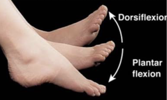
pointing the foot downward
Plantarflexion

Turning the foot inward
Inversion
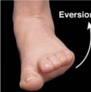
Turning the foot outward
Eversion

lifting the body part upward
Elevation

moving the body part downward
Ex.) Mandible and Scapula
Depression
freely movable joints; all synovial joints
Diarthrosis
functionally immovable joints, providing maximum stability; cranial sutures; immovable joint
Synarthroses
slightly moveable joints
Amphiarthroses

Origin: posterior, superior tibia and fibula
Insertion: Navicular tuberosity, plantar surface of the tarsals and metatarsals 2-4
Action: Inversion (primary) & plantarflexion (secondary) of the ankle
Innervation: Tibial Nerve
Posterior Tibialis
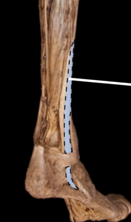
Origin: Posterior surface of the tibia
Insertion: Base of the distal phalanges of the 2nd-5th toes
Action:
Flexion of the lateral four digits
Plantarflexion and inversion of the ankle
Innervation: Tibial Nerve
Flexor Digitorum Longus

Origin: Midshaft of fibula
Insertion: Distal phalanx of 1st toe
Action:
Flexion of the 1st toe
Ankle plantar flexion and inversion
Innervation: Tibial Nerve
Flexor Hallucis Longus
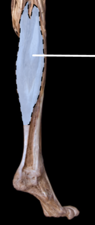
Origin:
Lateral Head: Lateral condyle of the femur
Medial Head: Medial condyle of the femur
Insertion: Posterior calcaneal tubercle
Action:
Plantar Flexion of ankle when knee is extended (Primary)
Knee flexion when ankle is dorsiflexed (secondary)
Innervation: Tibial Nerve
Gastrocnemius

Origin: Posterior aspect of the head of the fibula, superior tibia, and interosseous membrane
Insertion: Posterior calcaneal tubercle
Action: Plantarflexion of the ankle
Innervation: Tibial Nerve
Soleus

Origin: posterior femur above lateral condyle, lateral supracondylar line of the femur
Insertion: Medial aspect of the posterior calcaneus
Action: Assists gastrocnemius ankle plantar flexion and knee flexion (synergist only)
Plantaris

Origin: Inferior, anterior surface of the fibula and interosseous membrane
Insertion: Base of the 5th metatarsal, dorsal side
Action: Dorsiflexion & Eversion of the ankle
Innervation: Deep Fibular Nerve
Fibularis Tertius

Origin: Head and superior, lateral surface of the fibula
Insertion: 1st metatarsal and medial cuneiform (plantar surface)
Action:
Eversion of ankle
Plantarflexion of the ankle (weak)
Innervation: Superficial Fibular Nerve
Fibularis Longus

Origin: distal fibula shaft
Insertion: tuberosity at base of 5th metatarsal
Action:
Eversion of ankle
Plantarflexion of the ankle (weak)
Innervation: Superficial Fibular Nerve
Fibularis Brevis

Origin: Lateral Condyle of tibia, superior medial surface of the fibula and interosseous membrane
Insertion: Middle and distal phalanges of the lateral four digits
Action:
Extends lateral 4 digits
Dorsiflexes the ankle
Innervation: Deep fibular nerve
Extensor Digitorum Longus
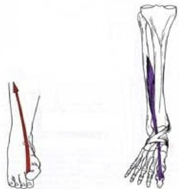
Origin: Middle part of the anterior surface of the fibula and interosseous membrane
Insertion: Distal phalanx of the great toe
Action:
Extends the great toe
Dorsiflexes the ankle
Innervation: Deep Fibular Nerve
Extensor Hallucis Longus
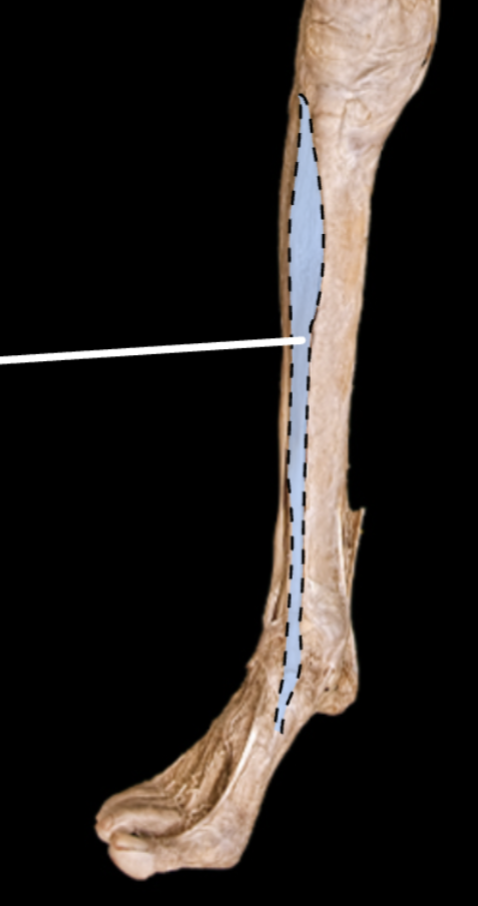
Origin: Lateral condyle and upper ½ of the tibial shaft
Insertion: inferior surface of medial cuneiform and first metatarsal bone
Action: Dorsiflexion (primary) & inversion of the ankle (secondary)
Innervation: Deep Fibular Nerve
Anterior Tibialis

Origin: anterior inferior iliac spine and ilium superior to the acetabulum
Insertion: Base of the patella and by patellar ligament to tibial tuberosity
Action: knee extension, assists in hip flexion
Innervation: Femoral Nerve
Rectus Femoris

Origin: Intertrochanteric line and medial lip of the linea aspera of
the femur
Insertion: base of the patella and by patellar ligament to tibial tuberosity
Action: Extends the leg at the knee joint
Innervation: Femoral Nerve
Vastus Medialis
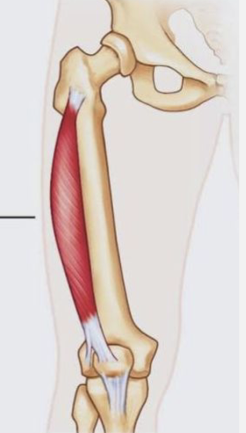
Origin: Greater trochanter and lateral lip of the linea aspera of the femur
Insertion: base of the patella and by patellar ligament to tibial tuberosity
Action: Extends the leg at the knee joint
Innervation: Femoral Nerve
Vastus Lateralis
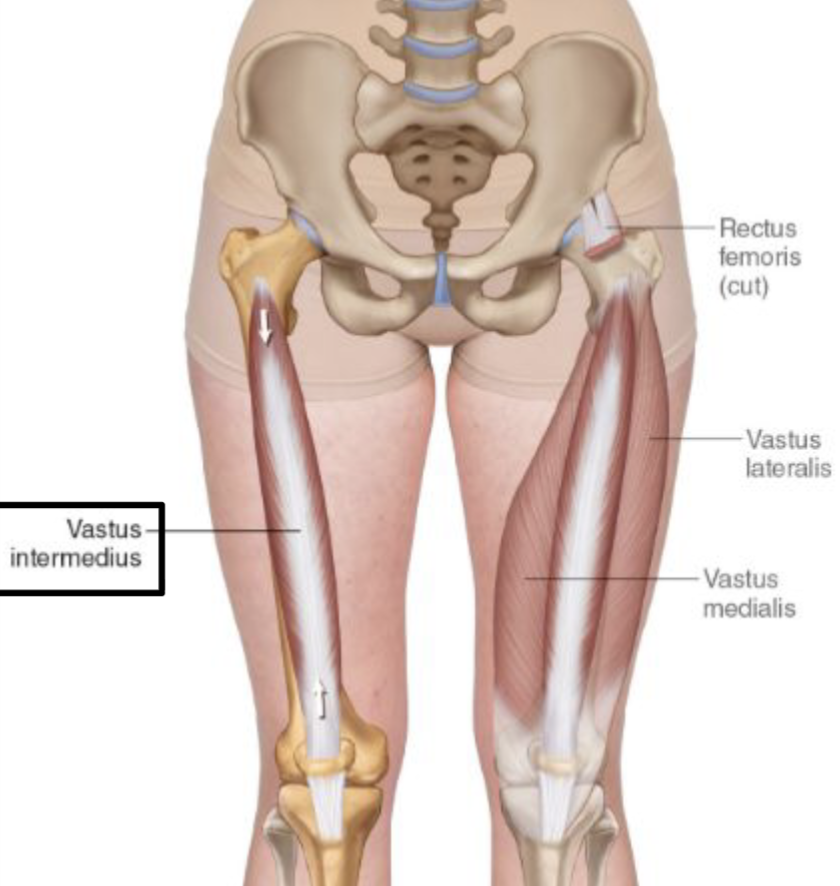
Origin: Anterior and lateral surface of the body of the femur
Insertion: base of the patella and by patellar ligament to tibial tuberosity
Action: Extends the leg at the knee joint
Innervation: Femoral Nerve
Vastus Intermedius

Origin:
Long Head—Ischial Tuberosity
Short Head—Linea aspera and lateral supracondylar line of the femur
Insertion: lateral side of the head of the fibula
Action: Flexes the knee and extends the hip
Innervation: Sciatic
Biceps Femoris

Origin: Ischial tuberosity
Insertion: Posterior aspect of the medial condyle of the tibia
Action: extends hip, knee flexion
Innervation: Sciatic
Semimembranosus

Origin: Ischial Tuberosity
Insertion: Medial aspect of the superior part of the tibia (pes anserine)
Action: Extends hip; knee flexion
Innervation: Sciatic
Semitendinosus

Origin: body and inferior ramus of the pubis
Insertion: Superior aspect of the medial surface of the tibia (pes anserine)
Action: Hip adduction and internal rotation; knee flexion
Innervation: Obturator Nerve
Gracilis

Origin: Anterior superior iliac spine
Insertion: Superior part of the medial surface of the tibia (pes anserine)
Action: Flexion, abduction and external rotation of the hip; knee flexion
Innervation: Femoral nerve
Sartorius

Origin: lateral condyle of femur
Insertion: posterior- medial surface of the tibia
Action: “unlock the knee joint” by externally rotating the femur on the tibia during standing
Innervation: Tibial Nerve
Popliteus
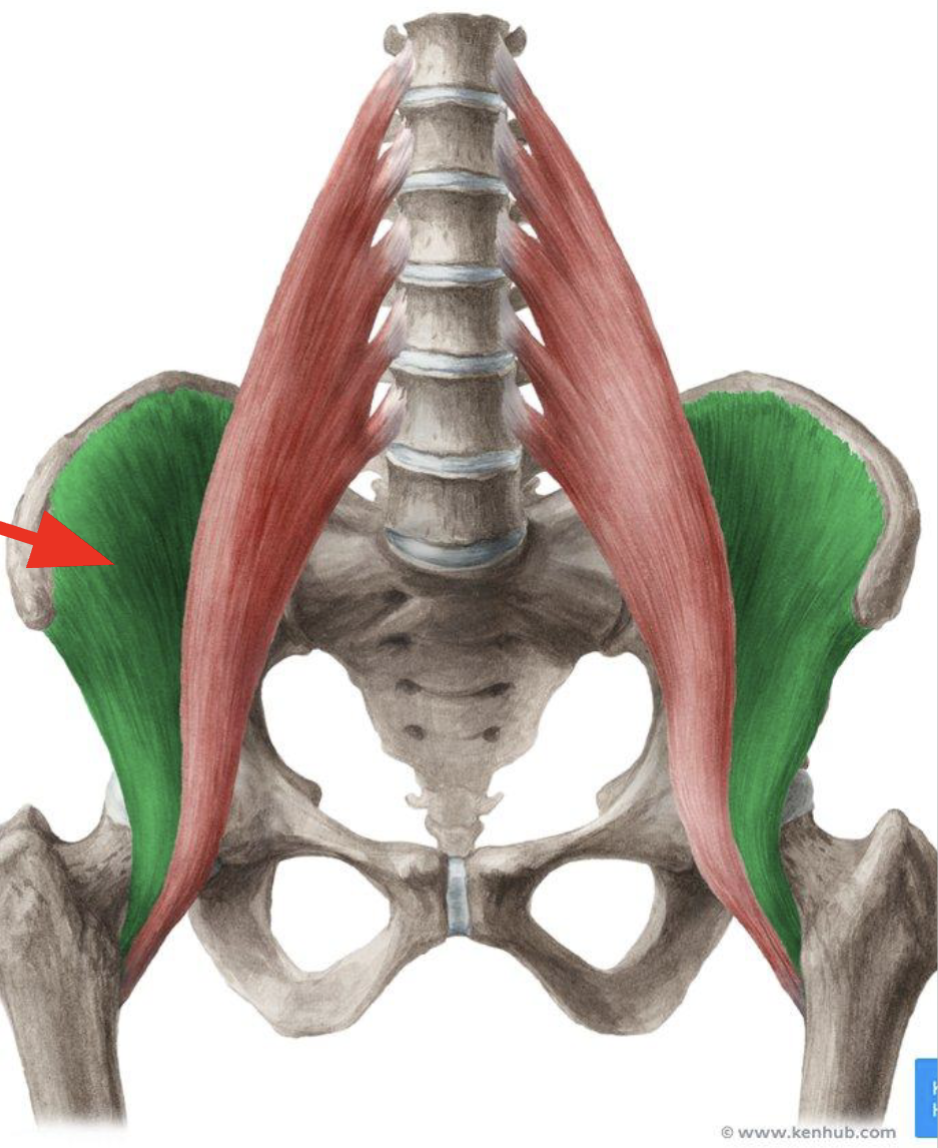
Origin: Iliac fossa, iliac crest
Insertion: Lesser trochanter of femur
Action: Hip Flexion
Innervation: Femoral Nerve
Iliacus

Origin: Transverse processes of lumbar vertebrae (T12-L5)
Insertion: Lesser trochanter of femur
Action: Hip flexion
Innervation: Lumbar Nerves (L1-L3)
Psoas Major
Psoas Major
Origin: Sides of T12-L5 vertebrae and discs between them; transverse processes of all lumbar vertebrae
Insertion: Lesser trochanter of femur
Action: Act conjointly in flexing thigh at hip joint and in stabilizing this joint. Postural muscle that helps control deviation of trunk and is active during standing.
Innervation: Ventral rami of lumbar nerves (L1, L2, and L3)

Origin: Dorsal ilium, sacrum, and coccyx
Insertion: Iliotibial tract, gluteal tuberosity of femur
Action:
Hip extension (primary)
adduction & external rotation (secondary)
Innervation: Inferior Gluteal Nerve
Gluteus Maximus

Origin: Gluteal surface of ilium
Insertion: Greater trochanter
Action: Hip abduction and internal rotation
Innervation: Superior Gluteal Nerve
Gluteus Medius

Origin: External gluteal surface of ilium (deep to gluteus medius)
Insertion: Greater trochanter
Action: Hip abduction and internal rotation
Innervation: Superior Gluteal Nerve
Gluteus Minimus
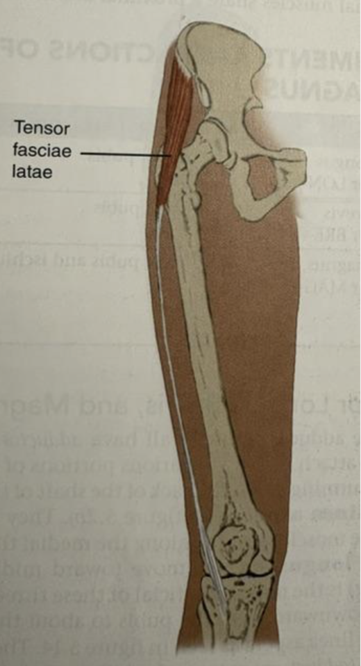
Origin:
Insertion: Gerdy’s Tubercle
Action:
Innervation:
Tensor Fascia Latae

Origin: Pubis & Ischial Tuberosity
Insertion: Gluteal Tuberosity, Linea Aspera, Adductor Adductor Tubercle
Action: Hip adduction, flexion, internal rotation (primary)
Innervation: Obturator nerve
Adductor Magnus

Origin: Near pubic symphsis
Insertion: Linea Aspera
Action: Hip adduction, flexion, internal rotation
Innervation: Obturator Nerve
Adductor Longus

Origin: Body and Inferior Ramus of the Pubis
Insertion: Linea Aspera
Action: Hip adduction, flexion, internal rotation
Innervation: Obturator nerve
Adductor Brevis
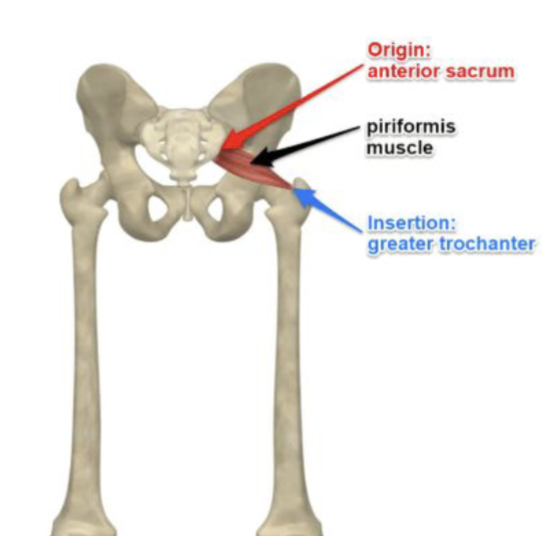
Origin: Anterior sacrum
Insertion: Greater Trochanter
Action: Hip External Rotation
Innervation: L5, S1, S2
Piriformis
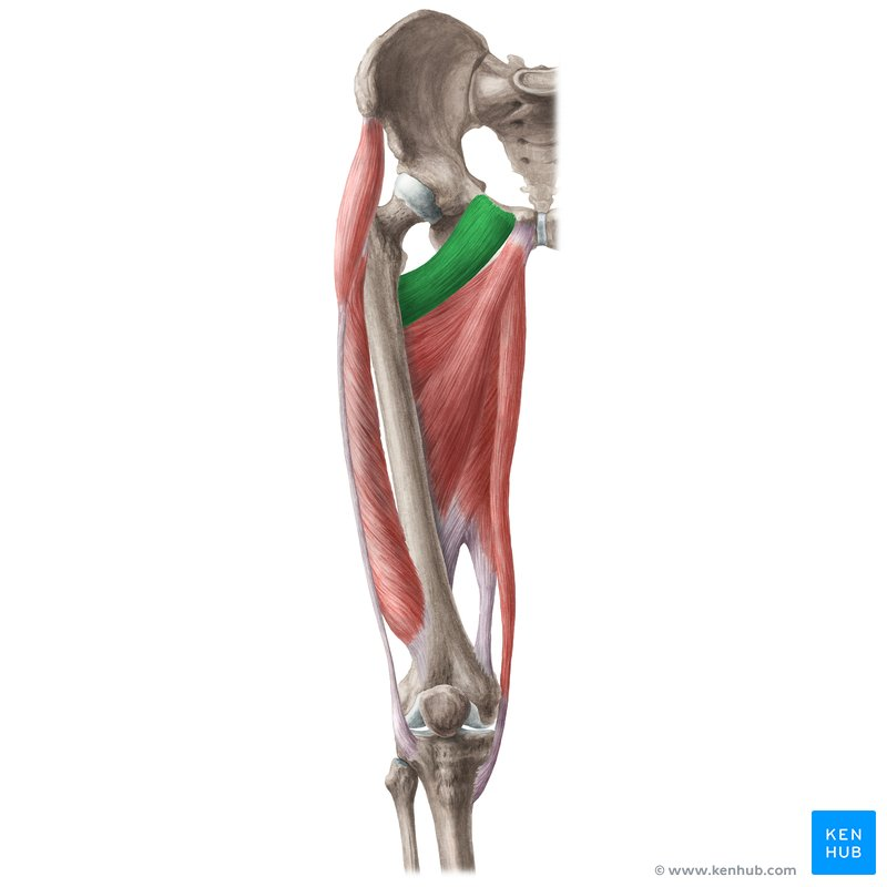
Origin: Pubis
Insertion: Lesser Trochanter
Action: Hip adduction, flexion, internal rotation
Innervation: Femoral nerve
Pectineus

Origin: pubis and ischium
Insertion: trochanteric fossa
Action: Hip External Rotation
Innervation: obturator nerve
Obturator Externus

Origin: Obturator membrane & sciatic notch
Insertion: Greater trochanter
Action: Hip External Rotation
Innervation: L5, S1
Obturator Internus

Origin: Ischial spine
Insertion: Greater Trochanter
Action: Hip External Rotation
Innervation: L5, S1
Gemellus Superior

Origin: Ischial tuberosity
Insertion: Greater trochanter
Action: Hip External Rotation
Innervation: L5, S1
Gemellus Inferior
Origin:
Insertion:
Action:
Innervation:
Anterior Talofibular Ligament
Origin:
Insertion:
Action:
Innervation:
Posterior Talofibular Ligament

prevents neutral inversion
Origin:
Insertion:
Action:
Innervation:
Calcaneaofibular Ligament
Ant. Tib. tal
Tibionavicular
P. Tib tal
Tibiocalcaneal
Collectively 4 ligaments support the medial ankle; make up the Deltoid Ligament. Medial Ligamentous support TCJ.
Deltoid Ligament
Origin:
Insertion:
Action:
Innervation:
Plantar Fascia
Origin: sides of metatarsals
Insertion: bases of the proximal phalanges and the extensor expansion
Action:
Palmar: adduction
Dorsal: abduction
Innervation: lateral plantar nerve
Interosseous Membrane
Origin:
Insertion:
Action:
Innervation:
Anterior Tibiofibular Ligament (ATFL)
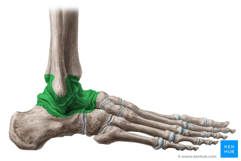
Consist of the tibia, fibula, and talus
Synovial hinge joint
Talocrural Joint (TCJ)

side to side
Subtalar Joint (STJ)
Retinacula
Posterior Tibiofibular Ligament
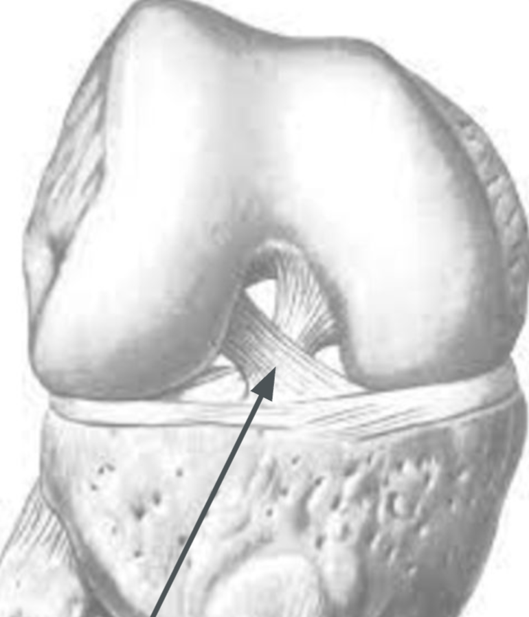
Primary anterior stabilizer of the knee
3 strands twisted together
Attachments:
anterior-medial tibia
posterior-lateral femoral condyle
Prevents anterior displacement of tibia
Anterior Cruciate Ligament

Primary posterior stabilizer of the knee
strongest ligament in the knee
Attachments:
posterior tibial plateau
medial femoral condyle (near intercondylar notch)
Prevents posterior displacement of tibia & hyperextension of the knee
Posterior Cruciate Ligament

Round, rope-like
Runs from lateral epicondyle to the head of the fibula
Protect from varus forces (forces that gap the outside of the knee)
Lateral Collateral Ligament