Lec 4 ANSC
 Call Kai
 Learn
Learn Practice Test
 Spaced Repetition
 Match
Match Flashcards
 Knowt Play
1/49

There's no tags or description
Looks like no tags are added yet.
Name | Mastery | Learn | Test | Matching | Spaced | Call with Kai | Chat |
|---|
No analytics yet
Send a link to your students to track their progress
50 Terms
what is neural tube
= hollow, embryonic precursor structure to the entire central nervous system.
Location: Forms along the dorsal surface of the early embryo during early development.
Purpose/Function: The solid walls of the tube differentiate into the brain and spinal cord, while the hollow core becomes the ventricles and central canal.
Analogy: A long piece of hollow plumbing dough; the dough bakes into the solid pipe wall while the hollow inside lets water flow through.

what is cerebral cortex, why is it important
= outermost layer of gray matter capping the cerebrum.
Location: The wrinkled outer surface layer of the brain.
Purpose/Function: Serves as the ultimate processing layer for conscious awareness, abstract thinking, and voluntary actions.
Analogy: The outer surface crust of a planet where all civilization, cities, and communications are active.
what is basal ganglia/basal nuclei
= Clusters of grey matter nuclei buried deep within the white matter of the cerebrum.
Location: Positioned deep inside the cerebral hemispheres, surrounding the thalamus.
Purpose/Function: Helps coordinate smooth, voluntary physical body movements and suppresses unwanted muscle twitches.
what is limbic system , what are 2 main units?
= ancient brain network responsible for emotions, basic survival drives, and memory formation.
Location: A ring of structures looping around the center of the diencephalon.
Purpose/Function: The Amygdala
drives fear and emotional responses; the Hippocampus handles the consolidation of long-term memories
corpus callosum
= thick bridge of white matter tracts that links the two halves of the brain.
Location: Positioned deep along the midline, bridging the left and right cerebral hemispheres.
Purpose/Function: Allows the left and right sides of your brain to rapidly share data and coordinate processing.
what are the 4 lobes of brain and main functions?
Frontal is the forehead; Parietal is the top-back; Occipital is the base-back; Temporal is the side/temple.
Purpose/Function:
Frontal handles motor/reasoning; Parietal handles touch input; Occipital processes vision; Temporal processes hearing/smell.
Analogy: Dividing a massive corporate office floor into four dedicated departments (e.g., accounting, marketing, shipping, and design).
Somatosensory vs. Corticospinal Tracts (and Pyramids)
=Somatosensory tracts are ascending sensory lines; routes skin/body sensations up to the cortex
= Corticospinal tracts are descending motor lines that delivers voluntary muscle commands down from the cortex.
cross over at the brainstem bumps called pyramids.
Location: Travel vertically through the white matter columns of the brainstem and spinal cord.
what does brainstem contain?
=foundational stalk of the brain containing the medulla, pons, and midbrain.
Location: Sits at the absolute base of the brain, directly above and continuous with the spinal cord.
Purpose/Function: Controls automatic, essential survival functions (breathing, heart rate) and acts as the transit route for cranial nerves.
what are cranial nerves, what is vagus nerve?
=set of 12 peripheral nerve pairs that originate directly from the brainstem instead of the spinal cord.
Location: Exit the brainstem through specific holes in the base of the skull.
Purpose/Function: Carry sensory and motor information directly for the head, neck, face, and vital thoracic organs.
VAGUS=prominent cranial nerve that carries the vast majority of the body's parasympathetic autonomic signals.
Location: Originates in the brainstem and travels downward, branching widely across all thoracic and abdominal visceral cavities.
Purpose/Function: Acts as a two-way street, routing sensory data from organs up to the brain, and delivering rest-and-digest commands down to the heart and gut.
reticular formation
= interconnected network of interneurons running through the core of the brainstem.
Location: Weaves vertically through the middle of the midbrain, pons, and medulla.
Purpose/Function: Acts as the master arousal and consciousness regulator, filtering background noise and waking up the cerebral cortex.
spinal reflexes, what makes them unique?
= Automated, fast motor responses that are integrated directly within the spinal cord without needing brain input.
Location: Synapses directly inside the gray matter segments of the spinal cord.
Purpose/Function: Allows the body to react immediately to pain or danger (e.g., pulling back from a hot stove) to prevent tissue damage.
Analogy: A local automated sprinkler system that triggers immediately when local heat hits, without waiting for a command from city hall.
dorsal vs ventral roots, what are Dorsal Horns vs. Ventral Horns (plus Lateral Horns)
= two functional pathways that a spinal nerve splits into right before entering the spinal cord.
Location: Dorsal root branches onto the backside of the cord; Ventral root branches off the front side.
Purpose/Function: Separates incoming sensory lines (dorsal carries sensory info) from outgoing motor lines (ventral carries motor info).
HORNS:
= the regional gray matter sections of the internal spinal cord layout.
Location: Inside the inner butterfly-shaped gray matter core of the spinal cord.
Purpose/Function: Dorsal horns house sensory nuclei; Ventral horns contain somatic motor cell bodies; Lateral horns contain autonomic visceral nuclei.
Analogy: Dividing a sorting warehouse into three distinct zones: inbound sorting (dorsal), outbound heavy cargo (ventral), and automated facilities (lateral).

spinal nerves: what system do they belong to and where do they branch from?
= bundled and mixed peripheral nerves that branch symmetrically off each segment of the spinal cord.
Location: Exit the vertebral column through gaps between adjacent vertebrae.
Purpose/Function: Carry both incoming sensory data and outgoing motor commands between the body and the spinal cord.
Analogy:
The individual bundle cables that break off a main power line to hook up power to a specific house.
what are Cervical, Thoracic, Lumbar, Sacral
The four anatomical regions of the spinal cord and vertebral column.
Location: Cervical is the neck; Thoracic is the chest; Lumbar is the lower back; Sacral is the pelvic base.
Purpose/Function: Organizes the spinal cord into functional levels associated with specific structural nerve branches.
Analogy: Dividing a multi-story office skyscraper into four distinct regional zone levels (e.g., floors 1-5, 6-10, etc.).
what is vertebral column/ vertebrae
= flexible bony spine column (vertebral column) composed of individual interlocking bones (vertebrae)
Location: Extends downward from the base of the skull along the midline of the back.
Purpose/Function: Forms a protective bony tunnel (spinal canal) that prevents the delicate spinal cord from being crushed or severed.
what is medulla oblongata
= lowest, most vital section of the brainstem continuous with the spinal cord.
Location: Transition zone at the base of the skull, connecting the pons directly to the spinal cord.
Purpose/Function: Acts as the primary autonomic homeostatic control center for breathing, heart rate, and blood pressure.
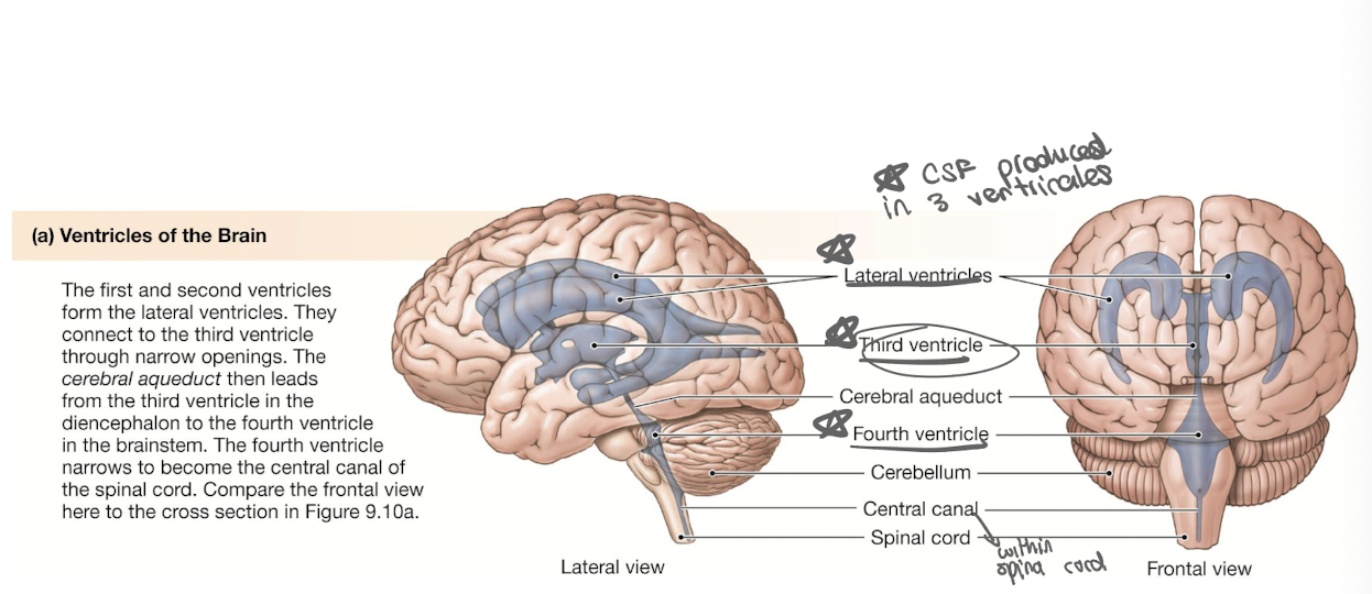
what are ventricles, what is central canal
= continuous network of four hollow, interconnected fluid-filled chambers deep inside the brain.
Location: Left/right lateral ventricles sit in the cerebrum; the third ventricle is in the diencephalon; the fourth ventricle lies between the brainstem and cerebellum.
Purpose/Function: Continuously manufactures and routes cerebrospinal fluid (CSF) through the central nervous system.
Analogy: The internal plumbing pipes and water chambers running through a large apartment complex.
CENTRAL CANAL:
= microscopic, fluid-filled tube running down the center of the spinal cord.
Location: Positioned in the exact center of the spinal cord's gray matter core.
Purpose/Function: Allows cerebrospinal fluid to flow downward through the length of the spinal cord.
Analogy: The narrow internal straw running down the middle of a long cylindrical stick of rock candy.
what is pons
= bulbous segment of the brainstem that acts as a structural and functional bridge.
Location: Positioned on the brainstem directly above the medulla and below the midbrain.
Purpose/Function: Relays information between the cerebellum and cerebrum, and houses secondary control centers for breathing.
what is cerebrum
= largest, most prominent division of the adult brain responsible for higher-level cognitive processing.
Location: Occupies the entire upper cranial vault, draping over the brainstem.
Purpose/Function: Controls conscious thought, abstract reasoning, memory storage, sensory perception, and voluntary motor movements.
Analogy: The master executive board room inside corporate headquarters where the CEO and managers calculate future strategies.
what is diencephalon , what does it contain?
= core major division of the forebrain containing the thalamus, hypothalamus, and epithalamus.
Location: Tucked deep inside the center of the brain, directly beneath the cerebral hemispheres.
Purpose/Function: Acts as the primary homeostatic control center (water balance, temperature) and sensory relay station.
Analogy: The busy central post office switchboard that filters, sorts, and forwards all incoming letters to different departments.
what is cerebellum
= highly folded structure at the back of the brain responsible for motor coordination.
Location: Attached to the back of the brainstem, sitting underneath the occipital lobe of the cerebrum.
Purpose/Function: Coordinates body movement, balance, posture, and fine-tunes physical muscle accuracy.
what is forebrain (prosencephalon)? midbrain (mesencephalon)? hindbrain?
FOREBRAIN= primary anterior brain vesicle that differentiates into the higher processing centers of the mature brain.
Location: The absolute top/front region of the developing embryonic brain.
Purpose/Function: Divides into the telencephalon (which becomes the cerebrum) and the diencephalon.
Analogy: The front nose-cone compartment of a rocket ship where the advanced guidance computers are stowed.
MIDBRAIN= One of the seven major divisions of the CNS that forms the upper part of the brainstem.
Location: Positioned between the diencephalon and the pons.
Purpose/Function: Coordinates involuntary reflex responses to auditory and visual stimuli while housing the cerebral aqueduct.
Analogy: A regional traffic junction guard who ducks and reacts to sudden flashing lights or loud alarms.
HINDBRAIN= primary posterior brain vesicle that differentiates into the lower, vital survival structures of the brainstem.
Location: The base/back region of the developing embryonic brain stem.
Purpose/Function: Divides into the metencephalon (pons and cerebellum) and myelencephalon (medulla oblongata).
Analogy: The foundational basement of a factory where the water pumps and main electrical generators are anchored.

what is ependyma / ependymal cells
= specialized layer of simple glial epithelium that lines the fluid cavities of the CNS.
Location: Lines the interior walls of the brain ventricles and the central spinal canal.
Purpose/Function: Features rhythmic, beating cilia that actively circulate cerebrospinal fluid (CSF) and acts as a source of neural stem cells.
Analogy: Miniature water-wheels installed along a canal to keep the current moving steadily in one direction.

adults structurs of brain (6) vs ventricles (6)
Adult structures are the final, fully formed tissues of the mature nervous system, while ventricles are the hollow, fluid-filled spaces left inside them.

GRAY VS WHITE MATTER in CNS VS PNS: what is it made of?
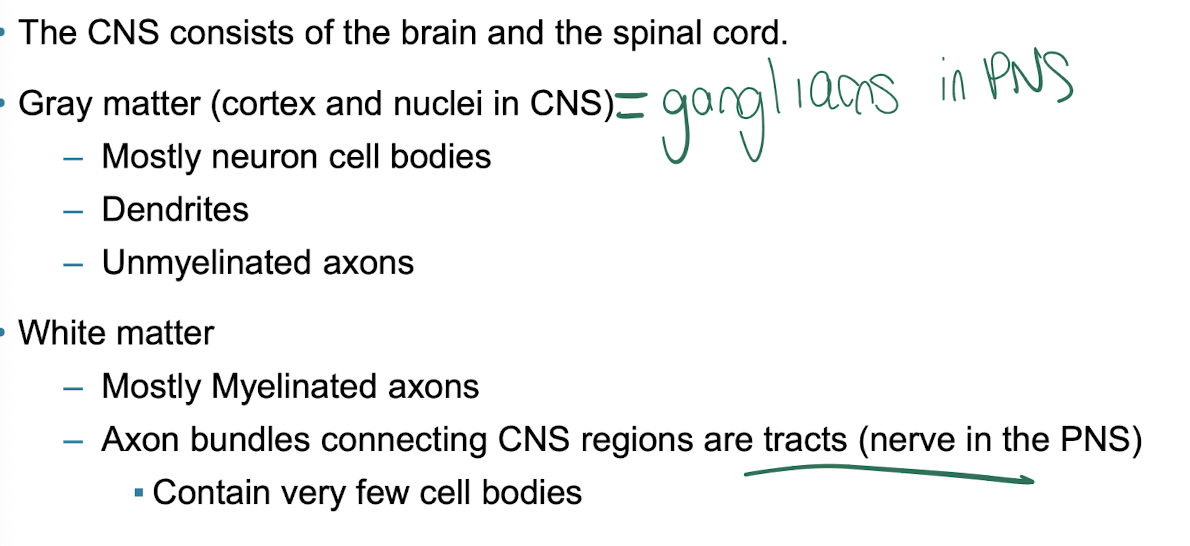
What are the layers of meninges? What is the autonomic ganglion?
autonomic= a cluster of nerve cell bodies located outside the central nervous system.

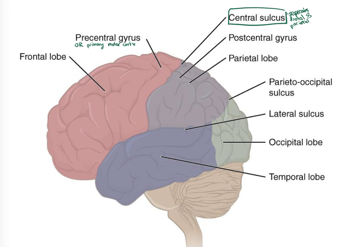
side view of brain

what are gyri vs sulci vs fissures, longitudinal fissure?

9 sections of brain
what are 4 main protections of the brain?
skull
cranial meninges
cerebrospinal fluid
BB barrier?
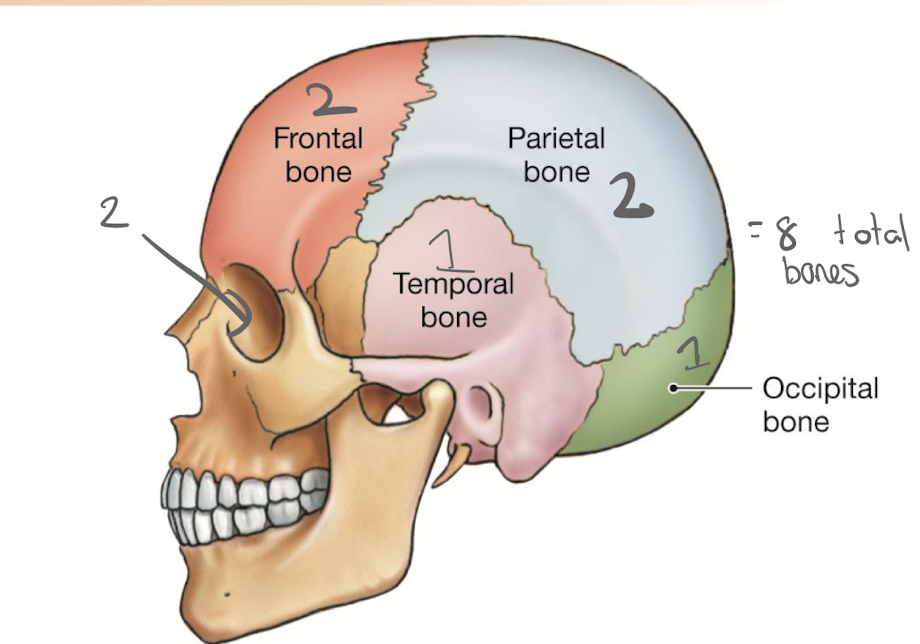
how many total bones in skull?
human skull consists of 22 total bones, divided into 8 cranial bones that protect the brain and 14 facial bones that shape the face
jaw bone is the only one that can ‘move’, all others are fused together
what are mininges (3) and what do they do? what is subarachnoid space and what does it contain ( and what comes in contact w brain and spinal cord surface )
Meninges= protective membranes, lie between bone and tissues to stabilize neural tissue and protect CNS from bruising against bone
– Dura mater- The thickest, toughest, outermost layer of the meninges.
– Arachnoid membrane = spiderweb-like middle layer of the meninges.
– Pia mater = The ultra-thin, delicate, innermost layer of the meninges. Adheres directly flush to every fold (gyri) and groove (sulci) of the brain and spinal cord surface.

cerebral spinal fluid: what is it made up of? what produces it? how is the total ~150mL distributed?
TOTAL = 125mL
25ml in ventricales
125 in subarachnoid

what are the 3 ventricles of brain and what is produced within them?
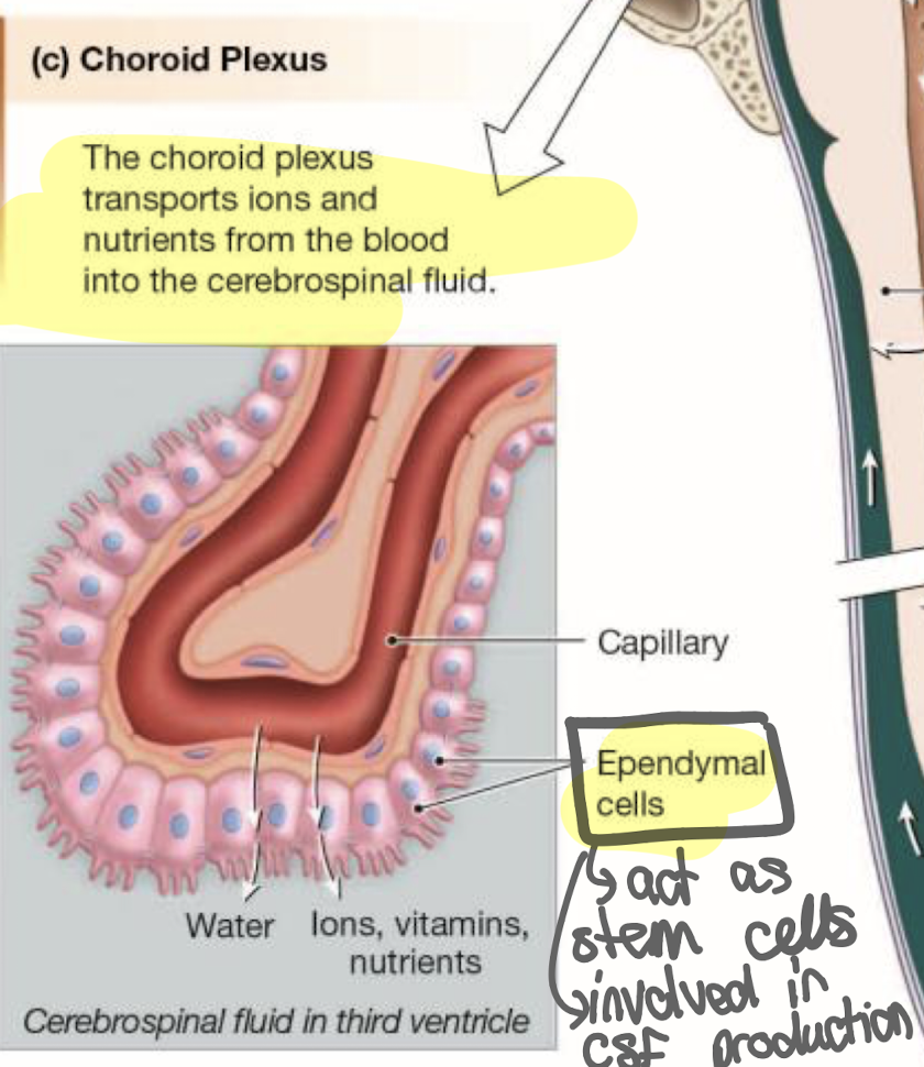
what is choroid plexus? what is it made of?
= Highly specialized clusters of vascular capillaries that manufacture cerebrospinal fluid.
Location: Projecting directly into the open floors of the brain ventricles.
Purpose/Function: Selectively pumps ions, vitamins, and water out of blood plasma into the ventricles to create fresh CSF.
present in all 4 ventricles, it transports ions and nutrients from blood into CSF
made of ependymal cells
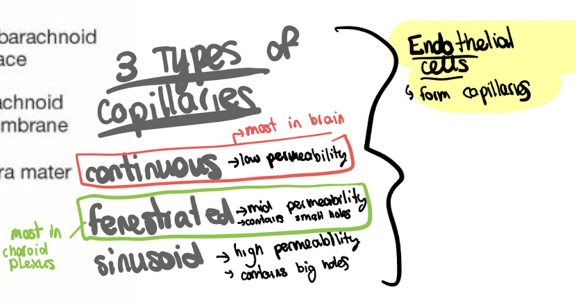
what are the 3 types of capillaries, how does their permeability vary?
alltogether what types of cells do they form?
alltogether form endothelial cells
how is CSF reabsorbed? what are arachnoid villus?
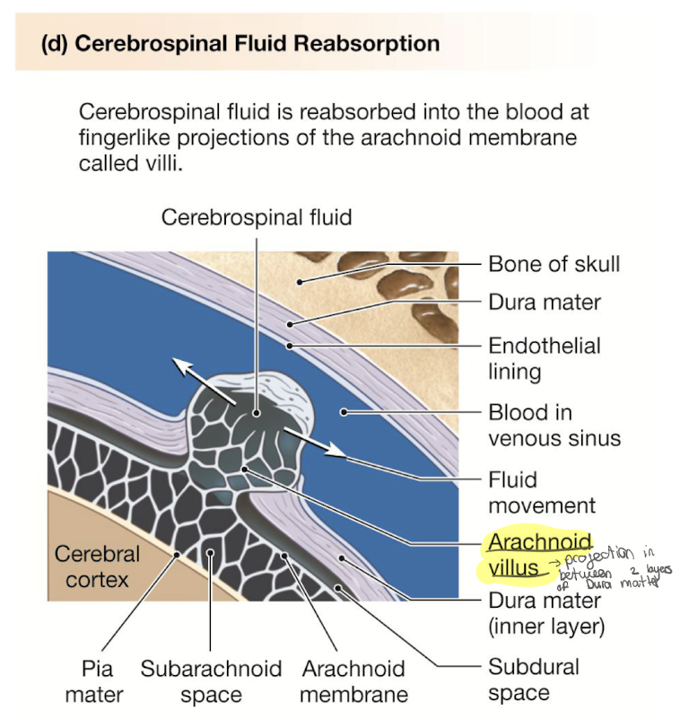
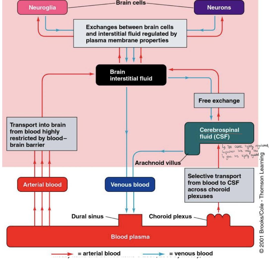
diagram of fluid flow around brain, in CSF: what is important about ions, proteins and glucose? what are 3 main roles of CSF?
• 1. Cushions and insulates delicate nervous tissue.
• 2. Gives Buoyancy to the brain (“floats” in CSF).
• 3. Exchange of gases (O2 and CO2), nutrients and
wastes
BBB: what is it formed of? what does it protect brain of? what can pass through it?
formed of:
Endothelial cells: The cells that line the inside of your brain's blood vessels.
Tight junctions: Extremely tight seals between those endothelial cells that block open gaps.
Pericytes: Small, supportive cells wrapped around the outside of the blood vessels.
Astrocyte end-feet: Extended parts of star-shaped brain cells (glia) that wrap a protective layer around the vessels.
Basement membrane: A thin, structural sheet of tissue holding the layers in place.
Highly selective permeability of brain capillaries!
• Astrocytes foot processes promote tight junctions between endothelial cells
• Protects brain from toxic water-soluble compounds and pathogens
• Small lipid-soluble molecules cross the blood-brain barrier


spinal cord: how is it branched? what does dorsal root carry and contain vs ventral root carry
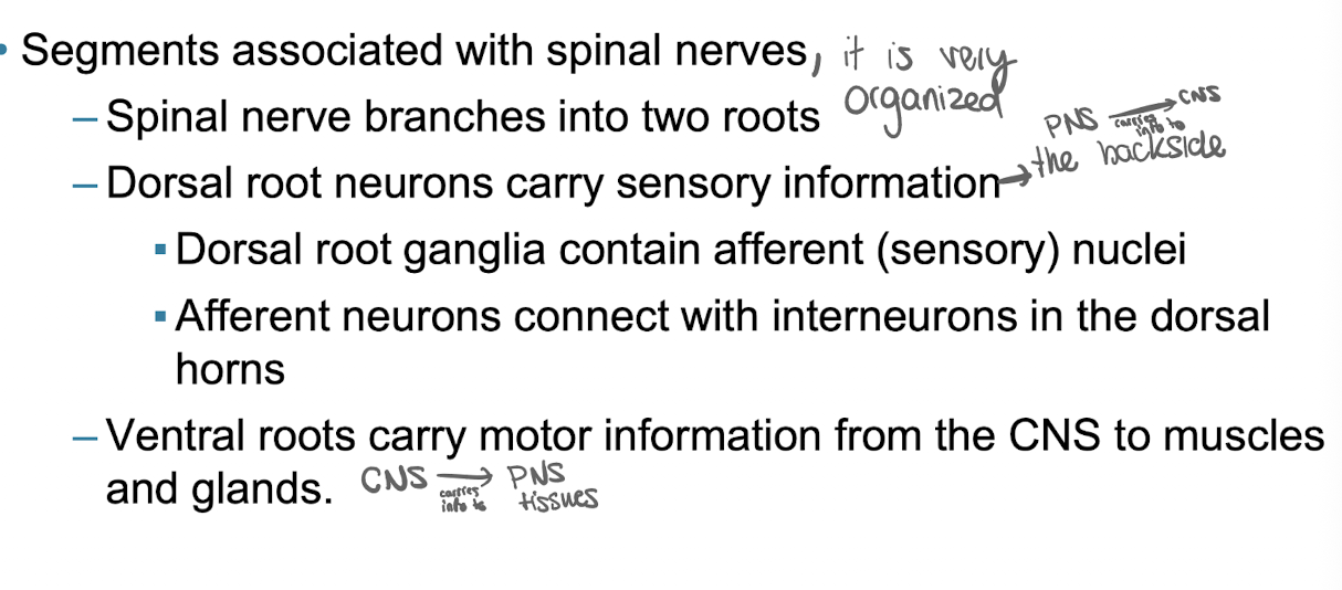
the spinal cord: GRAY matter—> what (3) components does it contain?
Posterior/dorsal horn: Receives and processes incoming sensory signals from the body, contains visceral and somatic sensory nuclei
Anterior/ventral horn: Contains motor neurons that send motor commands to voluntary skeletal muscles, and contains somatic motor nuclei
Lateral horn: Houses autonomic neurons that control involuntary functions, present only in the thoracic and upper lumbar regions, contains visceral motor nuclei
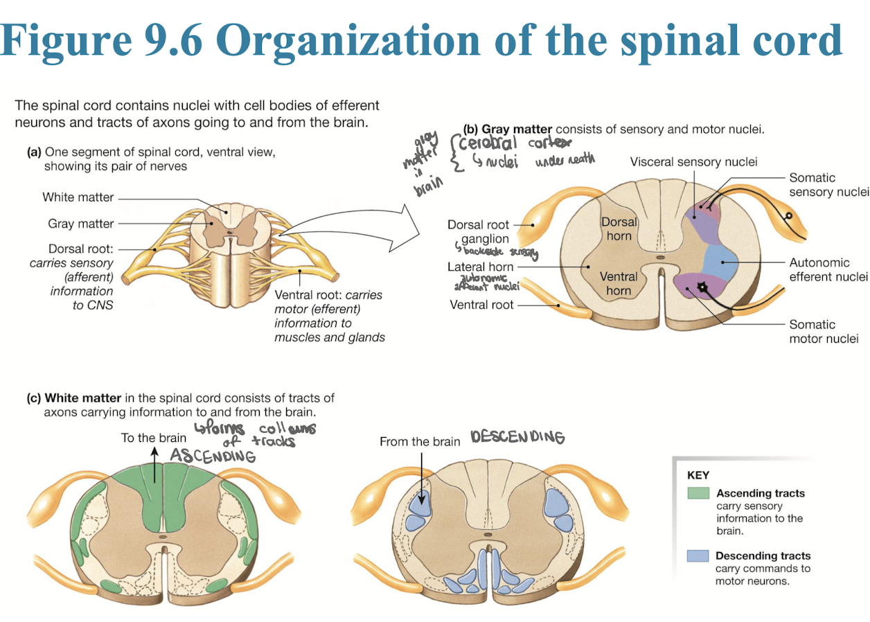
the spinal cord: WHITE matter—> how is it divided? which parts carry info TO the brain vs FROM the brain? ( ascending vs descending)
Divided into columns of tracts
– Ascending tracts take sensory information to the brain
– Descending tracts carry motor signals from the brain.
– Propriospinal tracts stay in the cord
white vs grey matter: composition, color, functions, location in brain, location in spincal cord
Composition: Grey matter consists of cell bodies, dendrites, and axon terminals; white matter consists of myelinated axons (nerve fibers).
Color Source: Grey matter gets its pinkish-grey color from dense cell structures and capillaries; white matter is white due to the fatty myelin sheath wrapping the axons.
Primary Function: Grey matter handles information processing, thinking, and control; white matter handles signal transmission and connectivity.
Location in Brain: Grey matter forms the outer layer (cortex); white matter sits on the inside (subcortex).
Location in Spinal Cord: Grey matter forms the inner butterfly shape (horns); white matter wraps around the outside (columns).
organization of WHITE vs GREY matter of spinal cord
Describe how a hollow neural tube develops into the ventricles and seven major divisions of the CNS.
The hollow center of the embryonic neural tube stays hollow and expands to become the fluid-filled ventricular system and the spinal central canal.
The solid walls of the tube multiply and differentiate into the tissue layers of the seven major adult divisions: Cerebrum, Diencephalon, Midbrain, Pons, Cerebellum, Medulla Oblongata, and Spinal Cord.

Starting at the skull and moving inward, name the membranes and other structures that enclose the brain (5)
Skull (Cranium): The rigid bony outer shell.
Dura Mater: The thickest, leather-like outermost meningeal membrane.
Arachnoid Membrane: The web-like middle membrane.
Subarachnoid Space: The fluid-filled gap containing circulating Cerebrospinal Fluid (CSF) and major blood vessels.
Pia Mater: The ultra-thin innermost membrane adhered flush to the brain's wrinkles.
Explain the formation, distribution, and functions of cerebrospinal fluid (3)
Continuously produced by the choroid plexus (capillary clusters) within the brain ventricles.
Distribution: Flows from the ventricles into the subarachnoid space around the brain and spinal cord, and down the central canal. Reabsorbed into venous blood via finger-like villi.
3 Key Functions:
Physical Cushioning: Insulates and protects tissue.
Buoyancy: Lessens effective brain weight so it floats rather than crushing itself.
Chemical/Nutrient Exchange: Cycles oxygen, nutrients, and waste materials.
Describe the structure and functions of the blood-brain barrier.
Structure: Brain capillaries feature a highly selective, non-leaky endothelial lining. Astrocyte foot processes wrap around them to signal and promote the creation of tight junctions between cells.
Function: Shields delicate brain neurons from dangerous water-soluble toxins, pathogens, and wild systemic hormone fluctuations. Only small, lipid-soluble molecules (like oxygen, carbon dioxide, and anesthesia) can passively diffuse right through
Describe the organization and major functions of the spinal cord.
The Roots (Somatic Split): Spinal nerves split into a dorsal root (carries incoming sensory info via the dorsal root ganglion to the back horns) and a ventral root (carries motor commands outward from the front horns to muscles).
The Horns (Gray Matter Processing): Dorsal horn = sensory nuclei; Lateral horn = autonomic motor; Ventral horn = voluntary somatic motor cell bodies.
The Tracts (White Matter Transmission): Ascending tracts carry sensory inputs up to the brain; descending tracts route motor signals down to tissues.
Describe the organization and major functions of the cerebral cortex.
deeply wrinkled outer layer of gray matter capped with structural folds (gyri) and grooves (sulci) to optimize processing surface area.
The Four Major Functional Lobes:
Frontal Lobe: Manages conscious reasoning, voluntary motor commands (via the precentral gyrus), and behavior.
Parietal Lobe: Primary somatosensory integration area (via the postcentral gyrus) handling touch, pressure, and body tracking.
Occipital Lobe: Dedicated master processing hub for vision and visual imagery.
Temporal Lobe: Coordinates primary processing centers for hearing and smell.