NMSK - Behaviour a movement
1/146
There's no tags or description
Looks like no tags are added yet.
Name | Mastery | Learn | Test | Matching | Spaced |
|---|
No study sessions yet.
147 Terms
What are 2 components when considering welfare?
Physiological components and behavioural components
Why is understanding behaviour important?
provides an insight into an animal’s emotional state and welfare state
law and legal obligations
client, practice and human-animal bond
clinical and surgical success
minimising negative experiences of a vet clinic
What 2 behavioural competencies should graduate vets show
Do no harm
Apply ‘behavioural first aid’
How do we assess welfare
Behaviour, physiology, longevity and reproduction
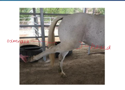
How can we use locomotion as an indicator?
•evaluate what is normal/abnormal
•assess gait patterns
•identify gait adaptations - e.g. lameness
•performance indicators
•welfare indicators
what are some locomotive indicators of pain
time spent lying down, no. times food stamped, no. ear flicks and time walking (restless)
list some examples of the impact of behaviour on NMSK system
stereotypical behaviour (e.g. weaving leading to worn feet/muscle injuries and awkward weight bearing leading to musculoskeletal system remodelling itself in response)
Scales to Assess Paint and Quality of life in companion animals
Success of operations - pre/post-op considerations and prognosis, ensuring gentle exercise
what are some things to consider before assessing locomotion
space available
surface conditions (firm (can listen to foot falls and assess gait pattern), level, non-slip)
age of animal
any medical conditions
handler
speed of gait (slow or fast)
How do you assess locomotion
visual observation, analysis equipment (high speed treadmills, video cameras, data analysis software, force plates) - vets don’t tend to use these
We can assess quality and divergence from the ‘norm’.
Compare using human observation to technical equipment
Human observation:
subjective, biased
frame rate - can miss detail
low technical effort and convenient
requires experience
low cost
Technical equipment:
measurable
objective, less bias
required dedicated equipment, space, resources, personnel
higher cost
What do we as vets need to do for assessing locomotion?
Get good at observing it and recognising what’s normal/abnormal
observe lots of animals
familiarise yourself with the processes
EXPERIENCE
be able to describe patterns of locomotion
What do we mean by gait? Give an overview
The specific patterns of footfalls during locomotion - walk, trot, canter
They change with speed
Characteristic sequences
Strides take place within a gait
Define ‘stride’
Complete cycle of movement
what are the 2 phases of a stride
stance phase (weight bearing limb)
Swing phase (non-weight bearing limb)

Describe the walk gait
- How many beats ?
-Symmetry ?
-Sequence of foot-falls
4-beat
symmetric
RH-RF-LH-LF
never more than 3 or less than 2 limbs weight-bearing at any one time
centre of gravity always been a triangle of weight bearing feet.
Describe the trot gait
- How many beats ?
-Symmetry ?
-Sequence of foot-falls
2 beat
very ancient pattern seen in horses
symmetric
diagonal gait
body supported alternately by left and right diagonal pairs (LH and RF, RH and LF)
period of suspension b/w successive stance phases
Marked axial twisting (resisted by design of axial system - evolution)
Describe the canter gait
- How many beats ?
-Symmetry ?
-Sequence of foot-falls
3 beat
asymmetric
rocking horse motion:
RH RF+LH LF
LH LF+RH RF
one moment of suspension, after single forelimb leaves the ground, prior to single hindlimb contact
lead leg - L/R
one diagonal pair
2 limbs out of phase
Describe the gallop gait
- How many beats ?
-Symmetry ?
-Sequence of foot-falls
fast, 4 beat
asymmetric gait
horses usually lead with their inside leg around a turn (called the lead leg)
moment of suspension
What is meant by the moment of suspension?
period when no feet are in contact with the ground e.g. fast trot, canter or gallop
normally one per cycle
greyhounds and cheetahs have 2 per gallop cycle
what are the 3 kinds of gallop
Transverse - LH RH, LF RF* (*one moment of suspension
Rotary - RH LH* LF RF* (*two moments of suspension)
Counter rotary - anticlockwise footfalls LH RH, RF, LF (asymmetric motion)
Who uses each type of gallop
Transverse - dogs at low speed, horses (odd-toed ungulates) and cattle (large even-toed ungulates)
Rotary - all cats, dogs at high speed, gazelle, antelope (small, even-toed ungulates), running rodents - in a horse it’s called a disunited canter
Counter-rotary - greyhounds on the track (race anticlockwise)
Why may gait change?
physically necessary (limb flied off the ground - pendulum effect, centrifugal force acting upwards and froude number = ratio of inertial force: gravitational force (>1 = suspension phase)
Metabolic advantages - optimal speed for each gait at which energy cost is minimal. (treadmill study - measured O2 consumed at each gait). Respiratory pattern and locomotion/respiratory coupling
Mechanical advantages (reduced bone strain)
Explain the neurological basis of limb coordination
sensation: vision, vestibular system (inner ear, balance orientation), mechanoreceptors (touch), nociceptors (pain), proprioceptors (body position)
Motor response to stimuli: Nervous system (in/voluntary control and reflexes), central pattern generators, brainstem, cerebellum (balance), constant monitoring of muscle length and tension (muscle spindles and golgi tendon organs)
how are equines adapted to high speed locomotion?
increase stride length: distal limb elongates, mobile scapula (increase length of limb, whiplash effect (small motion of the upper limb > flick of the lower limb)
Minimise limb mass - most of the work done involves accelerating and decelerating limbs, muscles positioned proximally (near pivot-point), reduced number of bones in limb.
Adaptions decrease mass (inertia) of lower limb.
Conservation of energy - whiplash effect, long tendons (transfer load, shock absorbers and energy store), stable joints (limit range of movement but little extra support is required).
Relatively rigid spine/sacroiliac junction: large gut, large body mass, transfer of energy from powerful hind quarters which minimised up and down movements of the body during locomotion.
how are canines adapted to high-speed locomotion
elongation of limbs (mass proximal)
digitigrade (claws for catching prey, claws may assist grip)
flexible back (arches and straightens which increases stride length), no gut restriction
tail - assists with balance when out of balance
what are some clinical consequences of adaptions in equines
low safety margins - bones and tendons (fractures and tendon strains)
little soft tissue covering distal limbs - poor wound/fracture healing
little soft tissue to absorb impact loads (joint injuries/osteoarthritis
Outline the evolutionary adaptions of horses
survive on prairies and grasslands
flight not flight
Detect dangers = good hearing, sight and smell
Sleep standing up: stay apparatus
Speed and endurance: cardiac, musculoskeletal and respiratory
Survive on large volumes of poor-quality roughage
what well developed senses do horses have?
large eyes, far back on an elongated head = high visual acuity and wide field of view
excellent hearing - mobile ears
accomplished sense of smell
how are horses adapted to sleep standing up?
stay apparatus
uses tendons, ligaments and muscles with high fibrous content
very few muscles used = lower energy demand
If the muscles were used = lots of energy needed, which would be disadvantageous
Outline what is involved in the passive stay apparatus of both the fore- and hindlimbs
Forelimb: serratus ventralis, check apparatus
Hindlimb: patella locking and reciprocal apparatus
BOTH: suspensory apparatus
Outline the suspensory apparatus
suspensory ligament (interosseus muscle in dogs), splits into the sesamoidian ligaments) at the sesamoid bones
distal sesamoidian ligaments resist carpal and fetlock extension without muscular effort
prevents movement
what sesamoidian ligaments are there?
Intersesamoidian
Short sesamoidian (sesamoid to P1)
Oblique sesamoidian (sesamoid to bottom of P1)
Straight sesamoidian (sesamoid to P2)

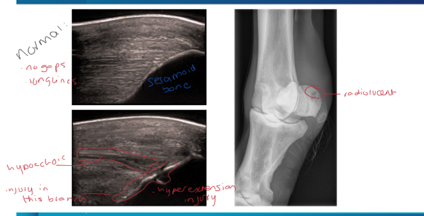
How may suspensory branch desmitis/sesamoiditis appear on an ultrasound/radiograph
Ultrasound:
no clear ligament/bone barrier
hypoechoic areas within the long white lines
broken bone/ligament border
Radiograph:
can see radiolucent areas on the bone
see ostephytes
abnormal bone growth
Outline the serratus ventralis and the role it has in the stay apparatus
only in forelimb
forms a link between the costal side of scapula and the ribs and vertebrae
contains a tendinous layer
suspends the thorax when the muscles relax - due to tendinous quality, prevents the trunk from sinking
two parts of the muscle: cervical and thoracic.
outline the check apparatus and its role in the stay apparatus
forelimb only
Superior check ligament = accessory ligament of the SDFT
palmar radius to the SDFT in the distal radius
resists carpal and fetlock extension without muscular effort
Inferior check ligament = Accessory ligament of DDFT
palmar carpus to the DDFT mid metacarpus
resists fetlock extension without muscular effort
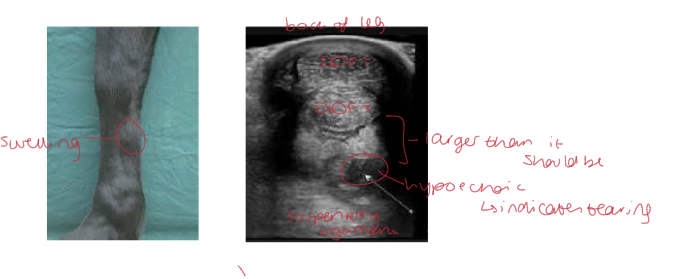
How may we identify check ligament desmitis?
On observation:
swelling may be visible on the limb where theses ligaments are located
Ultrasound:
may appear larger than normal with evident hypoechoic areas which indicate tearing

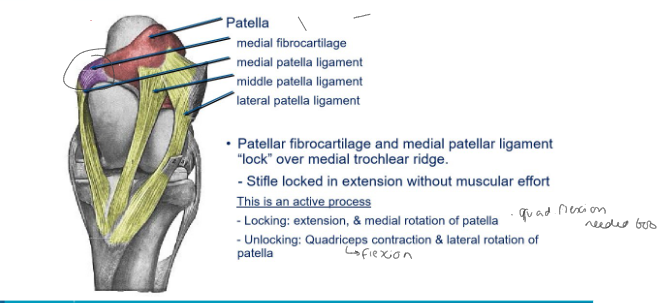
Outline patella locking
When relaxed:
the patella is free to slide within the trochlear groove as stifle moves
When locked:
medial patellar ligament sits over the proximal medial trochlear ridge
Stifle can no longer bend
Patella is locked in position without muscular effort


is patella locking active? how is it controlled and undone?
yes it’s active
Controlled: - quadriceps extends, patella rotates medially
Unlocked: Quadriceps flexes, patella rotates laterally

What is upward fixation of the patella?
when the patella locks into place and won’t unlock
Horse will struggle to walk, with stifle and hock extended
How can we treat upwards fixation of the patella?
cut the medial ligament
over time it fuses again, but will be shorter and fatter, which prevents unwanted locking
in what kind of horses is upwards patella fixation more likely?
Horses with a straight hindlimb conformation e.g. minatures and shetlands
Weak quadriceps - older and non-athletic horses and breeds.

Outline the reciprocal apparatus
Only in the hindlimb
two tendinous cords cause united movement of the stifle and hock
peroneus tertius - cranial
superficial digital flexor - caudal
Powerful proximal muscles act upon distal limb
When stifle is locked, hock is also immobile:
stifle extends, so must hock (proximal SDF is pulled, so is proximal calcaneus)
stifle flexes, so does hock
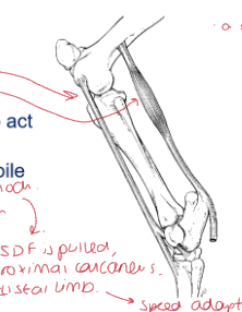
How can we identify a ruptured peroneus tertius?
upon examining hindlimb
stifle is flexed
hock remains extended

How are horses adapted for speed?
Increased stride length:
long legs (especially distally)
mediolaterally flattened thorax (legs can slide easily along the thorax body)
scapula positioned laterally and free to swing
vestigial ulna (increases lever action of the triceps)
Increase stride rate:
reduced distal limb mass (single digit)
muscle weight proximal
long tendons (pulley effect)
Minimise energy wastage:
single plane of movement of limbs
stretched tendons store kinetic energy as heat - can act as springs
what two things factor into speed of an animal?
stride length
stride rate
Outline some factors of the thoracic limbs in comparison to the hindlimbs
carry more static body weight - translate horizontal velocity provided by hindlimbs, minimise wasteful vertical energy
shorter and straighter than hindlimbs (no bone articulation to skeleton - act as suspension)
broader, more rounded hoof
energy storage (tendons)
Outline some factors of the hindlimbs in comparison to the forelimbs
provide forward impulsion of the horse
direct bone articulations
narrower, more pointed hoof
What is meant by the power train
Other adaptions of the horse for high speed
heart
lungs
muscles
How do horses have efficient airflow?
less complicated nasal passage
easy access to trachea
muscles can dilate the nostrils
straight line from nostrils to trachea
what affects cardiac output?
CO = SV x HR
stroke volume x heart rate
why are horse hearts so well adapted for speed and high CO?
2% body weight
SV is closely related to heart size
SV only slightly increases during exercise, HR increases a lot
What is:
Heart size (% body)
Resting HR
Max HR
Increase in HR
resing CO (L/min)
max CO (L/min)
increase in CO
In a horse
a) 2%
b) 32 bpm
c) 240 bpm
d) 7.5
e) 35L/min
f) 280 L/min
g) 8 x increase in CO
why do horses have so many muscles?
create lots of KE
store KE as heat - act as springs - return on the next stride?
what do we need to be careful of with horses due to their adaptions?
horses have been adapted to the very limits of optimal function
they’re easily pushed past the limit
Easily injure:
Stress fractures
tendon tears
why do tendons tear?
Hyperthermia
very few cells and blood vessels
Due to high KE, high temperatures reached, more prone to injury
Common locations:
Half way down cannon bone in the middle of the SDFT
Name 4 other adaptions of horses
long teeth continually erupt - hyposodont
(large grinding surface to reduce stem length)
long GI tract - hind gut fermenters, can digest poor quality roughage
Excellent thermoregulators - efficient sweat glands
Breed variations: size, shape, coat length, dietary requirements, sensitivity to medications.
what features have been highly conserved during evolution?
Relative position of CNS and GIT:
invertebrates: CNS below GIT
Vertebrates: CNS above GIT
4/6/8 legs
segmentation
head-tail orientation
why do vertebrate animals e.g. dogs and cats have the digits/forelimbs they have?
Forelimbs:
change in weight placement
Limbs have moved further from the head - as head has extended away - now have necks
Digits:
tend to be teh same length: due to weight placement and the fact limbs are under the body - not on the sides
Outline evolutionary adaptions in the cat and dog in the pectoral girdle
Scapula:
now a limb bone
laterally flattened
Coracoid:
fused and part of the scapula in mammals = coracoid process
Clavicle:
many variations in mammals
not in dogs, present in cats (very small/cartilaginous)
Interclavicle:
derived from skin armour, now on the cranial end of the sternum
what are some general cursorial specialisations?
anatomical (evolutionary) adaptions for running along the ground
longer limbs (there is an optimal length)
reduced lower limb mass
recruitment of scapula as a limb bone
loss of clavicle
give 2 adaptions of the proximal forelimb for cursorial specialisation
no clavicle (or rudimentary)
allows scapula to become an extra limb bone
Scapula vertical against lateral thorax:
acromion is less prominent than primates and fossorial animals
shoulder joint constrained by muscles to pro/retraction
what is an important rule of evolution?
You can’t change the nerve supply
nerve that supplies something will move with it during its adaption
name some cursorial specialisations in the carnivore distal forelimb
limited pronation/supination in dogs, more in cats
reduces muscle work since antebrachium is more stable
cats can do this better because they’re ambush predators, therefore grappling with their prey is more important
Radio-carpal joint has limited abduction/adduction:
far less than primates, more in than in horses
Digitigrade:
accessory pad becomes redundant
plantigrade: tarsus/metatarsus/phalanges on ground
digitigrade = phalanges on the ground
unguligrade = only P3 on the ground
what is meant by a)plantigrade b)digitigrade c)unguligrade
plantigrade: tarsus/metatarsus/phalanges on ground
digitigrade = phalanges on the ground
unguligrade = only P3 on the ground
what are some cursorial specialisations in the carnivore hindlimb?
small abdomen = larger hip movement
hip joints are more mobile
strong stifle joint, doesn’t lock
large muscles for acceleration and high speed
limb joints move independently
strong gastrocnemius supported by long hip extensors
compare the hip joint in carnivores and herbivores
Carnivores:
very good RoM
angled femoral neck
Large herbivores:
much more supportive
asymmetric femoral head
femoral neck is more compact and vertical
limited RoM
Outline some evolutionary features of dogs
able to survive hot and cold - adverse conditions
diet - anatomically carnivore, actually omnivore
ability to locate prey - hunt at dawn/dusk/night, good senses
stalk prey - soft footed, posture, controlled MSK movements
catch prey: work as a team, run to fatigue hunting strategy, turn quickly
kill prey: strong jaws/teeth
outline some MSK adaptions for function in dogs
muscles - fast twitch fibres for speed
slow twitch fibres for endurance
mixed type fibre IID
rely more on fat stores than glycogen for muscle energy
laxity and flexibility of joints for posture and manoeuvrability
light body frame
long limbs - jumping leverage
tail for balance
flexible neck = wide range of vision
muscles to ears to locate sound
breed conformation and differences
Outline cat evolutionary factors
evolved from territorial, solitary, hunting cat
able to survive dry conditions (water retention, urine conc)
obligate carnivores
good senses and hunt at night/dawn/dusk
stalk prey: posture, soft footed, controlled MSK movements
catch prey: jumping, standing start to fast running, turn quickly, flexibility, claws
kill prey: claws, jaws
equipped to defend territory: marking, signalling, fighting
why can’t cat species run for a long time like dogs?
heat generated can’t be removed quickly enough
run very quickly but for short periods of time.
Outline some MSK adaptions of cats (1)
type Ii fibres for speed - short bursts, anaerobic
IIA fatigue resistant
IIB - power but fatiguable
laxity/flexibility in joints - 3% elastin content (1% in dogs)
light body frame - wider medullary cavities and thinner cortices in long bones.
long limbs - more cylindrical than dogs
tail for balance
flexible ligament - no nuchal ligament = greater range of motion
Outline some MSK adaptions in cats (2)
muscles to ears to locate sound
retractable claws to secure prey
greater range of movement in the spine and hsoulder
longer and more slender vertebrae
broader and shorter scapula
weight bearing is more evenly shared b/w front and back limbs (in comparison to dogs)
antebrachium range of pronation is 45-55 degrees
masseter muscle = strongest depending on definition.
what is the scapula equivalent to
the pelvis
how have the pelvis and scapula developed differently in evolution?
pelvis: wasn’t attached, now is
scapula: was attached, now isn’t
to axial skeleton
what is the humerus the equivalent of in the hindlimb and what is its latin name?
femur
stylopodium
what is the radius/ulna the equivalent of in the hindlimb and what is its latin name?
tibia/fibula
zeugopodium
what is the latin name for the carpus/tarsus, metacarpus/tarsus and phalanges?
autopodium
Outline anatomical difference in dog and cat triceps brachii
Dog: 4 heads: accessory, medial, lateral, long
Cat: 5 heads (lateral head is in 3 parts), no accessory
Outline anatomical difference in dog and cat radius and ulna
dogs can’t supinate as much as cats
Outline anatomical difference in dog and cat manus/Pes
Digit I small/absent
digits III and IV longer than II and V
paired sesamoid bone at each McPh/MtPh joint
Cats:
claw retraction by hyperextension of DIP joint
natural position is retracted — dorsal elastic ligament
extended by pull from DDFT
Outline anatomical difference in dog and cat shoulder/elbow
Cats:
Acromion has 2 processes - hamate is distal, suprahamate is flat and proximal
Coarcoid process - beak-like, extends craniomedially from glenoid cavity (risk of fracture)
Vestigial clavicle: small ossicle
Supracondylar foramen: proximal to medial epicondyle (medial nerve and brachial artery pass through)
Ulnar nerve: lies under the medial head of triceps
No supratrochlear foramen in olecranon fossa (less likely to fracture distal epiphysis)
Incomplete ossification of humeral condyle
Outline anatomical difference in dog and cat brachioradialis
cats:
well developed - elbow flexor supplied by radial nerve
because cats climb trees.
Outline anatomical difference in dog and cat hip/thigh
round ligament supplies significant blood supply
muscles around the hip are broader
tensor fasciae lata needs longer incision and vastus lateralis greater subperiosteal elevation
caudofemoralis muscle is caudal to gluteal group, cranial to and acts with biceps femoris
sartorius muscle: single in cat (2 in dog), lat approach to shaft of femur
sacrotuberous ligament - absent in cats (sacrum to tuber ischium in dogs)
acetabulum is load-bearing central and caudal thirds (dog is cranial third)
Outline anatomical difference in dog and cat stifle
cats:
poorly or unmineralised sesamoid bones
CrCl is larger than caudal
Outline anatomical difference in dog and cat tarsus
Cats:
no long component to medial and lateral collateral ligaments just two short components
Outline anatomical difference in dog and cat tibialis cranialis and extensor digitorum longus
united in cats
Outline anatomical difference in dog and cat flexor digitorum profundus
tibilais caudalis stays separate with its own tendon
Outline anatomical difference in dog and cat soleus
origin with DDR, inserts with gastrocnemius in cats
not really present in dogs
Outline anatomical difference in dog and cat flexor digitorum superficialis (SDF)
cats have extra muscle fibres distal to tarsus
Outline anatomical difference in cats compared to dogs: axial system:
Flexible lumbar region: increases stride length
Lower gut mass: increased RoM of hips
Nuchal ligament reaches C2 in dogs: not to skull, no laminar part, same function to suspend head and neck in front of the body
No nuchal ligament in cats - lots of fast muscle fibres instead
why are the thoracic spinal processes angled?
applies weight downwards
enables the head and neck to be supported
Outline anatomical difference in dog and cat neck and spine
Neck:
no nuchal ligament in cats- prone to ventroflexion when weak
Dogs nuchal ligament runs from C1 to T1 spinous process
Spine:
more flexibe IV discs in cats - 20% length (15-17% indogs)
longer cord in cats relative to vertebral column in dogs
what is meant by a congential disease?
exists at or before birth usually through heredity as a disorder.
acquired at bird/during uterine development usually as a result of environmental influences
what is meant by a hereditary disease
genetically transmitted from parent to offspring
what is meant by development of orthopaedic conditions
normal at birth and the condition develops during growth (usually early post-natal period)
what is meant by dysplasia
abnormality of development
what are some factors that predispose cats/complicate MSK diseases in cats
Lifestyle:
climbers
fighting
sleeping under cars
running across roads
Diet:
excess/insufficient nutrient intake
all meat = nutritional secondary hyperparathyroidism
ventroflexion of neck with thiamine deficiency
multiple extoses in hypervitaminosis A
Obesity
Ageing changes
Metabolic problems - neck ventroflexion in hypokalaemia
How do developmental MSK diseases compare in cats to dogs?
rare
congenital luxated patella and pectus excavatum due to taurine defiency
How does treatment of ulna and radius fractures differ in cats and dogs
Cats need 2 plates, as radius and ulna aren’t fused
Only 1 plate in dogs because R +U are linked by fibrous tissue, providing stability.
what factors predispose/complicate MSK diseases in dogs
size/weight - biomechanical forces
genetic conformation
activity
diet
obesity
trauma
ageing changes