Biochem I Exam 1
 Call Kai
 Learn
Learn Practice Test
 Spaced Repetition
 Match
Match Flashcards
 Knowt Play
1/206

Earn XP
Description and Tags
Ch. 2, 4, 5, 6, 7, 10
Name | Mastery | Learn | Test | Matching | Spaced | Call with Kai | Chat |
|---|
No analytics yet
Send a link to your students to track their progress
207 Terms
zwitterion
neutral molecule with one positive and one negative charge; happens when amino acids are at pH 7
amino acid structure
tetrahedral; alpha carbon always chiral
aliphatic & aromatic
non-polar amino acids
Proline
cyclic, least hydrophobic aliphatic amino acid

Proline codes
Pro, P
Glycine
the only non-chiral amino acid

Glycine codes
Gly, G
Alanine
side chain is a methyl group

Alanine codes
Ala, A
Valine
side chain is an isopropyl group

Valine codes
Val, V
Leucine
side chain is an isobutyl group

Leucine codes
Leu, L
Isoleucine
contains 2 chiral carbons, most hydrophobic of the aliphatic amino acids

Isoleucine codes
Ile, I
aromatic amino acids
VERY hydrophobic, absorb UV at 280 nm
Phenylalanine
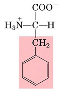
Phenylalanine codes
Phe, F
Tyrosine
OH group has a pKa of 10.5
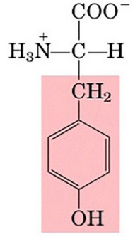
Tyrosine codes
Tyr, Y
Tryptophan
bi-cyclic indole ring, NH group has a pKa of 16-17
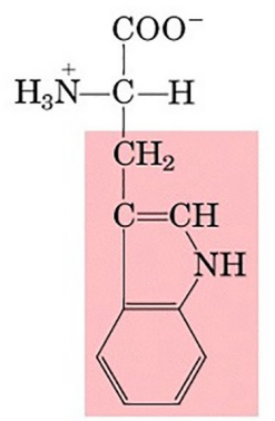
Tryptophan codes
Trp, W
Cysteine
important in disulfide linkages, weak acid (R group pKa = 8.3)

Cysteine codes
Cys, C
Methionine
“start” amino acid in majority of proteins, VERY hydrophobic, sulfur present in thioester linkage

Methionine codes
Met, M
acidic amino acids
contain carboxyl group, negatively charged at physiological pH, present as conj. bases
Aspartate
R group pKa = 3.9

Aspartate codes
Asp, D
Glutamate
R group pKa = 4.3

Glutamate codes
Glu, E
basic amino acids
hydrophilic nitrogenous bases, positively charged at physiological pH
Lysine
di-amino acid, protonated at pH 7 (R group pKa = 10.5)

Lysine codes
Lys, K
Arginine
most basic amino acid (R group pKa = 12.5)

Arginine codes
Arg, R
Histidine
the only amino acid that functions as a buffer in physiological range (R group pKa = 6.0)

Histidine codes
His, H
polar uncharged amino acids
polar side groups, hydrophilic
Serine
looks like Alanine with hydroxyl group, protonated at pH 7 (R group pKa = 13)
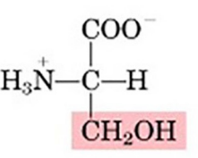
Serine codes
Ser, S
Threonine
2 chiral carbons, protonated at pH 7, (R group pKa = 13)

Threonine codes
Thr, T
Asparagine
amide of aspartic acid
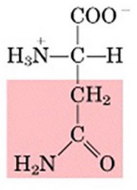
Asparagine codes
Asn, N
Glutamine
amide of glutamic acid

Glutamine codes
Gln, Q
Selenocysteine
natural amino acid, selenol group (SeH), pKa = 5.2
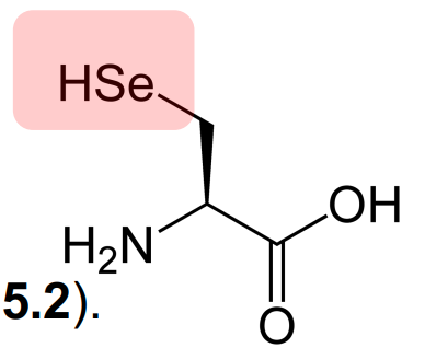
Selenocysteine codes
Sec, U
Pyrrolysine
found in some methanogenic archaea & bacteria, but not humans, pyrroline side chain, pKa unknown
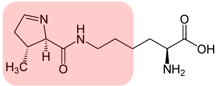
Pyrrolysine codes
Pyl, O
weak polyprotic
type of acid that amino acids are
alpha-carboxyl group
pKa = 2
alpha-amino group
pKa = 9
asymmetric because the alpha carbon of the amino acid is a chiral center
is the polypeptide chain symmetric or asymmetric?
trans to each other
carbonyl oxygen & amide hydrogen relationship in peptide backbone
peptides
short polymers of amino acids
residue
unit of amino acid
dipeptide
2 residues
tripeptide
3 residues
oligopeptide
12-20 residues
polypeptide
20+ residues
monomeric protein
one polypeptide chain
multimeric protein
more than one polypeptide chain
homomultimer
one kind of polypeptide chain
heteromultimer
2 or more different polypeptide chains
fibrous proteins
relatively simple, regular, linear proteins

globular proteins
proteins that are roughly spherical in shape

primary protein structure
amino acid sequence numbered from N-terminus to C-terminus
secondary protein structure
local structures stabilized by H-bonds, represent 3-dimensional arrangement of polypeptide
alpha helix & beta strand
the 2 major secondary structures of proteins
tertiary protein structure
overall 3-dimensional shape of a protein
quaternary protein structure
subunit organization of protein
prosthetic group
the non-amino acid part of the structure of a protein, tightly linked to apo-protein
primary structure
determined by covalently linked amino acid residues in polypeptide backbone
secondary and higher structures
determined by noncovalent & weak forces
H bonds
peptide backbone forms these bonds, side chain can form these bonds on the surface of a protein with water molecules
hydrophobic interactions
nonpolar side chains of amino acids cluster in nonpolar environments; forming of these bonds minimizes interactions of nonpolar residues with water and is highly favorable
ionic interactions
arise as electrostatic attractions between opposite charges or repulsions between like charges; side chains can carry positive charges or negative charges
on the protein surface because they can interact optimally with the water solvent
where are ionically charged residues usually located? why?
van der waals interactions
includes both attractive & repulsive forces; individual interactions are weak, but many occur in a protein
dipole-induced dipole interactions that arise from fluctuations in the electron charge distributions of adjacent nonbonded atoms
what causes attractive forces in van der waals interactions?
bond linking the alpha carbon & the carbon of the peptide bond (psi)
bond linking the nitrogen of the peptide bond & the adjacent alpha carbon (phi)
rotations allowed in secondary structure of protein
alpha helix of secondary structure
forms b/c of each carbonyl forming a H bond w/ the amino group of the 4th residue ahead in the sequence; 3.6 amino acid residues per turn; amphiphilic (hydrophilic + hydrophobic sides)
1.5 A
rise per residue in alpha helix
5.4 A (3.6 residues per turn x 1.5 rise per residue)
rise per turn (pitch) in alpha helix
backbone loop
closed by any H bond in alpha helix
beta sheet of secondary structure
sheet of polypeptides; parallel & antiparallel stable arrangements
parallel beta sheet
adjacent strands run in same direction
antiparallel beta sheet
adjacent strands run in opposite direction
fibrous protein
mechanically strong, insoluble, play structural role
fibroin & beta-keratin
form extensive beta sheets
collagen
triple helix structure, principal component of connective tissue
tropocollagen
basic unit of collagen, 3 intertwined polypeptide chains, composed of hydroxylysine & hydroxyproline
globular proteins
more abundant than fibrous proteins, large functional diversity, composed of domains
globular proteins
broad section of antiparallel beta-sheets, a few beta-turns, several peptide sections without defined secondary structure (random coil)
ribonuclease
globular protein w/ hydrophobic core & hydrophilic surface
globular protein structure
surface structure includes water molecules, alpha-helices on a surface are usually amphiphilic
protein domains
compact, folded protein structures that are usually stable by themselves in aqueous solution
multi-domain proteins
possess the sum of functional properties & behaviors of their constituent domains, 90% are duplicated in other proteins, many proteins contain multiple copies of the same