Thoracic Wall, Pleura, and Lungs
1/18
There's no tags or description
Looks like no tags are added yet.
Name | Mastery | Learn | Test | Matching | Spaced |
|---|
No study sessions yet.
19 Terms
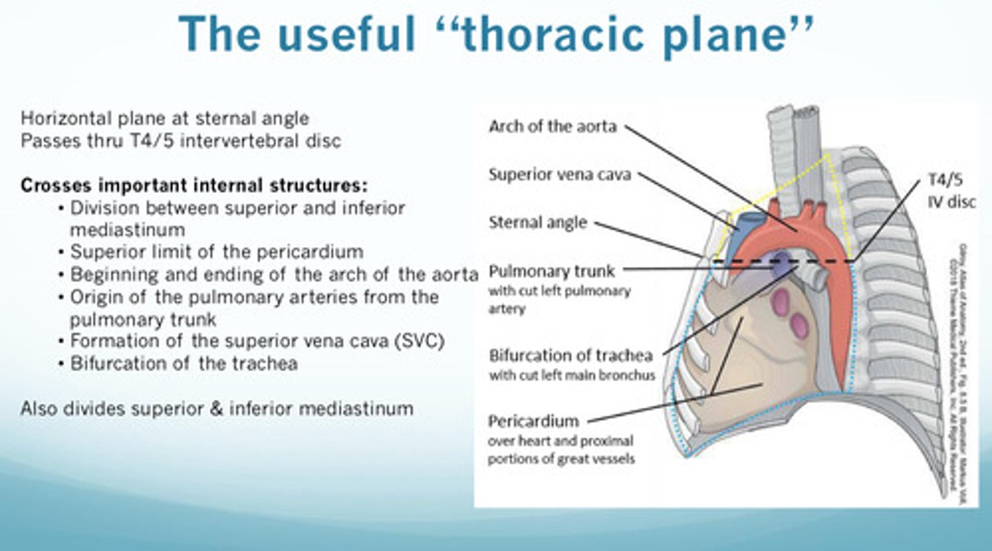
what is the sternal angle/thoracic plane and where is it located?
1) the thoracic plane is the horizontal plane at the sternal angle that passes through T4/5 intervertebral discs
2) the sternal angle/thoracic plane is where the 2nd rib attaches (first palpable rib)
why is the sternal angle/thoracic plane an important anatomical landmark?
important because it divides the superior and inferior mediastinum
what are the structures and landmarks that are found at the level of the sternal angle?
1) superior limit of the pericardium
2) beginning and ending of the arch of the aorta
3) origin of the pulmonary arteries from the pulmonary trunk
4) formation of the superior vena cava (SVC)
5) bifurcation of the trachea
what are the roles of the intercostal muscles (external and internal) during quiet respiration
1) all stabilize the thoracic wall
2) external intercostals assist in elevation during quiet breathing
3) internal intercostals and innermost intercostals depress the ribs as accessory muscles of exhalation
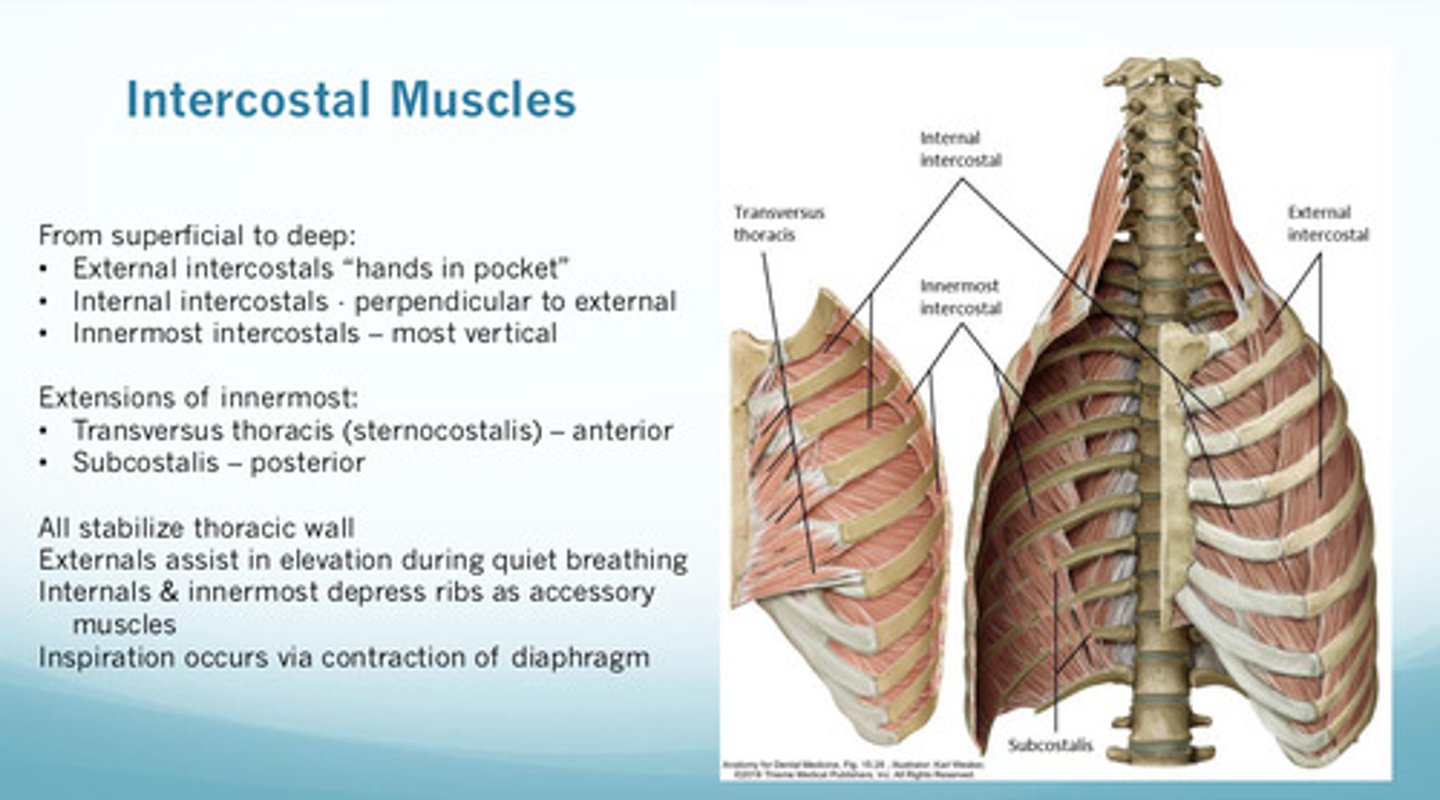
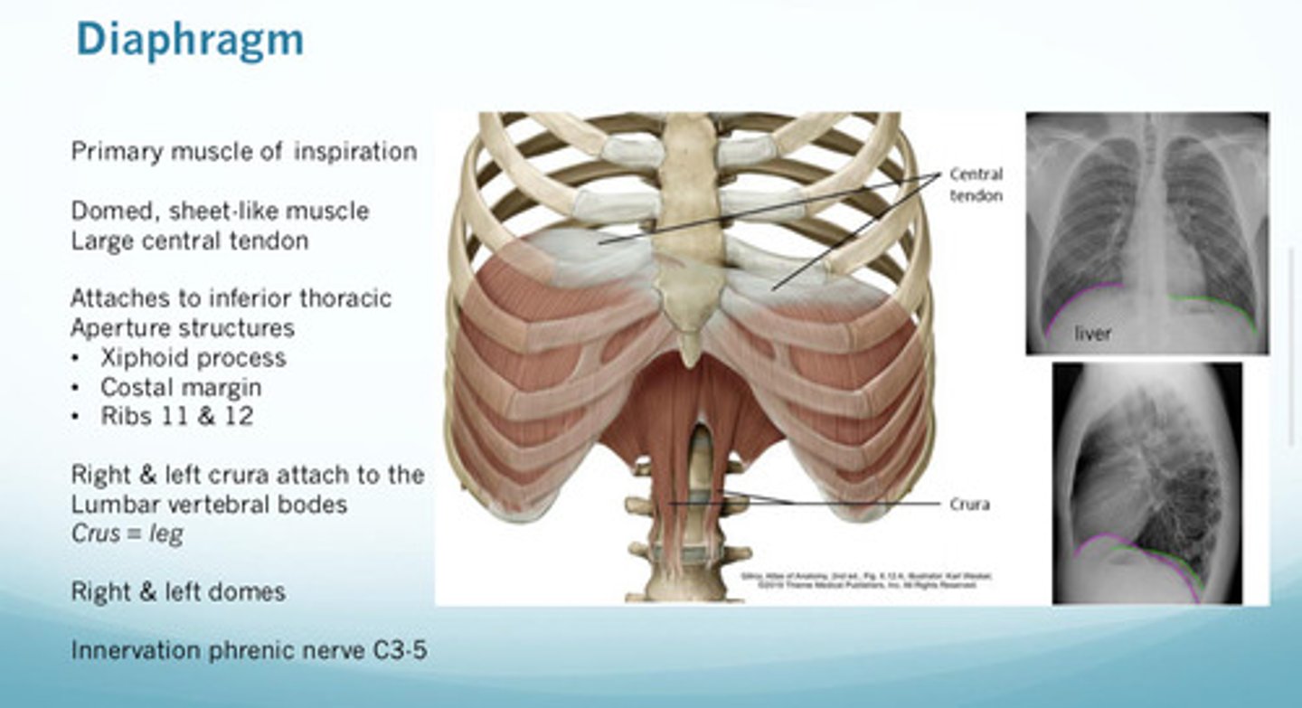
inspiration occurs via contraction of which muscle
diaphragm: primary muscle of inspiration (domed, sheet like muscle; large central tendon)
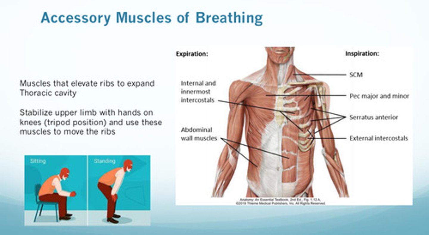
why are the accessory msucles of breathing important? (why and when)
these are important because they are the muscles that elevate the ribs to expand the thoracic cavity; people often use these during respiratory distress
what are the accessory muscles of inspiration
1) SCM
2) scalenes
3) serratus anterior
4) pectoralis
5) external intercostals

what are the accessory muscles of expiration
1) internal and innermost intercostals
2) obliques (both external and internal)
3) rectus abdominus
4) transverse thoracis
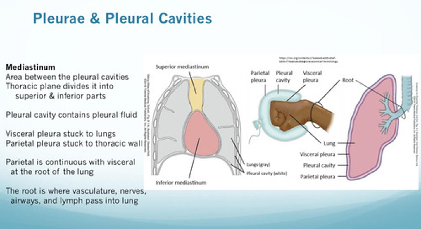
what is the mediastinum
the central compartment of the thoracic cavity; it lies between the 2 pleural cavities, which contain the lungs (the thoracic plane divides the mediastinum into superior and inferior parts)
the parietal pleura is named differently based on where it is _________?
located; costal parietal pleura if next to ribs, diaphragmatic parietal pleura if sitting on diaphragm, mediastinal parietal pleura if next to mediastinum
note that the 2 layers are continuous at the ____ of the lung
root! (the root is where vasculature, nerves, airways, and lymph pass into the lungs)
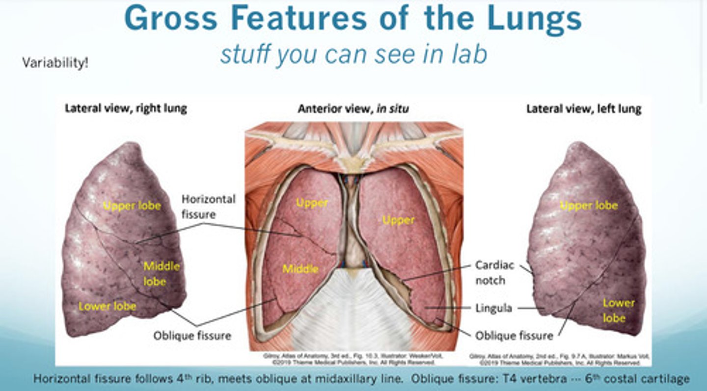
the horizontal and oblique fissures of the right lung divide which areas?
horizontal divides upper and middle lobes (follows the 4th rib, meets the oblique fissure at midaxillary line)
oblique divides middle and lower lobes (at the level of T4 vertebra, follows the 6th costal cartilage) —> same as in the left
what are other gross anatomical features of the left lung that the right lung does not have
1) cardiac notch in upper lobe (where heart sits)
2) has lingula in upper lobe
what is a pleural cavity and how does it relate to the 2 serous layers of the pleural sac: visceral and parietal?
1) visceral pleura is the inner layer that is stuck to the lungs
2) parietal pleura is the outer later that is stuck to the chest/thoracic wall (to the endothoracic fascia which is deep to the innermost intercostals)
3) in between the 2 layers is the pleural cavity, which contains the pleural fluid
what are the major gross anatomical differences between the right and left lung in terms of lobes and fissures?
right lung: has 3 lobes —> upper, middle, lower and 2 fissures —> horizontal and oblique
left lung: has 2 lobes —> upper and lower and 1 fissures —> oblique
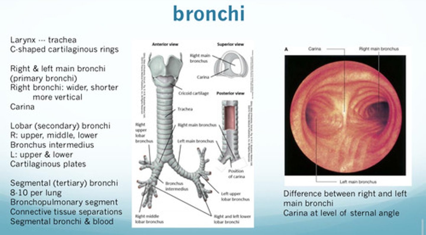
how are differences in the right and left main bronchus important in relation to aspiration?
because the right main bronchus is wider, shorter, and more vertical, this is where aspirated objects will typically sit as it is a straight shot down from the trachea; most aspirated objects end up in the right lower lobe
what are the major gross anatomical differences between the right and left lung in terms of main bronchi and lobar bronchi?
1) right primary/main bronchi are wider, shorter, more vertical than left primary/main bronchi
2) right lobar secondary bronchi: upper, middle, lower (right lung has bronchus intermedius that divides the upper and middle/lower lobar bronchus)
3) left lobar secondary bronchi: upper and lower
how is the innervation of the parietal and visceral pleura different?
innervation of visceral pleura —> visceral, insensitive to pain
innervation of parietal pleura —> somatic, pain transmitted by intercostal and phrenic nerves
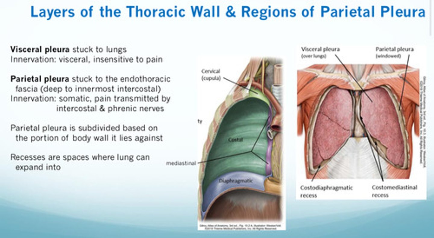
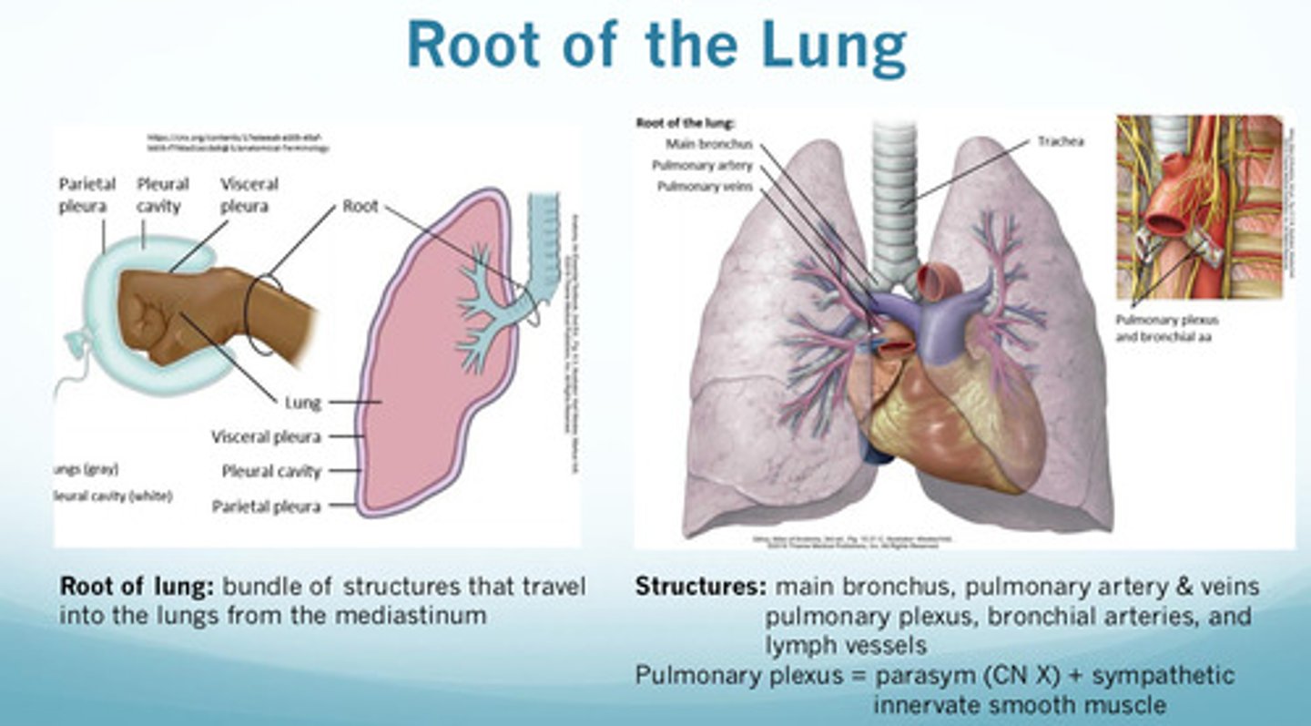
what are the structures found at the root of the lung?
at the root of the lung, there is a bundle of structures that travel into the lungs from the mediastinum
1) main bronchus
2) pulmonary artery and veins
3) pulmonary plexus (parasympathetic (CN X) + sympathetic innervate smooth muscle)
4) bronchial arteries
5) lymph vessels