Molar Anatomy
 Call Kai
 Learn
Learn Practice Test
 Spaced Repetition
 Match
Match Flashcards
 Knowt Play
1/52

There's no tags or description
Looks like no tags are added yet.
Name | Mastery | Learn | Test | Matching | Spaced | Call with Kai | Chat |
|---|
No analytics yet
Send a link to your students to track their progress
53 Terms
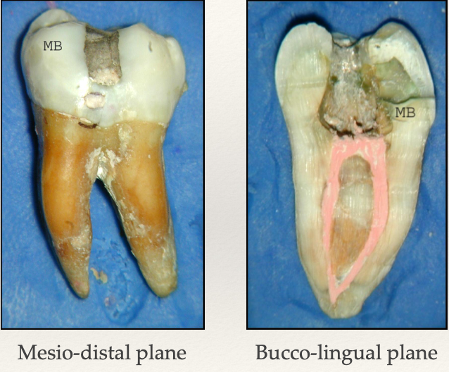
How many roots do mandibular molars have?
2 roots: mesial and distal
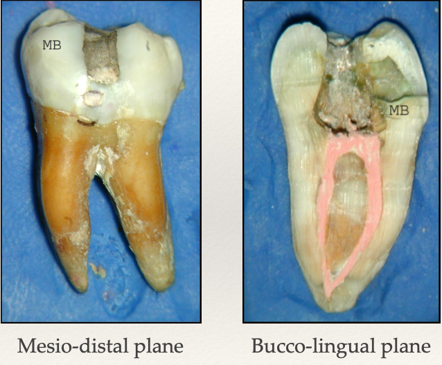
Which direction do mandibular molars tend to curve?
Distally and slightly lingual
64% of mandibular 1st molars have ______ canals?
3! The most common
What % of mandibular molars have their mesial root with 2 canals and 2 separate foramina?
60%
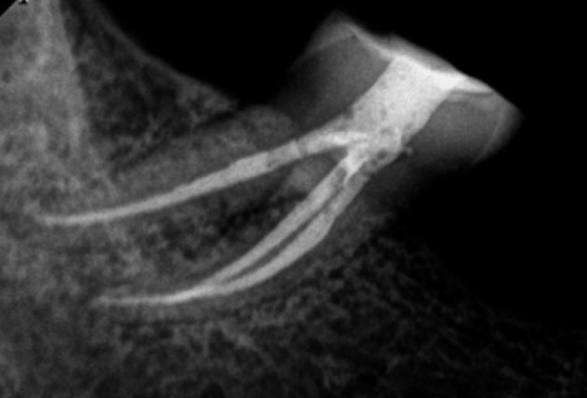
What % of mandibular molars have their mesial root with 2 canals and 1 foramina (canals join)?
40%
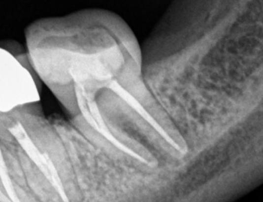
What % of mandibular molars have 1 canal for their distal root?
70%
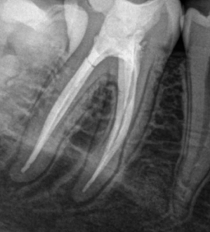
What % of mandibular molars have 2 canals, and 1 foramina for their distal root?
60%
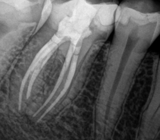
What % of mandibular molars have 2 canals, and 2 foramina for their distal root?
40%
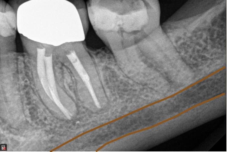
What is this anatomical image you should see in a mandibular molar radiograph?
Mandibular canal
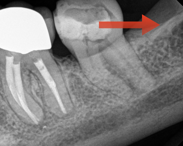
What is this anatomical image you should see in a mandibular molar radiograph?
External oblique ridge
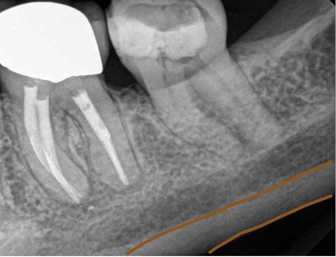
What is this anatomical image you should see in a mandibular molar radiograph?
Inferior border of the mandible
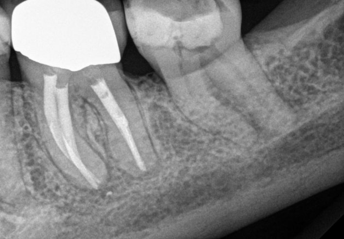
What is this anatomical image you should see in a mandibular molar radiograph?
Bone trabeculation
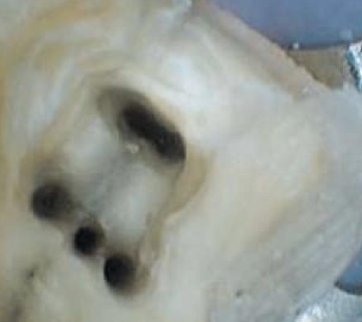
What are some variations you may see in the mesial canal?
3% of 1st molars may have a mid-mesial canal
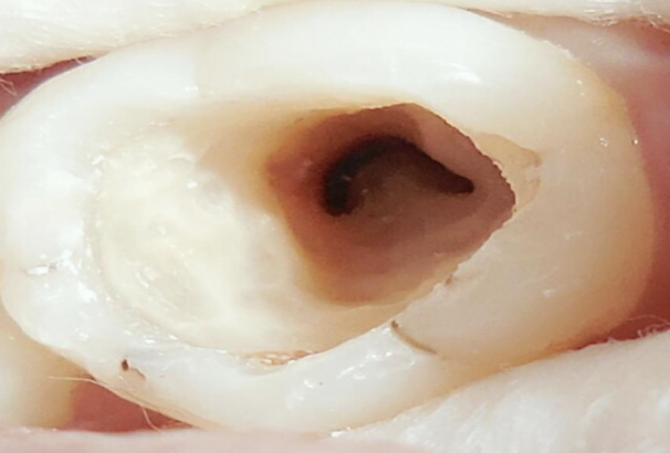
What % of mandibular molars have a c-shaped canal?
2.7-8% (usually 2nd molars)
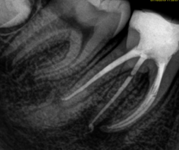
What is an anatomical variation where an extra, third root is present on a mandibular molar, typically the first molar?
Radix, seen in 15% of asian populations
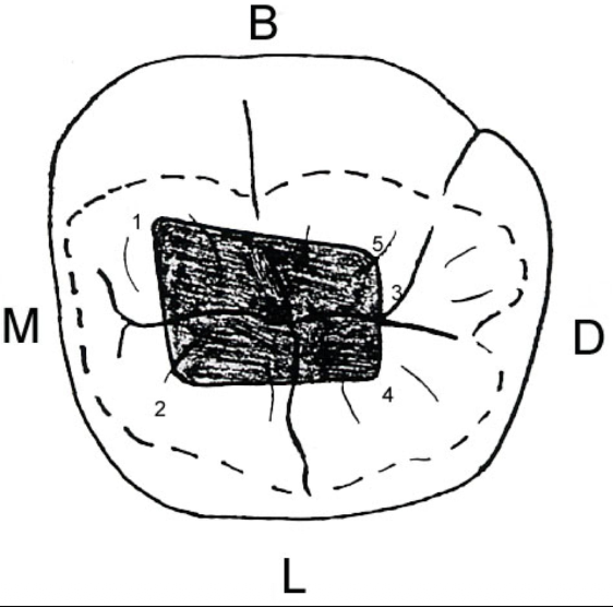
What should your access look like for a mandibular molar?
3/4 way between central groove and MB cusp tip
1.5mm lingual to the central groove on a line running parallel to mesial marginal ridge
2 mm distal to junction of the central groove and lingual groove
1.5mm lingual to #3
1.5mm buccal to #3
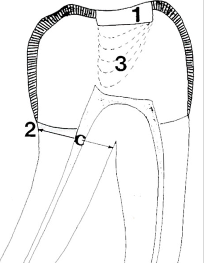
The first step to creating an access for a mandibular molar is to prepare the cavosurface outline and cut through enamel into dentin. How should you find the roots?
Use a perio probe to locate the edge of the mesial and distal roots
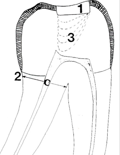
After locating the edge of the mesial and distal roots of a mandibular molar, where should you cut?
Towards the center of the chamber
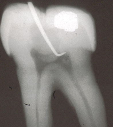
How should you locate and refine the pulp chamber of a mandibular molar when accessing the canal?
Use an endo-explorer to find the chamber
Use a G2 explorer to locate the roof of the chamber
Use an Endo-Z bur to unroof the chamber
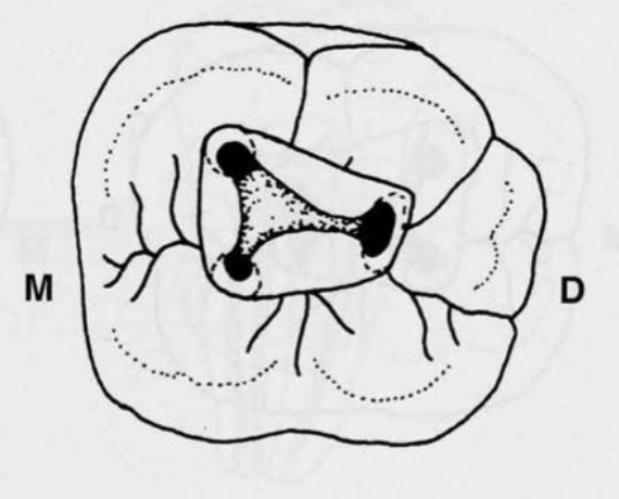
Mandibular molar access for 3 canals
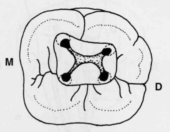
Mandibular molar access for 4 canals
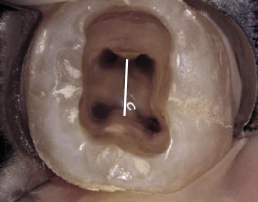
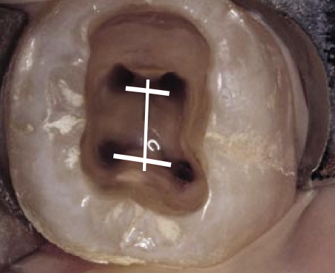
What is the 1st Law of Symmetry?
“Except for the maxillary molars, the orifices of the canals are equidistant from a line drawn in a mesiodistal direction through the pulp chamber”
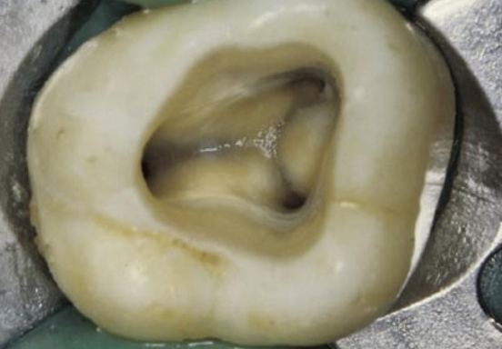
What is the 2nd Law of Symmetry?
“Except for maxillary molars, the orifices of the canals lie on a line perpendicular to a line drawn mesiodistally across the center of the floor”
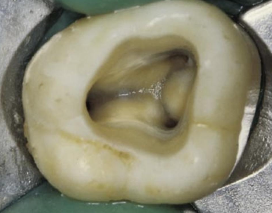
What is the law of color change?
“The color of the pulpal floor is always darker than the chamber walls
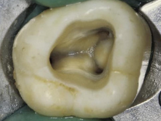
What is the Law of Orifice Location 1?
The orifi are always located at the junction of the pulpal floor and the wall
What are dentinal maps?
A dark line running between each orifice
Darkest part of the pulpal floor
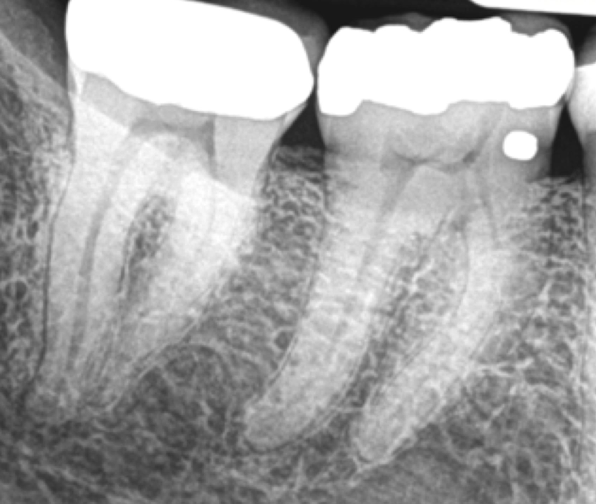
What are the steps in instrumentation of mandibular molars?
Coronal shaping
Working length
Glide path
Crown down
Final instrumentation
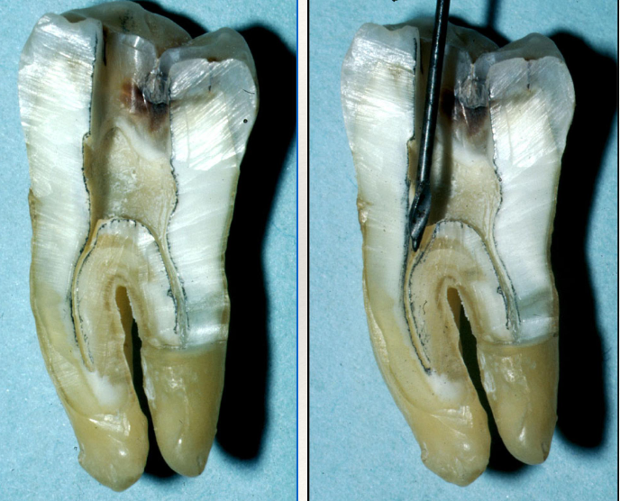
What is the benefit of using Gates Glidden, Hedstrom, or Orifice Openers for coronal shaping?
Provides straight-line access to canals, reduces risk of file breakage, and avoids the “danger zone”
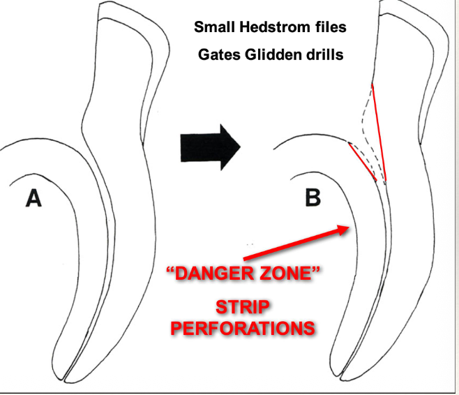
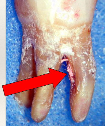
What is the “danger zone”?
Strip perforations- a severe breakdown of the dentinal wall caused by over-instrumentation or excessive shaping in curved or thin canals
When determining your working length for a mandibular molar, which file do you start with?
#10K file, then #15k file for WL film
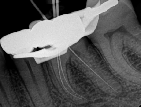
What tool can you utilize to separate the MB and ML canals?
21mm of a 25mm file, pick an easily replicable reference point and use the SLOB Rule to separate the MB and ML canals (or a combo of K-file and Hedstrom files)
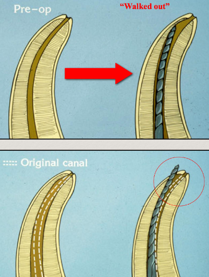
How do you establish a Glide Path?
Start with a #10K file
Instrumental 1mm beyond WL
Work file until loose
#15 file to 1mm past WL
Work file until loose
What can the SLOB Rule be used for?
To separate canals, length verification, identifying additional anatomy
What are the 3 roots of maxillary molars?
Mesio-buccal, disto-buccal, and palatal
What are the 4 roots of maxillary molars?
MB1, MB2, DB, P
Which direction do maxillary molars curve?
Roots tend to be either straight or curve slightly to distal
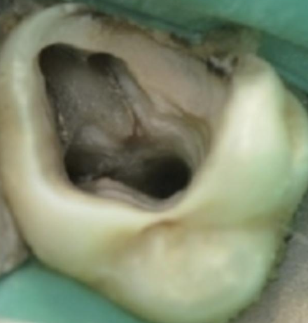
What does the mesiobuccal root look like?
Broad buccolingually, narrow mesiodistally
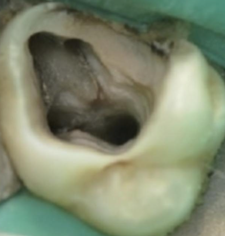
In what % of teeth is the MB2 canal present in maxillary molars?
96% of 1st molars, and 93% of 2nd molars
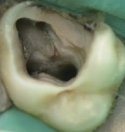
How far away is MB2 from MB2?
An average of 1.82mm
How many canals does the distobuccal root have?
One round canal that is usually the straightest. It can curve mesial or distal and the foramen tends to exit lateraly
How many canals does the palatal root have?
One canal that is usually the longest and largest. The foramen tends to exit buccally (85%)
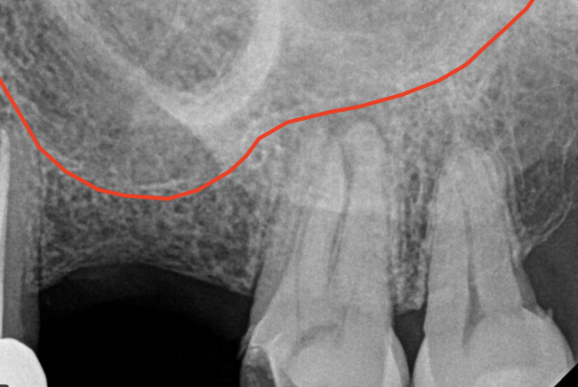
What is the closest root to the maxillary sinus?
MB root of the maxillary 2nd molar (0.83mm), can be superimposed with the apices. “projection effect” can occur and it can obscure EAL readings
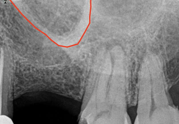
What can overlap with root apices?
Zygoma
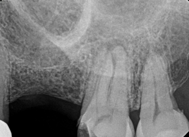
What can obscure PA lesions causing difficulty to see PDL and lamina dura?
Bony Trabeculation
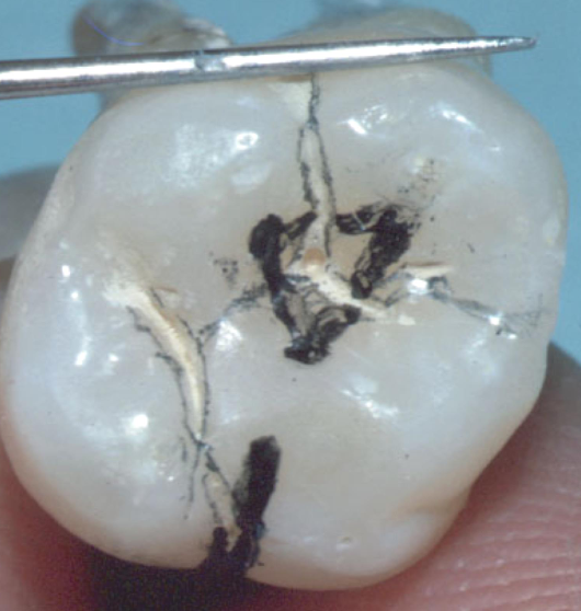
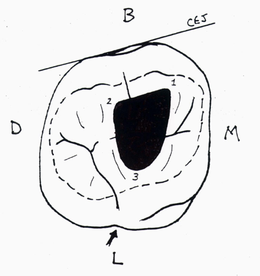
Maxillary molar access
The palatal root of a maxillary first molar commonly curves towards the
Buccal
Which root of a maxillary first molar should always be assumed to have 2 canals?
MB
What percent of mandibular first molars contain TWO canals in the distal root
30%
When removing the coronal canal curvature out of the pulp chamber (the mousehole) of the MB and ML canals of a mandibular molar using files and GG drills, the orifi are to be moved toward
Their respective line angles
Which canal in a mandibular molar should always be considered to have a fin or fins until proven otherwise?
D
Which of the following anatomic areas is considered the “danger zone”
Distal aspect of mesial root of mandibular molars
What anatomic structures may make it difficult to see the apices of a maxillar molar on a radiograph due to superimposition?
Zygoma
Floor of max sinus
Hard palate
Which root of a maxillary first molar should always be assumed to have 2 canals
MB