Introduction to Bacteriology (Bacteria, Shapes & Stains, Bacterial Culture, Special Growth Requirements)
 Call Kai
 Learn
Learn Practice Test
 Spaced Repetition
 Match
Match Flashcards
 Knowt Play
1/157

There's no tags or description
Looks like no tags are added yet.
Name | Mastery | Learn | Test | Matching | Spaced | Call with Kai | Chat |
|---|
No analytics yet
Send a link to your students to track their progress
158 Terms
Prokaryotes vs Eukaryotes
- Prokaryotes have no nucleus or membrane bound organelles
- Eukaryotes have a nucleus and membrane bound organelles
- Both can reproduce and respond to the environment

Which type of cells are eukaryotic?
- Plant and animal cells, fungi (modern forms of life)
Which type of cells are prokaryotic?
- Bacteria and Archaea (very old forms of life)
Bacteria
- Single-celled organisms that lack a nucleus; prokaryotes
What is a key component of bacteria? What is its function?
Its cell wall:
- Offers general support
- Maintains osmotic pressure
What component of bacteria is targeted by the immune system and antibiotics?
- Bacterial cell wall
Gram Stain
- A staining method that distinguishes between two different kinds of bacterial cell walls
- Classifies bacteria as either gram +/-
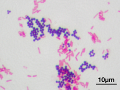
Gram Positive Bacteria
- Bacteria that have a thick peptido glycan cell wall, and no outer membrane. They stain very darkly (purple) in Gram stain.
Take up gram stain → purple

Gram Negative Bacteria
- Bacteria that have complex cell walls with less peptidoglycan but with lipopolysaccharides. Very toxic and hard to treat.
- Take up less of gram stain → pink

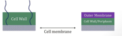
How do the cell walls of gram + and gram - bacteria differ?
- Gram +: Much thicker cell wall (takes up more gram stain); have molecules called Lipoteichoic acid
- Gram -: Much thinner cell wall (+ periplasm) as well as an outer membrane

Lipoteichoic Acid
What is unique to gram - bacteria?
- Outer membrane
What is the major structural component of bacterial cell walls?
- Peptidoglycan: Polymer sheets of sugars and peptides; sheets cross linked with other sheets

What are the sugars found in Peptidoglycan?
- N-acetylglucosamine (NAG)
- N-acetylmuramic acid (NAM)
NAM : NAG : NAM : NAG etc...

Are the peptides found in peptidoglycan attached to NAM or NAG?
- Attached to NAM → allows for cross linking
Layer upon layer of NAG : NAM : NAG (linked by peptides) → builds cell wall

Penicillin and Cephalosporin MOA
- Target peptidoglycan in bacterial cell walls → means they will only kill bacteria and not human cells
How many sheets of peptidoglycan are found in Gram +/- bacteria?
- (+): Up to 40 sheets (50%+ of cell wall)
- (-): Very few sheets (5-10% of cell wall)
Which bacteria do not gram stain well? Why?
- Mycoplasma: No cell wall → can't pick up gram stain
- Mycobacteria: Contains mycolic acid → does not gram stain well
- Chlamydia: Lacks muramic acid → can't gram stain
How is the cell wall of mycoplasma unique?
- Cell membrane has sterols for extra stability, makes up for lack of cell wall
What type of stain is used to visualize mycobacteria?
- Ziehl-Neelsen
Cell membrane is found where in bacteria?
- Below cell wall; is a phospholipid bilayer
- Present in Gram +/- bacteria
The cell membrane in bacteria is used for what?
- ETC and oxidative phosphorylation (occurs in cell membrane bc/ they lack mitochondria)
- Has enzymes and carrier molecules that are important for bringing stuff into cell
What is a unique feature of Gram + Bacteria?
- Molecules called lipoteichoic acid (LTA) are found on the surface
What is a major surface antigen that stimulates immune reaction?
- Lipoteichoic acid (LTA) in Gram + → induces cytokine release → binds antibodies and activates the complement cascade

What does LTA induce in animals?
- Arthritis
- Uveitis
- Meningeal inflammation
- Cascades for septic shock & multi-organ failure
What features are unique to gram - bacteria?
- Outer membrane
- Periplasmic space
Periplasm
- The gel-like material that fills the region between the cytoplasmic membrane and the outer membrane of Gram-negative bacteria.

What is located inside the periplasm of gram - bacteria?
- Many enzymes
- MOST IMPORTANT: β-Lactamase → used to inactivate antibiotics
Outer Membrane (Gram -)
- Unique to gram (-); contains lipopolysaccharide (LPS)

Lipopolysaccharide (LPS)
- Molecule found on surface of Gram (-) → MAJOR immune trigger → patients who have Gram (-) bacteremia get VERY sick
Do patients get more sick in the setting of Gram (+) or Gram (-) Bacteremia?
- Gram (-) d/t LPS
What are the components of Lipopolysaccharides?
- Polysaccharide
- Lipid A
- O Antigen
What is the role of lipid A in LPS?
- Highly toxic → component that makes ppl very sick in gram (-) bacteremia
- Triggers cytokine release
What is the role of O antigen in LPS?
- Target for antibodies
Overview of the differences between Gram +/- Bacteria
- (+): Cell wall and membrane; Lipoteichoic Acid (LTA)
- (-): Cell Wall/Periplasm + Outer membrane; Lipopolysaccharide (LPS)
Bacterial Capsules
- Sticky, gelatinous layer secreted by bacteria → helps it attach to host cells and protects against phagocytosis

What is the composition of bacterial capsules?
- Mostly water with some polysaccharides
How is Bacillus Anthracis (Anthrax) an exception to the normal composition of bacterial capsules?
- Its capsule is protein (in contrast to polysaccharides) made of d-glutamate → acts as a major virulence factor that allows unimpeded growth
What is the most important encapsulated bug?
- Strep Pneumonia
What are other examples of encapsulated bugs?
- H. influenza
- N. Meningitidis
- E. coli
- Salmonella
- Klebsiella
- Group B strep
Hungry nurses eat spicy Korean grub
How does the body protect itself from encapsulated bacteria?
- B cells secrete capsular antibodies (IgG) → binds capsules; phagocytes recognize Fc receptors on antibodies and swallow em up
Antibodies secreted from B cells that bind capsules are also useful for what?
- Can activate the complement cascade; formation of MAC → death
- Part of cascade (activated) is C3b, can tag pathogens for phagocytosis via opsonization
What happens if you lose antibodies/B-cells/complement?
- Causes recurrent encapsulated bacterial infections
Patients with Asplenia are at risk of what?
- ↑ Risk of sepsis from encapsulated bacteria d/t loss of splenic phagocytes
I.e removed spleen or sickle cell anemia
Which component of bacteria is a basis for many vaccines?
- The capsule; polysaccharides within capsule are often weakly immunogenic
Polysaccharides within capsule, which are targeted by vaccines, are often weakly immunogenic. What is done to the vaccine in order to elicit a larger immune response?
- The polysaccharides within vaccines are "conjugated" to a protein that DOES stimulate an immune response:
1. Diphtheria toxoid, tetanus toxoid, meningococcal outer membrane
Ex: Polysaccharide is weak. Add a toxoid to the polysaccharide within the vaccine → stronger immune response
What are some bacteria that are treated with conjugated vaccines?
- Neisseria meningitides
- Streptococcus pneumonia
- H. Influenza type b
Glycocalyx
- Sugar coat made of polysaccharides (similar to capsule) that is used to adhere to surfaces (i.e. S epidermis → biofilms on catheters)
Some bacteria have capsules, some have glycocalyx

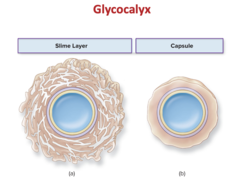
Glycocalyx vs Capsule
- Glycocalyx: Irregular, slimy fuzz layer used to adhere to surfaces
- Capsule: Distinct, firmly attached gelatinous layer
What is the role of the glycocalyx for S. Epidermis?
- Glycocalyx allows it to adhere to surfaces → able to form biofilms on catheters
If you have a central IV line in a patient made of a polymer, S. Epidermis can form biofilm → infection
Biofilm
- A microbial community that usually forms as a slimy layer on a surface
Pili and Fimbria
- Structurally similar to flagella, made of proteins
- Basically an arm/appendage for bacteria → allows them to adhere to surfaces (ordinary pili) // or to other bacteria for conjugation (sex pili)
What is the role of pili in E. coli?
- Allow them to attach to urinary treat → causes UTIs
(Ordinary pili)
What is the role of pili in Neiserria Gonorrhea?
- Pili exhibit a feature known as antigenic variation → bacteria can change the makeup of the proteins in their pili
What is the consequence of Neiserria Gonorrhea's pili exhibiting antigenic variation?
- If a pt has gonorrhea infection, they are at risk for subsequent gonorrhea infections bc/ the antibodies and immune system memory to the first infection does NOT apply to the second
Plasmids
- Small circular DNA molecules that replicate separately from the bacterial chromosome
- Can contain genes for antibiotic resistance, can synthesize toxins, and can be transferred from one bacteria to another

Flagellum
- Long protein arms used for bacteria mobility, pushes them from place → place

Why is it beneficial that bacteria ribosomes have a different structure compared to humans?
- Allows selective toxicity of antibiotics to bacteria, won't kill animal cells
Tetracyclines target what?
- Antibiotics that target the 30S subunit of bacterial ribosomes
Bacteria ribosomes: 50S + 30S
Aminoglycosides target what?
- Antibiotics that interfere with 30S protein synthesis
Bacteria ribosomes: 50S + 30S
What is the dormant state of bacteria?
- Some can enter a dormant state called a spore
Bacterial spore
- A dormant state for some bacteria with no metabolic activity
- Can survive a long period of starvation
- Resistant to dehydration, heat, chemicals
Basically like a seed, in the right condition, can "germinate" and become active

What are the components of a bacterial spore?
- Coat
- Cortex/core wall
- Diplicolinic acid

What is the role of the coat in the spore?
- Outermost layer made of keratin-line protein
- Impermeable to many chemicals + bacterial agents
What is the role of dipicolonic acid in the spore?
- Large amounts are found inside spores
- May help with heat resistance
What are the key spore forming bacteria?
- Bacillus Anthracis/Cereus
- Clostridium Perfringens/Tetani/Botulinum
How are bacteria typically identified?
- Their shape
- What color they stain
- Special tests
Common Bacterial Shapes
- Coccus: Spherical
- Bacilli: Rod shaped
- Coccobacillus: Oval
- Spirilli: Spiral-shaped

Cocci vs Diplococci vs Streptococci vs Staphylococci
- Cocci: 1 ball
- Diplococci: 2 balls
- Strep: Many balls in chain
- Staph: Bunches/clusters of balls

Streptococci vs Staphylococci
- Strep: Cocci in chain
- Staph: Bunches of cocci (grapes, yum!)

Are most cocci gram + or -?
- Most are gram (+)
- i.e strep and staph
Which gram - bugs are cocci?
- Neisseria (meningitides/gonorrhea)
- Moraxella Catarrhalis
Most cocci are gram (+)
Are most rods/bacilli gram + or -?
- Most are gram (-)
Which rods are gram +?
- Corynebacterium
- Clostridium
- Listeria
- Bacillus (anthrax)
Most rods are gram (-)
Branching/Filamentous bacteria: appearance and common ones
- Long/branching (resemble fungi)
- Actinomyces, Nocardia

Sphirochetes
- Spiral shaped bacteria

Which bacteria are spirochetes?
- Treponema (Syphilis)
- Borrelia (Lyme disease)
- Leptospira (leptospirosis)

Vibrio: appearance and common ones
- Comma shaped bacteria
- Vibrio cholerae

Pleomorphic Bacteria
- Can assume several shapes

What are key pleomorphic bacteria?
- Rickettsia
- Chlamydia
Why are Rickettsia and Chlamydia often grouped together?
- Two intracellular pathogens (obligate intracellular)
Simple Stains
- Utilized just to see bacteria under microscope (can see # of bacteria and their shapes)
- I.e. Methylene blue, safranin, crystal violet
Stains all bugs the same, nonspecific

What is the gram stain?
- Helps differentiate between gram +/- bacteria
- Purple → Gram +
- Pink → Gram -
Stain based on characteristics of their cell wallq

What are the steps of gram staining?
1. Fixation
2. Crystal violet
3. Iodine
4. Decolorization: Washes away purple from everything except Gram +
5. Safranin (Counter stain)
Gram (+) pick up crystal violet → purple
Gram (-) washes away crystal violet, stained by safranin (counter stain) → pink

Why do gram + bacteria pick up crystal violet?
- Because of their thick cell walls (peptidoglycan)
Why does Treponema (Syphilis) not gram stain well?
- Cell wall is too thin
Why do mycobacteria (tuberculosis) not gram stain well?
- Contains mycologic acid in cell wall (waxy, makes them impermeable to gram stain)
Acid fast stains used instead
Why do Mycoplasma not gram stain well?
- No cell wall (have cell membrane instead)
Why do Rickettsia/Chlamydia/Legionella not gram stain well?
- They are intracellular bacteria; grow inside other cells
Giemsa Stain
- Chlamydia, Borrelia, Rickettsia, Trypanosomes, Plasmodium
USED TO STAIN INTRACELLULAR BACTERIA

Ziehl-Neelsen Stain
- Use to stain Acid-fast bacteria (Nocardia, Mycobacterium)

Silver Stain
Special stain for 3 organisms:
- Pneumocystis Pneumonia (fungal infections seen in HIV/AIDS)
- Legionella (pnemonia, contaminates water → outbreaks in nursing homes)
- H Pylori (causes gastric ulcers)

India Ink
- Negative stain (makes the background dark, not the organism)
- Used in fungi/yeast (Cryptococcus neoformans)

Some bacteria can produce what as they grow?
- Pigments, can give colonies unique colors
Staph Aureus color
- Golden/yellow color
Aura = glowing = yellow
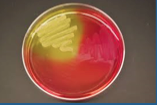
Pseudomonas Aeruginosa Color
- Blue-green
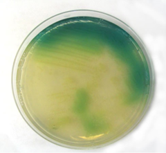
Serratia Color
- Red
Serratia = Sriracha = RED

Actinomyces
- Long filamentous bacteria that "cements" together
- Colonies have yellow/orange appearance

Where is Actinomyces commonly found?
- Normal oral flora but can form abscess infections in jaw

Agar Plate
- A petri dish filled with a jello-like nutrient material used to grow bacteria upon
Bacteria don't usually consume agar (from seaweed), can grow on top of it
