ENDO 1 - Main points
 Call Kai
 Learn
Learn Practice Test
 Spaced Repetition
 Match
Match Flashcards
 Knowt Play
1/43

There's no tags or description
Looks like no tags are added yet.
Name | Mastery | Learn | Test | Matching | Spaced | Call with Kai | Chat |
|---|
No analytics yet
Send a link to your students to track their progress
44 Terms
What are the consequences of short-term hyperglycemia and thier MOAs?
Symptoms of early diabetes (more common in T1DM → destruction of pancreas)
1) Polyuria (excess urination)
Hyperglycemia = large glomerular filtration load of glucose = saturation of SGLT transporters in the proximal tubule
SGLT2: Normally responsible for 90% of the reabsorption of glucose
SGLT1: Contributes more (up to 50%) when glucose load increases
1) Even with this extra help, the transporters cannot reabsorb all the filtered glucose → remains in tubular fluid + acts as an osmotic diuretic → retains water in the tubule = increased urine production
2) Polydipsia (increased thirst)
Polyuria leads to dehydration + low BP → thirst → increased drinking
Increased drinking is a compensatory response to volume loss from polyuria.
3) Weight loss + polyphagia (increased hunger)
Loss of insulin function (T1DM) → Inability to store glucose + increase glucose loss in urine = low energy stores + weight loss + poor glucose utilization (despite having high glucose concentrations in the blood)
Bad glucose utilization = increased hunger and polyphagia
Depletion of energy stores = weight loss
Explain the mechanism of early kidney nephropathy in patients with hyperglycemia. What medications help this effect?
Early: RAAS based
Late: Vasoconstriction and age/rage process
Saturation of SGLT transporters = increased reabsorption of glucose + Na + Cl (paracellularly) → decreased Na/Cl sensed by the macula densa = activation of tubuloglomerular feedback, leading to an INCREASE in GFR (increases filtration pressure) to “defend” perceived low GFR → renal damage in diabetes!
Hyperfiltration
Podocyte damage
Protein in urine
SGLT blockers indirectly help (because they allow more Na/Cl to travel to macula densa), inhibit the tubuloglomerular feedback, decrease glomerular filtration pressure, and decrease RAAS!
What is the blood glucose concentrations at which polyuria develops?
Typically quite high (above 250 mg/dl) in an otherwise healthy kidney
What is ketosis?
Ketosis (alternative fuel utilization)
With inadequate insulin function, the body utilizes fatty acids for the metabolism/production of ketone bodies = ketosis
What is AGE and RAGE? What are they caused by and what do they lead to?
Hyperglycemic cellular injury = consequence of chronic hyperglycemia
Advanced glycation end products (AGE)
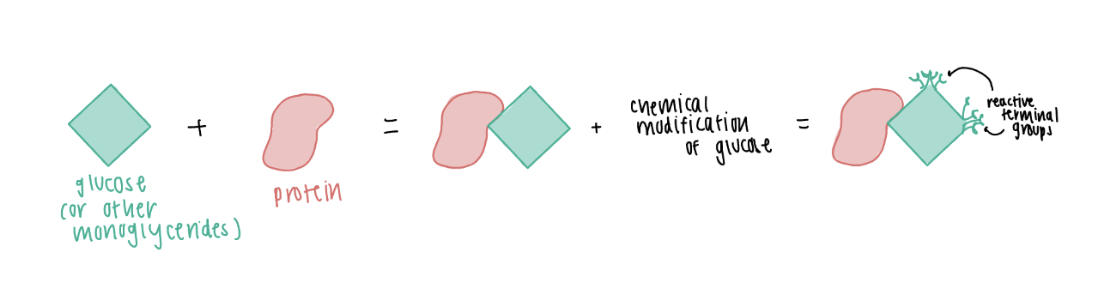
Produced through non-enzymatic attachment of glucose (or other monoglycerides) to proteins, followed by chemical modification of the glucose to form a reactive terminal group
Effect of the reactive terminal group:
1) Direct damage: Cross-linking proteins
Alteration in normal protein structure + function =
Damages/changes vascular proteins → endothelial cell damage + loss of capillaries
2) Activation of the receptor for advanced glycation end-products (RAGE)
RAGE - transmembrane protein is found on many cells (i.e., endothelial cells in blood vessels, immune cells, smooth muscle cells, kidney cells, neurons)
When bound to AGE leads to:
Increased production of ROS (oxidative damage)
Increased production of inflammatory mediators
Increased production of extracellular matrix (ECM) proteins = Basement membrane thickening (a preventative attempt to restrict bacterial infection)
Increased expression of RAGE
Decreased synthesis of nitric oxide (NO) + Increased synthesis of endothelin = vasoconstriction
Apoptosis (programmed cell death) of endothelial cells = loss of capillaries
Increased platelet and RBC aggregation microthrombosis
3) Oxidative damage
4) Inflammatory damage
What is an example of a glycated protein? What is it used for and what is the difference between this and an AGE?
Hemoglobin A1c is a glycated protein → it is not an AGE (does not undergo chemical modification)
Good representation of your glucose levels over 120 days (lifespan of RBCs)
Explain the mechanism of AGE/RAGE/long-term hyperglycemia’s effect in late-stage kidney nephropathy.
Early: RAAS based
Late: Vasoconstriction and age/rage process
RAGE activation leads to:
Decreased synthesis of nitric oxide (NO) + Increased synthesis of endothelin = vasoconstriction → hypoperfusion of glomeruli = glomerular ischemia → death
Increased production of extracellular matrix (ECM) proteins = Basement membrane thickening → prevent bacterium from coming in
Mesangial cell (tissue macrophages) increase → try to keep the bowman’s space clean but since they are part of the immune response = inflammatory response + damage to endothelial cells leading to…
Apoptosis of endothelial cells = loss of glomerular capillaries → decrease GFR
Explain the mechanism of how AGE/RAGE/long-term hyperglycemia affects nerves. What does it lead to?
Neuropathy (damage to the nerves outside your brain and spinal cord)
AGE/RAGE vascular damage → damage/blockage of vessels supplying nerves → inadequate blood supply → nerve infarcts → nerve damage → loss of sensation → limb injury → gangrene
Explain the mechanism of how AGE/RAGE/long-term hyperglycemia affects a patient’s eyes. What does it lead to?
Retinopathy
Damaged retinal vessels (microinfarctions) → bleeding
Vascular occlusion/ischemia → axonal swelling → cotton-wool spots
Leaky damaged capillaries → hard exudates
Ischemia → compensatory vascular proliferation/new vessel formation
Some parts of the eye are not receiving blood so the blood vessels that are still okay will proliferate
Can lead to blindness
Explain the mechanism of how AGE/RAGE/long-term hyperglycemia affects a patient’s vascular system (macrovascular disease). What conditions is this like? What medication does this process warrent?
AGE-RAGE → endothelial damage → lipid deposition in the subendothelium
Diabetes-associated dyslipidemia → inflammatory system activation → atherosclerosis → CAD/PVD
Same MOA as HTN and dyslipidemia but different origin
Explains why if patient is diabetic and has NORMAL LDL levels they still need a statin
Explain the mechanism of how AGE/RAGE/long-term hyperglycemia affects a patient’s liver. What conditions is this like? What medication does this process warrent?
Dyslipidemia
Non-alcoholic faatty liver disease:
Decreased/loss of insulin function =
Increased lipolysis
Increased fuel delivery to liver (too much fuels in liver)
Increased triglyceride synthesis
Fatty acid oxidation
Increased fat storage in hepatocytes = fat accumulation in liver (contributing to Non-alcoholic fatty liver disease)
Where is insulin released from? Where does it bind? What/where are its effects? What stimulates it?
Insulin
Released from beta cells (hepatocytes) → binds to insulin receptors on cell surfaces = conformational change → phosphorylation → intracellular messenger/signaling pathways = metabolic effect
Anabolic (build/store) effect:
Pancreas: Inhibits glucagon release
Hepatocytes: activates pathways of fuel storage (glycogen synthesis + lipogenesis), inhibits pathways of fuel mobilization (gluconeogenesis)
Glycogen synthesis:
Decrease glucose levels in liver = increase glucose transport into liver (GLUT2) = decreased glucose levels in bloodstream
Skeletal muscle: ↑ glucose uptake through GLUT4, ↑ glycogen synthesis, ↑ and protein synthesis
Adipocytes: ↑ glucose uptake through GLUT4, ↑ triglyceride synthesis, ↓ and triglyceride breakdown/lipolysis
Stimulus: rising BG, fed state (0-4 hours after a meal) → helps to decrease BG towards normal
Where is glucagon released from? Where does it bind? What/where are its effects? What stimulates it?
Released from alpha cells → binds to GPCR on cell surfaces
Catabolic effect (breakdown):
Hepatocytes:
Glycogenolysis:
Conversion of glycogen to glucose = increased glucose in liver = glucose travels down gradient (in GLUT2) into blood stream
Gluconeogenesis
Stimulus: Falling BG, fasting state (more than 4 hours after meal) → helps to increase BG towards normal
What is the difference between fed and fasting states in terms of the main fuels source and over goal?
Fed (0-4 hours after meal)
Increased insulin
Main fuel source = exogenous fuel from meal (glucose)
Skeletal muscle: glucose used for ATP, excess glucose stored as glycogen
Liver: glucose used for ATP, excess glucose stored as glycogen
Adipocytes: glucose used for ATP, excess glucose and fatty acids stored as triglycerides
Goal: Use and store fuel
Fasting (>4 hours after meal)
Increased glucagon and counter-regulatory hormones (I.e., epinephrine, cortisol) → mobilize fuel
Main fuel source = stored endogenous fuel (shift towards fatty acids)
Stored fuels (glycogen, TG) broken down, glucose and fatty acids released from other tissues → most cells use fatty acids for energy
Skeletal:
Glycogen broken down, glucose used for ATP
Fatty acids taken up for ATP as well
Liver:
Glycogen broken down, glucose released
Fatty acids take up from bloodstream for ATP
Adipocytes - TG broken down, fatty acids used for ATP and released
Goal: Mobilize fuel and maintain BG
What is the main purpose of the TCA cycle?
To take acetyl coA derived from fuels → use its energy through byproducts (NADH, and FADH2) which are used in oxidative phosphorylation (where most ATP is produced)
Major input (acetyl-CoA) → Major outputs (3 NADH, 1 FADH2, 1 GTP, 2 CO2)
What are the irreversible steps within the TCA cycle? What are the rate limiting aspects of this cycle?
Irreversible steps:
1) Citrate synthesis (acetyl coA → citrate)
2) Isocitrate dehydrogenase (isocitrate → alpha-ketoglutarate)
3) alpha-ketoglutarate dehydrogenase (alpha-ketoglutarate → succinyl coA)
Thiamine pyrophosphate (vit B1 required)
Rate limiting aspects:
Substrate availability → cycle needs substrates like acetyl coA
Product inhibition → accumulation of products slows pathway
Regulated steps slow when ATP and NADH are elevated
Explain the ATP and electron transport coupling chain
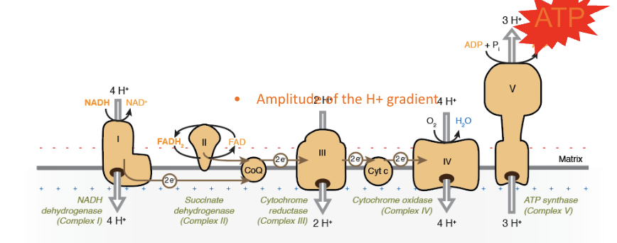
Connected through a proton gradient across the inner mitochondrial membrane
1) NADH → Complex I and FADH₂ → Complex II donate electrons.
2) Electrons travel:
Complex I/II → CoQ → Complex III → cytochrome c → Complex IV
Complexes I, III, and IV pump H⁺ from the mitochondrial matrix into the intermembrane space.
Complex II does NOT pump H⁺.
3) At Complex IV, O₂ is the final electron acceptor and combines with electrons + H⁺ → H₂O.
4) H⁺ accumulation in the intermembrane space creates an electrochemical gradient = potential energy.
5) H⁺ flows back into the matrix through ATP synthase (Complex V) → energy drives ADP + Pi → ATP.
Complex I = 4 H⁺
Complex III = 4 H⁺
Complex IV = 2 H⁺
Compare uncouplers of oxidative phosphorylation vs direct electron transport chain inhibitors vs ATP synthase inhibitors.
Uncouplers of oxidative phosphorylation
Allows H to cross the inner mitochondrial membrane without going through ATP synthase (out)
Electron transport continues but ATP synthesis decreases/stops →
ETC can continue running rapidly in an attempt to restore ATP synthesis = released as heat + speeds up oxygen consumption
Direct electron transport chain inhibitors
Direct electron transport chain inhibitors: prevent ETC from generating proton gradient
Stops whole process whereas the uncouplers allow ETC to continue but stop ATP synthesis
ATP synthase inhibitors
ATP synthase blocked → H cannot return through ATP synthase → ATP synthesis stops = build up of H gradient to the point where ETC eventually stops (loss of both ATP synthesis and ETC)
What happens during fatty acid oxidation, where does it happen, and how is it regulated?
TG → Fatty acids→ beta-oxidation (in mitochondria) → acetyl coA and NADH and FADH2
Acetyl-CoA → TCA cycle → ATP
NADH and FADH2 feed into the oxidative phosphorylation to generate ATP
Long chain fatty acids (chunky)
Long-chain fatty acid → carnitine shuttle → mitochondrial matrix → β-oxidation → acetyl-CoA + NADH + FADH2
Regulation of Fatty acid oxidation
Fed/high insulin → ↓ mitochondrial FA entry → ↓ β-oxidation
Fasting/low insulin → ↑ FA availability/entry → ↑ β-oxidation
What is glycolysis, where does it occur, what are the irreversible steps, and how is it regulated?
Glucose → pyruvate + ATP + NADH
Pyruvate: Can be converted into acetyl coA by pyruvate dehydrogenase (PDH)
Occurs in cytosol (does not require mitochondria) → all cells can perform glycolysis, including RBCs, which do not have mitochondria.
3 irreversible steps:
1) Hexokinase/glucokinase (HK/GK)
Glucose → glucose-6-phosphate
2) Phosphofructokinase (PFK)
Major regulated step within glycolysis
3) Pyruvate kinase (PK)
Final irreversible step producing pyruvate
Regulation:
High ATP → inhibits PFK and PK → ↓ glycolysis
Fed/high Insulin → ↑ glucokinase (GK) and PFK activity → ↑ glycolysis
How do glycolysis/PDH and β-oxidation change between fed and fasting states?
FED = burn glucose
↑ Insulin
↑ Glycolysis
↑ pyruvate dehydrogenase (PDH) activity
↓ β-oxidation
Glucose → pyruvate → acetyl-CoA → TCA
FASTING = burn fat
↓ Insulin
↓ Glycolysis/PDH
↑ β-oxidation
FA → acetyl-CoA + NADH + FADH₂ → ATP
What are the major fuel sources for ATP, and what fuels do RBCs and the brain use?
Glucose
Glucose → glycolysis → pyruvate → acetyl-CoA → TCA → NADH/FADH₂ → OXPHOS → ATP
Fatty acids
FA → β-oxidation (mitochondria)→ acetyl-CoA + NADH/FADH₂ → TCA/OXPHOS → ATP
Ketone bodies
Ketones → acetyl-CoA → TCA/OXPHOS (mitochondria)→ ATP
RBCs = glucose ONLY
No mitochondria
Cannot do TCA, β-oxidation, or OXPHOS
ATP comes from glycolysis only
Brain
Normal/fed: primarily glucose
Does not significantly use fatty acids
Prolonged fasting: increasingly uses ketone bodies to spare glucose
How does the body prevent hypoglycemia during fasting?
↓ Blood glucose → ↓ insulin + ↑ counterregulatory hormones
(glucagon, catecholamines, cortisol, GH)Response:
1) Increase glucose availability
Glycogenolysis: liver glycogen → glucose, especially early fasting
Gluconeogenesis: makes new glucose, increasingly important with prolonged fasting
2) Decrease the body's need for glucose (use of other fuels)
↓ Insulin + ↑ catecholamines → activation of hormone-sensitvie lipase (HSL) = lipolysis + ketogenesis
Adipose TG → free fatty acids + glycerol
Free fatty acids → β-oxidation → acetyl CoA
Glycerol → liver → gluconeogenesis
Ketogenesis → alternative fuel that uses acetyl CoA, especially for the brain during prolonged fasting
Note: Lipolysis can happen without ketogenesis, while ketogenesis cannot happen without lipolysis.
Explain the mechanism of action for insulin release from beta cells.
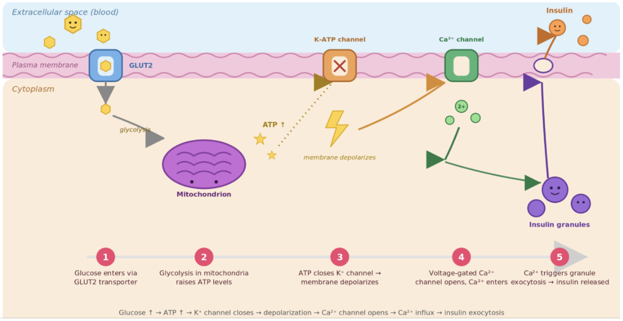
Increased blood glucose → enters through GLUT2 → glucose undergoes glycolysis = increased ATP → ATP-sensitive K channels close = decreased K leaving the cell → increased membrane depolarization
Because of the depolarization voltage gated Ca2+ channels open → Ca influx into the Beta cells → triggers insulin-containing vesicles to undergo exocytosis → insulin enters the blood
Explain the mechanism of how glucagon is released from alpha cells.
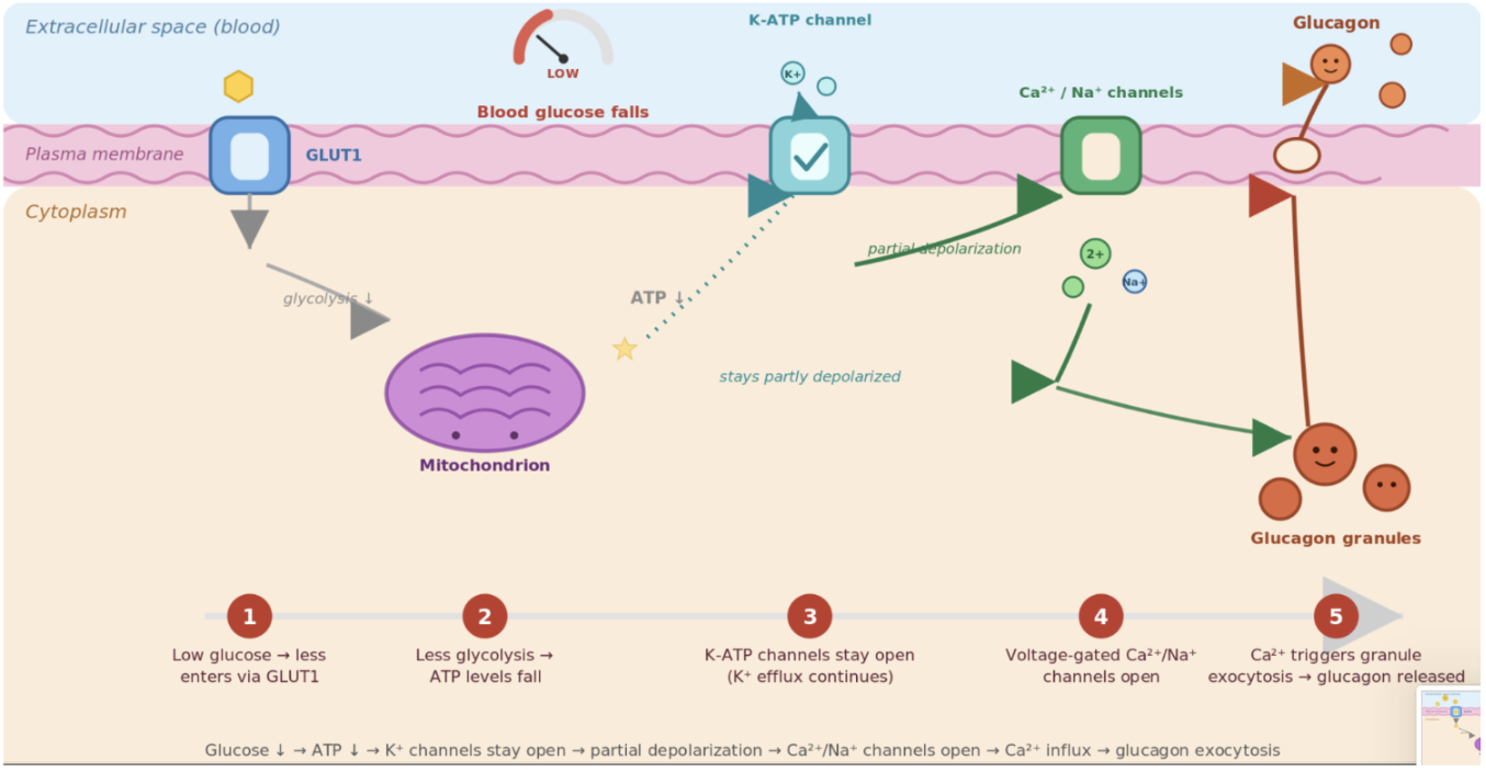
Low glucose → less enters alpha cells thorugh GLUT1 → less glucolysis → ATP levels fall → ATP-sensitive K channels stay open (K efflux continues) → decreased membrane depolarization
Ca/Na channels open → Ca influx into the Alpha cells → triggers glucagon-containing vesicles to undergo exocytosis → glucagon enters the blood
What is gluconeogenesis? What are the major carbon sources for gluconeogenesis?
Gluconeogenesis (GNG) = synthesis of glucose from non-carbohydrate precursors
1) Lactate → pyruvate → glucose
2) Glycerol → gluconeogenic intermediates → glucose
3) Glucogenic amino acids (especially alanine) → pyruvate/TCA intermediates → glucose
Protein is a major source of carbon for gluconeogenesis during fasting
Explain the energy requirements for gluconeogenesis and where it comes from. What are the processes that regulate GNG?
GNG is energy-expensive and requires ATP, GTP, and NADH.
β-oxidation of fatty acids is critical for providing the energy needed to drive gluconeogenesis.
TG → Fatty acids and glycerol → fatty acids go through beta-oxidation (in mitochondria) to produce Acetyl-CoA and NADH and FADH2
Acetyl-CoA → TCA cycle → ATP
lactate/glycerol/amino acids → gluconeogenesis → glucose
Regulation
1) Fasting/counterregulatory hormones → ↑ GNG
Glucagon + catecholamines + cortisol → activate key GNG enzymes
2) Fed state/insulin → ↓ GNG
3) ↑ β-oxidation → ↑ acetyl-CoA + energy
Acetyl-CoA activates pyruvate carboxylase, while β-oxidation supplies the large amount of energy required for GNG.
What is the difference between glycogenesis and glycogenolysis? What are their effects in the liver and muscle?
Glycogenesis = glucose storage as glycogen
Fed → insulin stimulates
Regulation in liver vs muscle
Liver: increased blood glucose → hepatocyte through GLUT2→ glucose-6-Phosphate → glucose-1-Phosphate → glycogen
Muscle: increased Insulin → GLUT4 moves to membrane → ↑ glucose uptake from blood → glucose → glucose-6-phosphate (enzyme hexokinase) → glycogen utilization and storage
Glycogenolysis = glycogen breakdown
Fasting/exercise → counterregulatory hormones/glucagon stimulate
Regulation in liver vs muscle
Liver: releasing glucose into the bloodstream
Glycogen → G1P → G6P —(glucose-6-phosphatase)→ glucose
Muscle: Provides glucose-6-phosphate for glycolysis
G6P → F6P → glycolysis → ATP for muscle contraction (personal reserve for that muscle)
Explain the transportation of glucose through different organs (Brain, RBCs, skeletal muscles, adipose, liver, kidney). What are they each independantly dependant on?
GLUT transporters: facilitated diffusion → down concentration gradients
Brain - GLUT3, RBCs - GLUT1
Glucose uptake is not insulin-dependent
Skeletal muscle and adipose: GLUT4
Insulin dependent
After a meal: ↑ Blood glucose → ↑ insulin → ↑ GLUT4-mediated glucose uptake into muscle/adipose
During fasting: ↓ insulin → ↓ GLUT4-mediated glucose uptake
Exercise → ↑ GLUT4-mediated glucose uptake independently of insulin
Liver: GLUT2
Not insulin dependent
GLUT2 is bidirectional
Fed: Blood glucose → GLUT2 → hepatocyte
Fasting state: Glycogenolysis + gluconeogenesis → G6P → glucose
Kidney/intestines: SGLTs
sodium-glucose cotransporters (SGLTs) → uses the Na⁺ concentration gradient to move glucose into cells
What happens to glucose when it enters most cells? Why does this happen
Once glucose enters most cells, it is phosphorylated:
Glucose → glucose-6-phosphate (G6P)
This helps maintain a low concentration of free intracellular glucose, favoring continued glucose movement from the blood into the cell.
What does a CGM measure? Which patients could benefit from use of a CGM?
CGM
Measures glucose in interstitial fluid (may have delay in readings/not accurate readings under 100mg/dL)
Use fingerstick when CGM reading doesn't match symptoms or is unreliable
Patients using insulin, T1DM, T2DM using insulin, frequent hypoglycemia, pregnant individuals
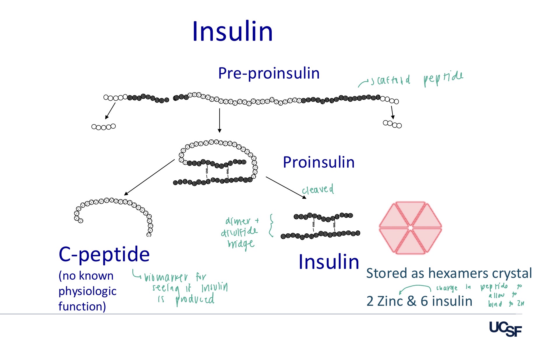
How is insulin made in pancreatic beta cells? How is it stored?
Preproinsulin (scaffold peptide) → Proinsulin (A chain, B chain, and C-peptides) is cleaved → Insulin + C-peptide (biomarker of endogenous insulin production)
A chain and B chains are held together by disulfides
Insulin is stored as hexamer crystals
Composed of 2 zinc and 6 insulin
This allows β cells to store insulin until a stimulus, primarily increased blood glucose, triggers secretion.
How is endogenous vs exogenous insulin cleared?
Endogenous:
Liver: ~60%
Kidney: ~40%
Exogenous:
Kidney: ~60%
Liver: ~40%
What happens if insulin levels are too high?
Hypoglycemia
→ palpitations, tachycardia, tremor, hunger
→ severe: confusion, weakness, seizure, coma if untreated
Hypoglycemic unawareness
Higher risk after frequent hypoglycemia
Confusion
Weakness
Seizures
Coma
What happens if insulin levels are too low?
Liver cells turn fatty acids into ketones
Ketones: alternative fuel source (bs no glucose in cell for energy)
Need insulin to uptake glucose into the cell
Ketones are acidic and can drop the pH of the blood
If left uncorrected, high ketone levels can lead to diabetic ketoacidosis (DKA), → life threatening condition
Other than low/high insulin, what are other AE’s that can ocur with use of insulin?
Immunopathology
Insulin allergy (rare with human insulin)
Immune insulin resistance (common)
Limited inactivation of insulin by IgG antibodies
Lipodystrophy/hypertrophy at the injection site
Dystrophy with older animal insulin
Hypertrophy with human (change sites/liposuction)
What is Exogenous glucagon used for? What does it depend on? What routes of administration exists for it?
Hypoglycemic emergencies
Particularly when the patient is unconscious/altered and cannot safely take oral glucose, and IV glucose is not possible or IV access is difficult.
Depends on the patient having hepatic glycogen stores
Glucagon → ↓ hepatic glycogen → ↓ glucose available for release → reduced glucagon response
Routes and formulations
SQ, IM injection, prefilled pen, Nasal spray (Baqsimi)
How does insulin aggregation affect its absorption and duration? What can aggregation be altered by?
Insulin can aggregate: Monomers → dimers → hexamers (2 Zn + 6 insulin)
More aggregation = more hexamers
Slower dissociation
Slower SC absoprtion
LONGER duration
Less aggregation = less hexamers
Faster absorption
Faster onset
Altering aggregation:
Low pH → more aggregation
Ions (Zinc) → more aggregation
How are lispro, aspart, and glulisine modified to produce rapid action?
Lispro
Switches B28 Pro + B29 Lys → B28 Lys + B29 Pro
Aspart
B28 Pro → Asp
Glulisine
B3 Asn → Lys
B29 Lys → Glu
B chain on insulin (B28 - Pro, B29 - Lys, and B30 - Thr) → help stabilize the overall dimer and hexamer structure
These changes affect dimer/hexamer stability → less aggregation but do not influence insulin binding to receptors
Compare and contrast T1DM and T2DM with regard to: the typical age of onset, body weight, the underlying pathophysiology of the hyperglycemia, predisposition to ketoacidosis, response to therapy: diet, exercise, insulin, and non-insulin glycemic agents.
T1
Age of onset: Child/adolescence + any age
Body weight: Lean/normal weight
Underlying pathophysiology: Autoimmune destruction of pancreatic beta cells
Ketoacidosis: High predisposition
Response to therapy:
Diet/Exercise: Can be helpful for glucose management but cannot replace insulin
Insulin: Required for survival
Non-insulin glycemic agents: Not primary treatment → fundamental problem is lack of insulin
T2
Age of onset: Adulthood + sometimes younger patients
Body weight: Overweight/obesity
Underlying pathophysiology: Insulin resistance + progressive β-cell dysfunction → relative insulin deficiency
Ketoacidosis: Much lower predisposition, through DKA can occur still
Response to therapy:
Diet/Exercise: Very important; lifestyle modification can improve insulin sensitivity and glucose control
Insulin: may eventually be needed, especially as beta cell function declines
Non-insulin glycemic agents: Major component of treatment
Including medications that improve insulin sensitivity, decrease hepatic glucose production, increase insulin secretion, increase urinary glucose excretion, etc.
How do you calculate a patient estimated average glucose? (eAG) Why is this used?
eAG (mg/dL) ≈ 28.7 × A1C − 46.7
EX: A1C = 7% = eAG of 154mg/dL
ADAG trial (A1C-Derived Average Glucose study) demonstrated a strong relationship between A1C and a patient's average glucose.
What are the ambulatory glucose profile (AGP)/CGM goals for most adults with diabetes?
Time below range (TBR) = Hypoglycemia
<70 mg/dL should be <4%
<54 mg/dL should be <1%
Time in range (TIR) = 70-180mg/dL >70% of the time
Time above range (TAR) = Hyperglycemia
>180 mg/dL: <25%
>250 mg/dL: <5%
What are the general ADA glycemic goals for many nonpregnant adults with diabetes?
A1C → <7%
Preprandial (pre-meal) glucose → 80–130 mg/dL
Postprandial glucose → <180 mg/dL
CGM Time in Range → >70%
Time Below Range <70 → <4%
Time Very Low <54 → <1%
What is diabetic ketoacidosis (DKA)?
DKA: profound insulin deficiency (associated with T1DM)→ hyperglycemia + ketone production + metabolic acidosis