ANAPHY SKELETAL SYSTEM PART 2
 Call Kai
 Learn
Learn Practice Test
 Spaced Repetition
 Match
Match Flashcards
 Knowt Play
1/122

Earn XP
Description and Tags
comprises The Appendicular Skeleton in Detail, They types of Joints ETC
Name | Mastery | Learn | Test | Matching | Spaced | Call with Kai | Chat |
|---|
No analytics yet
Send a link to your students to track their progress
123 Terms
Appendicular Skeleton
It is composed of 126 bones of the Appendages (Limbs)
Bone Structures of the Appendicular Skeleton
Pectoral Girdle, Upper Limbs, Pelvic Girdle, Thigh, Legs
Pectoral Girdle
-Connects the upper limbs to the axial skeleton
-Consists of 4 main bones, the 2 scapulae and the 2 clavicles
Clavicle
Also known as the collarbone, it is a long, S-Shaped Bone that lies horizontally across the front of the shoulder just in front of the first rib.
Its function is to act as a brace to hold the arm away from the top of the thorax and help prevent shoulder dislocation
Structurally has three main Anatomical Parts:
Medial (sternal) end
Lateral (Acrominal) end
Shaft
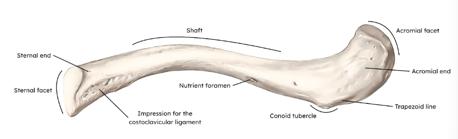
Sternal End of the Clavicle
Attaches to the Manubrium of the Sternum, forming the sternoclavicular joint
Acrominal End of the Clavicle
articulates with the Acromion of the Scapula, forming the acroclavicular joint
Shaft of the Clavicle
The prominent and visible part of the Clavicle—commonly known to be prone to be fractured.
Scapula
Also known as the Shoulder blade, it is a flat, triangular bone located on the posterior side of the shoulder surrouned by muscles. This allows it to move freely across the back of the Thoracic Cage.
Has:
Three borders (Superior, Medial, and Lateral) and;
Three Angles (Superior, inferior, and lateral)
Glenoid Cavity
Contained in the Lateral Angle of the Scapula, this is a shallow socket that articulates with the head of the humerus to form the glenohumeral joint
Supraglenoid and Infaglenoid tubercles
This is located above and below the Glenoid Cavity seving as attachment points for arm muscles.
Acromion Process
This is the enlarged lateral end of the spine of the scapula. It connects with the clavicle laterally at the acromioclavicular joint
Coracoid Process
This is a beaklike process pointing laterally over the top of he shoulder and anchors some of the muscles of the arm.
Contains the Supra-scapular Notch—a nerve passageway
Bones of the Upper Limbs
These are the 30 separate bones that form the skeletal framkework of each upper arm including the:
Arm
Forearm and;
Hand
Together, these bones create the framework for movement at the shoulder, elbow, and wrist, while also providing attachment sites for muscles that control the hand and arm
Humerus
This bone forms the entire arm. This is also the longest and largest bone of the upper limb.
Head of the Humerus
is the large, rounded head that faces upward, medially, and slightly backward—articulating with the glenoid cavity of the scapula to form the shoulder joint
Anatomical Neck of the Humerus Head
The immediately inferior section of the Humerus head which separates the head of the humerus from the rest of the bone as well as provides attatchment for the joint capsule.
The Greater Tubercle of the Humerus Head
This is the tubercle of the humerus head that is located laterally and has areas for muscle attachment
The Lesser Tubercle of the Humerus Head
This is the tubercle located on the anterior side of the humerus head, this also serves as a muscle attachment site.
Intertubercular Groove
known as the bicipital groove, this is located in between the greater and lesser tubercle that allows passage of the tendon of the biceps brachii muscle
Surgical Neck of the Humerus
This is located below the two tubercles—connecting the upper end of the humerus to its shaft. it is also a common site of fractures
Shaft of the Humerus
Is the long part of the humerus. It is cylindrical in its proximal section and becomes flatter towards the distal section
Deltoid Tuberosity
Along the lateral side of the shaft, this is a triangular and roughened area where the deltoid muscle attaches to.
Trochlea
Located in the distomedial end of the Humerus, it is a spool-like part that articulates with the ulna.
Capitulum
Located in the distolateral end of the humers, it is a ball-like part that articulates with the radius
Olecranon Fossa
On the posterior surface, at the distal section of the humerus, it is a deep, triangular depression that receives the olecranon of the ulna when the elbow is extended
Coronoid Fossa
Located on the anterior surface, at the mediodistal section of the humerus, it is a small depression that receives the coronoid process of the Ulna during flexion
Radial Fossa
Located on the anterior surface, at the laterodistal section of the humerus, it is a small depression that receives the Radial styloid process during flexion
Medial Epicondyle
Located at the mediodistal end of the humerus, this serves as the attachment site for forearm flexor muscles. It is also relatively bigger in size than its counterpart
Lateral Epicondyle
Located at the Laterodistal end of the humerus, it serves as the attachment site for forearm extensor muscles. it is also relatively smaller in size than its counterpart.
Ulna
The medial bone of the forearm
Proximal end of the Ulna
This is the thinck, hooklike, top part of the ulna that forms the elbow joint with the humerus
Olecranon
Located at the proximal end of the ulna, this is the large, prominent curved process that acts as the lever for the elbow joint—forming the visible tip of the elbow.
Trochlear Notch
This is the large, C-shaped depression created by the olecranon and the coronoid process. It is responsible for gripping and articulating the trochlea of the humerus.
Coronoid Process
This is the protruding process that forms the lower part of the trochlear notch. This is is what tucks into the coronoid fossa of the humerus during flexion.
Ulnar Tuberosity
Located right below the coronoid process, this projection is a rough area for muscle attachment such as the brachialis
Radial Notch
Located on the lateral side of the ulna, this projection is where the head of the radius articulates to form the proximal radioulnar joint
Shaft of the Ulna
The long portion of the ulna that gradually becomes smaller towards its distal end.
Interosseous Border of the Ulna
This is located at the shaft of the ulna where the interosseous membrane attaches and connects to the ulna to the radius
Styloid Process of the Ulna
It is a small, bony projection that anchors the crucial ligaments and cartilage needed to keep the pinky-side of the wrist joint stable.
Head of the Ulna
It is the rounded bump found at the distal end of the Ulna acting as a stationary pivot point that the radius sweeps around to follow forearm rotation
Styloid Process of the Ulna
This is the small, pointed peg-like projection on the distal end of the ulna. It is the visible bump on the pinky-side of the wrist. This anchors the ligaments and cartilage needed to keep the lateral side of the wrist joint stable
Radius
It is the lateral bone of the forearm, located on the thumb side. This bone has 3 surfaces and 3 anglles—allowing it to rotate around the ulna during forearm movements
Head of the Radius
This is a disc shaped head with a shallow depression on top, which articulates with the capitulum of the humerus, its outer surface is smooth and articulates with the radial notch of the ulna, forming the proximal radioulnar joint
Neck of the Radius
Separates the head of the radius to the shaft and the distal end of the radius
Radial Tuberosity
A rough oval-shaped projection that serves as a muscle attachment point located just bellow the neck of the radius
Shaft of the Radius
This is the long slightly curved section of the radius. It becomes wider towards its distal end
Interosseous border of the Radius
Located in the radial shaft, it provides attachment for the interosseous membrane that connects the radius to the ulna
Distal end of the Radius
It is the wide, large bottom portion of the radius on the thumb-side of the forearm that forms the primary connection point for your wrist joint
Ulnar Notch
Located on the medial side of the distal end of the radius, it articulates with the head of the ulna to form the distal radioulnar joint
Styloid process of the Radius
Located on the lateral side of the distal end of the radius, this is a pointed projection that provides attachments for ligaments supporting the wrist
Bones of the Hand
Consists of 8 carpal bones, 5 metacarpal bones, and 14 phalanges
Carpal Bones
These 8 bones make up the wrist and the base of the hand
Acronym for the 8 Carpal Bones
Some Lovers Try Positions That They Can’t Handle
(Scaphoid) (Lunate) (Triquetrum) (Pisiform) (Trapezium) (Trapezoid) (Capitate) (Hamate)
Proximal Row Carpal Bones
Scaphoid, Lunate, Triquetrum, and Pisiform
Distal Row Carpal Bones
Trapezium, Trapezoid, Capitate, Hamate
Scaphoid
Largest of the 4 proximal row carpal bones that is shaped like a boat
Lunate
one of the proximal row carpal bones that has a crescent shape
Triquetrum
one of the proximal row carpal bones that looks like a pyramid
Pisiform
one of the proximal row carpal bones that is small rounded and lies infront of the triquetrum
Trapezium
one of the distal row carpal bones that has a groove on its front surface and forms a saddle shaped joint with the first metacarpal
Trapezoid
The smallest of the distal row carpal bones and is also wedge-shaped
Capitate
Largest of the distal row carpal bones and sits in the center of the wrist
Hamate
The wedge shaped and has a hook like projection out of all the distal row carpal bones
Metacarpal bones
are the bones of the palm that are numbered 1 to 5 starting from the thumb. These bones also lie between the carpal bones and finger bones
First Metacarpal
the metacarpal bone belonging to the thumb. This is short, strong and more mobile than the other metacarpal bones
Second Metacarpal
The metacarpal bone belonging to the index finger
Third metacarpal
the metacarpal bone belonging to the middle finger
Fourth Metacarpal
The metacarpal bone belonging to the ring finger that helps with strong grippign actions
Fifth metacarpal
The metacarpal bone belonging to the pinky finger that helps with srong gripping actions
Base of the Metacarpal
Part of the metacarpals that connects with the carpal bones as well as sometimes with the neighboring metacarpals
Shaft of the metacarpal
Part of the metacarpals that are slightly curved with rough areas for muscle attachments
Head of the metacarpal
Part of the metacarpals that connects with the proximal phalanx of each finger and forms the knuckles
Phalanges
Bones of the fingers and thumb containing 14 bones
Number of Phalanges of the Thumb
2, proximal and distal
Number of Phalanges of Fingers 2-5
3, proximal, medial, distal
Interphalangeal Joints
The joints between the phalanges
Pelvic Girdle
This is the bones of the pelvis that form a largely immobile structure to function as a weight-bearing framework for the whole body.
Hip Bone
the large, curved bone that forms the lateral and anterior parts of the pelvis consisting of the:
-ilium
-ischium and;
-pubis
Acetabulum
Located on the lateral side of the hipbone, this is a cup-shape cavity where the ilium, ischium and the pubis meet. it aslo forms part of the hip joint
Obturator Foramen
A large opening located between the ischium and the pubis which serves as a site for muscle attachment
Ilium
The superior and largest part of the hip bone and hasa broad, fan-like shape—extending upward from the acetabulum and forms the lateral boundary of the greater pelvis.
Iliac Crest
The upper border of the ilium that is curved and can be felt along the waist
Iliac Fossa
A large smooth depression located at the internal surface of the ilium
Greater Sciatic Notch
Located on the posterior border of the ilium, it is a large indentation that helps form an opening for the passage of structures from the pelvis to the lower limb
Ischium
This is the posteroinferior part of the hip bone which plays and important role in supporting the body when sitting down
Ischial Tuberosity
The large roughened area of the ischium which bears the body’s weight in the seated position. This projection also serves as an attachment point for muscles.
Lesser Sciatic Notch
Located above the ischial tuberosity, this is a smooth, concave indentation on the posterior border of the ischium of the hip bone.
Pubis
This bone forms the anterior part of the hip bone.
Pubic Body
The medial portion of the pubis.
Pubic Symphysis
a slightly movable cartilaginous joint located at the front of the pelvis—connecting the left and right pubic bodies.
Superior Pubic Ramus
This connects the pubis to the ilium.
Inferior Pubic Ramus
This extends downward and joins the ischial ramus to form the ischiopubic ramus
Pelvis
Composed of 4 bones: the two hip bones, the sacrum, and the coccyx. Its main functions are to support the weight of the upper body, transfer this weight to the lower limbs, provide attachment for muscles, and protect the pelvic organs.
Greater Pelvis
The broad, upper part of the pelvis that is associated with teh abdominal cavity
Lesser Pelvis
The lower, narrower part of the pelvis that contains the pelvic Organs
Pelvic Brim
The boundary between the greater and lesser pelvis
The pelvic inlet
The upper opening of the lesser pelvis formed by the pelvic brim. is anterosuperior
Pelvic outlet
the lower opening of the lesser pelvis. is posteroinferior
Sacrum
The large, triangular bone formed by the fusion of the 5 sacral vertebrae located at the base of the vertebral column, It also forms the posterior part of the pelvis
The Sacral base
The upper end of the sacrum that articulates with the 5th lumbar vertebra