bio 315 lecture exam 1 study guide
 Call Kai
 Learn
Learn Practice Test
 Spaced Repetition
 Match
Match Flashcards
 Knowt Play
1/250

There's no tags or description
Looks like no tags are added yet.
Name | Mastery | Learn | Test | Matching | Spaced | Call with Kai | Chat |
|---|
No analytics yet
Send a link to your students to track their progress
251 Terms
How is the body organized with respect to cells, tissues, organs, and organ systems?
Cells and their functional subunits or organelles that perform specific cell functions make up the tissue level which is a group of cells working together to perform a common function. Tissues make up organs which are discrete structures made of up more than one tissue. Such organs then make organ systems which are organs working together for a common purpose.
What is a cell
It is the basic, structural, functional, and biological unit of all known living organisms.
What are primary tissues
Epithelial, connective, nervous, and muscle. They work together through specialized cells to provide covering, support, movement, and communication.
what is an organ
a discrete structure made up of more than one tissue. It is a collection of different types of tissues joined together in a structural unit to perform a specific, vital function.
what is an organ system
Organs working together for a common purpose. It is a group of physical parts that work together to complete a common job for the body.
What is a pluripotent stem cell
It is a cell that can divide endlessly and turn into almost any type of cell in the body aside from the placenta. They are specifically embryonic stem cells and are unspecialized cells that can divide indefinitely and turn into almost any cell.
What are the four primary tissues
Epithelial, connective, muscle, and nervous
What are the basic functions of epithelial tissue
Protection, absorption, secretion, filtration, forms slippery surfaces
What are the basic functions of connective tissue
To Support, connect, and protect other tissues and organs in the body
What are the basic functions of muscle tissue
Movement, posture and stability, generate muscle tension
What makes up the axial region of the body
Head, neck, and trunk
what makes up the appendicular region
limbs
what are the different body cavities
dorsal and ventral cavities
what organs are in the dorsal body cavity
Brain and spinal cord
what organs are in the ventral body cavity
organs in the thoracic cavity and the abdominopelvic cavity
What is the visceral serous membrane
It is the layer of serous membrane that wraps directly around and adheres to internal organs within the body’s ventral cavities
What is the parietal serous membrane
It is the outer layer of a serous membrane that lines the inner walls of closed body cavities. It attaches to and covers the walls of body cavities
What is the function of the visceral serous membrane
It protects the internal organs and reduces friction as they move. The membrane secretes a slippery, watery serious fluid into the space between the organ and body wall that allows the organ to glide smoothly during movement.
what is the function of the parietal serous membrane
It secretes a lubricating serious fluid that reduces friction. It anchors organs within the body alongs with forming a tough boundary that shields the internal organ from surrounding tissue and trauma.
what is dorsal or posterior
towards the back
what is ventral or anterior
towards the front
what is superior
towards the head
what is inferior
towards the feet
what is medial
towards the middle
what is lateral
away from middle
what is intermediate
in between medial and lateral
what is proximal
closer to point of attachment
what is distal
farther from point of attachment
what is superficial
towards surface
what is deep
away from surface
what is the coronal (frontal) plane
divides the body front and back (ventral and dorsal)
what is median (midsagittal) plane
divides the body left and right
what is transverse plane
divides the body superior and inferior
what are the steps necessary to prepare tissue for microscopic viewing
1) fixation
2) sectioned using microtome
3) stained
What chemical fixatives are used for light microscopy
formalin (formaldehyde and alcoho)
What chemical fixatives are used for electron microscopy
paraformaldehyde and glutaraldehyde
what is an acidic stain
Negatively charged dye molecules that bind to positive charges in tissue (proteins)
what is a basic stain
Positively charged dye molecules that bind to negative charges (nuclei)
what is the difference between light and electron microscopy
Light microscopy illuminates tissue with a beam of light where electron microscopy uses beams of electrons
How do hematoxylin and eosin bind to the tissues
Hematoxylin is a basic stain, which presents blue in color, and it colors negatively charged tissues whereas eosin is an acidic stain and presents as pink in color, so it colors proteins or other positively charged tissues.
How do X rays produce images of the body
It passes electromagnetic waves of very short lengths throughout tissues in order to create a shadow map based on density. Dense structures show up as white areas where less dense areas are black.
How do computed axial tomography (CT or CAT) scans produce images of the body
CAT scans use the same imaging technique as typical x rays do, however, cat scans take successive x rays around a persons full circumference and the images are viewed in the transverse plane which makes it easier to identify soft tissues that are nearby each other.
How do positron emission tomography (PET) scans produce images of the body
They produce images by tracking how active cells and tissues absorb a safe radioactive chemical substance. The radioactive isotope is injected into the body and isotopes are generally incorporated into sugars which accumulate in metabolically active tissues.
How do magnetic resonance imaging (MRI) scans produce images of the body
They produce images by using a powerful magnetic field and radio waves to interact with hydrogen atoms in the tissues. The magnets alter the spin of hydrogen atoms and when they are shut off, radio waves emitted by the atoms are detected and used to form images. Most high quality images of soft tissues.
How do sonography scans produce images of the body
They send high-frequency sound waves into the tissue and measure the returning echoes. The images are not as high resolution but a cheap method.
What are the three components of a typical cell
Plasma membrane, cytoplasm, nucleus
what are the components of the plasma membrane
Phospholipids, proteins, carbohydrates, and cholesterol
What is the structure of the plasma membrane
It is composed of a phospholipid bilayer with proteins, carbohydrates, and cholesterol interspersed throughout. The membrane consists of the phospholipids facing opposite directions, with the tails interacting with other tails and the heads interacting with the extracellular fluid. The carbohydrates are attached to membrane proteins, cholesterol is dispersed throughout the membrane, and proteins are either integrated in the membrane or attached to it.
what are the two types of membrane proteins
integral proteins and peripheral proteins
what are integral proteins
Proteins that are firmly imbedded in the lipid bilayer
what are peripheral proteins
proteins that attach to the membrane surface, mainly on the inner portion of the bilayer
what are glycoproteins
they are proteins with short carbohydrate chains covalently attached to them so that the sugar groups face the outside of the cell. They can be either integral or peripheral proteins.
what are the components of the cytoplasm
cytosol and organelles
what is the structure of the cytoplasm
It is the jelly like material that fills the inside of the cell. The organelles are suspended inside the cytosol.
What is the structure of the nucleus
It is a large, membrane bound organelle that stores genetic material and acts as the control center. It has a double membrane that separates the inside of the nucleus from the cytoplasm.
What are the primary functions of the nucleus
store, protect, and manage the cell’s genetic material
what are the primary functions of the rough ER
Synthesis of proteins to be secreted out of the cell
what are the primary functions of the smooth ER
lipid (fat synthesis), detoxifying enzymes, calcium storage
what are the primary functions of the golgi apparatus
It modifies, sorts, and packages proteins and lipids for transport to different locations inside or outside the cell.
what are the primary functions of lysosomes
They digest unwanted substances (intra and extracellular). the recycling center of the cell, breaking down waste materials, foreign invaders.
what are the primary functions of peroxisomes
It is the breakdown of fatty acids through oxidation and detoxification of harmful substances. It protects the cell and breaks down long chains of fatty acids, amino acids, and toxins
What are the components of the cytoskeleton
Microtubules, microfilaments, and intermediate filaments
How is the plasma membrane organized according to the fluid-mosaic model of cell
membranes?
It is a flexible double layer of lipids embedded with proteins, cholesterol, and carbohydrates. The outer part is the water loving heads of the phospholipids and the middle is the water fearing tails that point toward each other. Proteins are dispered throughout the layer or placed on the surfaces. The cholesterol is tucked inside and sits next to the lipid tails. Carbohydrates are attached to proteins on the surface and acts as a name tag so cells can identify each other.
What are the primary differences between phagocytosis and pinocytosis?
They are both forms of endocytosis, which is a mechanism by which molecules or particles are not able to pass through membranes. Phagocytosis is “cell eating” and occurs to larger particles. Pinocytosis is “cell drinking” and happens to smaller particles. Phagocytosis engulfs large, solid particles while pinocytosis is the intake of extracellular fluids and dissolved small particles
What are the steps involved in exocytosis
1) substance is enclosed in secretory vesicle
2) vesicle migrates to plasma membrane
3) proteins from the vesicles bind with membrane proteins
4) lipid layers from both membranes bind and the vesicle releases its contents to the outside of the cell
Simple squamous morphology
single layer of flat cells with disc shaped nuclei
Simple cuboidal morphology
Single layer of cube like cells with large, spherical central nuclei
Simple columnar morphology
single layer of column-shaped cells with centrally located oval nuclei
Pseudostratified columnar morphology
both tall and short columnar cells
all originate at basement membrane
always simple
stratified squamous morphology
many layers, flat in shape at apical surface
Stratified cuboidal morphology
two layers of cube shaped cells
stratified columnar morphology
several layers; basal cells usually cuboidal, superficial cells elongated
transitional epithelium morphology
basal cells usually cuboidal or columnar, superficial cells dome shaped
What are the differences between microvilli and cilia?
Microvilli are finger-like extensions of the plasma membrane that maximize surface area for absorption. Cilia are whip like hair structures that are designated for movement across the apical surface membranes
How is a cilium organized?
Cilium contains a core of 9 pairs of microtubules in one middle pair (doublet) They have dynein arms that interconnect between the doublets that allow for bending. the microtubules are arranged similarly to cytoplasmic centrioles
What is the basal lamina
It is a non-cellular supporting sheet between the epithelium and the connective tissue deep to it. It is a specialized layer of extracellular matrix. If there is no basal lamina, epithelial cells will not grow
What is a tight junction
They close off intercellular space. Interlocking proteins in membranes of two cells line up oppositely at the top of the cell. They are specialized connections between neighboring cells that form a watertight, impermeable seal to block fluid and molecules from passing through the space between cells.

What is a desmosome
Two disc-like plaques connected across intercellular space by protein chains

what is a gap junction
passageway between two adjacent cells that forms pores. it allows small molecules to move direction between neighboring cells. It directly links the cytoplasm of adjacent cells, allowing certain things to pass freely between them

What is the difference between simple and compound exocrine glands
Simple glands only have a single duct where compound glands have branching of ducts
Simple tubular
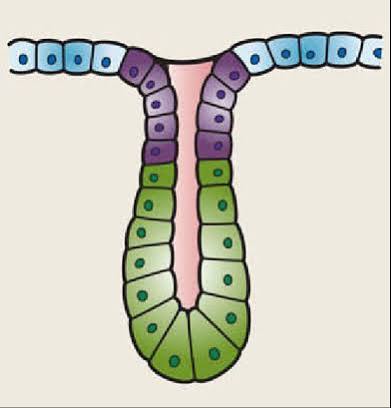
Simple branched tubular

simple alveolar

simple branched alveolar

compound tubular

Compound alveolar

Compound tubuloalveolar

What is the difference between exocrine and endocrine glands?
Exocrine glands carry the products of the glands to the epithelial surface whereas endocrine glands secrete substances directly into the bloodstream
What are the three features that are common to all types of connective tissue, except for blood?
1) cells are separated by large amounts of extracellular matrix that contains protein fibers
2) specialized cells that maintain the tissue rather than being tightly packed surface layers
3) Share a common embryonic origin: mesenchyme cell
what are the three types of loose connective tissue
areolar, adipose, reticular
what are three functions of areolar connective tissue
it is the main battlefield in fight against infection, it wraps and cushions organs, it holds and conveys tissue fluid, plays important role in inflammation
What cell type is responsible for producing the extracellular matrix and protein fibers of areolar connective tissues
Fibroblasts
What is the role of areolar connective tissue in fighting invading pathogens
The protector cells gather in loose connective tissue and they gather in the areolar C.T at sites of infection. Since it is located directly below epithelial layers, it is the first line of cellular defense when the outer protective barrier is breached
What cells are involved in areolar connective tissue fighting invading pathogens
Macrophages: phagocytosis
Plasma cells: antibodies
Mast cells: histamine
Neutrophils, lymphocytes, eosinophils: all protect against pathogens
What are the different types of cartilage
Hyaline cartilage, elastic cartilage, fibrocartilage
What does hyaline cartilage look like microscopically
The chondrocyte cells are floating in the extracellular matrix while being surrounded by lacuna. Protein fibers are not visible
What does elastic cartilage look like microscopically
The protein fibers are visible but go in random directions, but the chondrocytes are still just in the extracellular matrix surrounded by lacunae
What does hyaline cartilage look like microscopically
The fibers are not visible. The chondrocytes are in the extracellular matrix surrounded by lacunae
Where is hyaline cartilage found
costal cartilages
Where is elastic cartilage found
the ear