Anatomy Lecture 3,4, Lumbar Sacral Plexus
1/251
There's no tags or description
Looks like no tags are added yet.
Name | Mastery | Learn | Test | Matching | Spaced |
|---|
No study sessions yet.
252 Terms

what view is the left one from?
left anterior

what view is the right one from?
left posterior view

what is circle #1
medial condyle
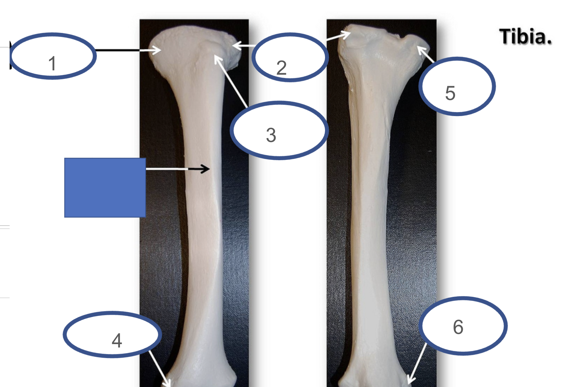
what is circle #2?
lateral condyle

what is circle #3
tibial tuberosity

what is circle #4 ?
medial malleolus

what is circle #5
medial condyle

what is buuble #6
medial malleolus
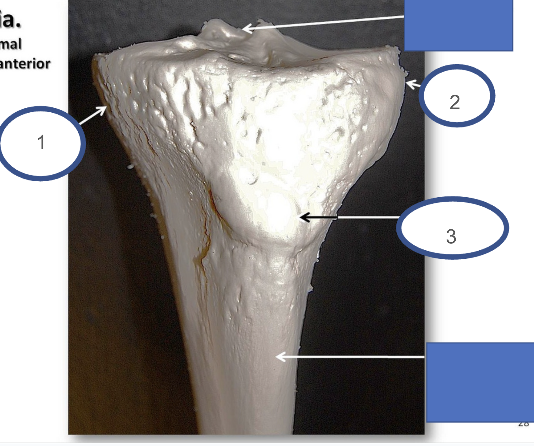
what bone and view is this?
superior anterior left tibia

what is bubble #1
medial condyle

what is bubble #2 ?
lateral condyle

what is bubble #3?
tibial tuberosity

what view is the left one from?
right anterior

what view is the right one from?
right posterior

what is bubble #1?
lateral condyle (tibia)

what is bubble #2
head of fibula

what is bubble #3?
fibula
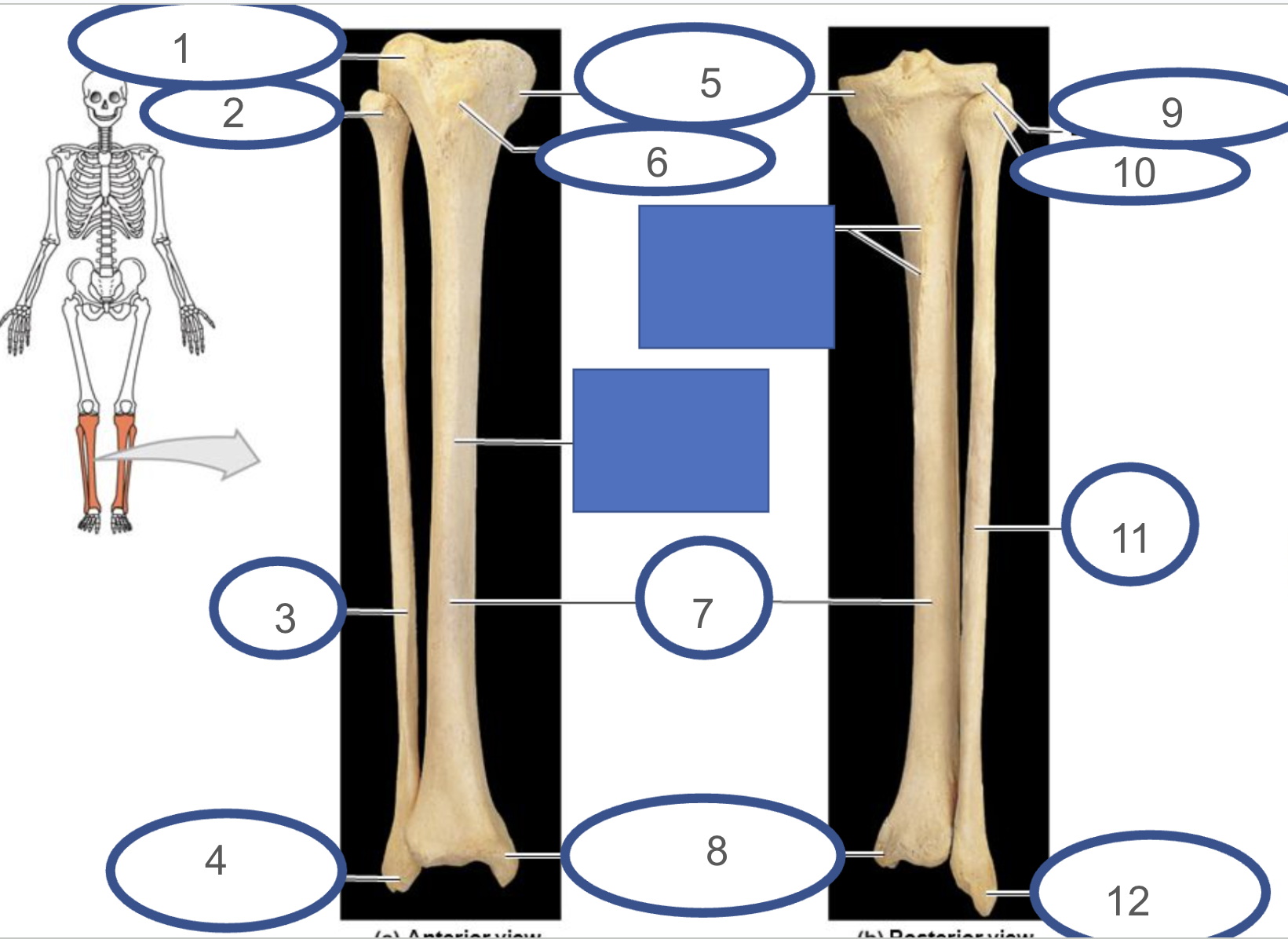
what is bubble #4
lateral malleolus (fibula)

what is bubble #5
medial condyle

what is bubble #6
tibial tuberosity

what is bubble #7
tibia

What is bubble #8
medial malleolus (tibia)

what is bubble #9
lateral condyle

What is bubble #10
head of fibula

what is bubble #11
fibula

what is bubble #12
lateral malleolus (fibula)
valgus
joint medial to the ones above and below it
varus
joint lateral to the joints above and below it
Quad Tendon
Quadriceps Femoris to Patella
Patellar Tendon
Continuation quad tendon
Patella to tibial
tubercle (tuberosity
Ligaments
Bone to Bone
Often Restrain Motion
Tendons
Muscle to Bone
Transfer Force for Movement
Ligaments of Knee
Anterior Cruciate Ligament (ACL)
Posterior Cruciate Ligament (PCL)
Medial Collateral Ligament (MCL)
Lateral Collateral Ligament (LCL)

what is bubble #1?
quadriceps tendon
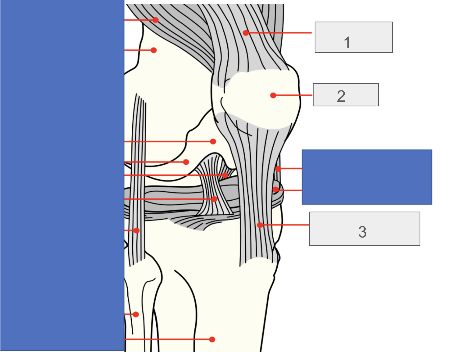
what is bubble #2?
patella

what is bubble #3?
patellar tendon
Anterior Cruciate Ligament (ACL)
Prevents ANTERIOR translation of tibia on femur
Posterior Cruciate Ligament
(PCL)
Prevents POSTERIOR translation of tibia on femur
Medial Collateral Ligament (MCL) (Tibial Collateral Ligament)
Prevents VALGUS instability of Knee
Lateral Collateral Ligament LCL (Fibular Collateral Ligament)
Prevents VARUS instability of knee
what is the soft tissue of the knee
Medial Meniscus
Lateral Meniscus

what are are you looking at and from what angle ?
the knee from the superior angle

what does bubble #1 represent?
patellar ligament

what does bubble #2 represent?
anterior cruciate ligament

what does bubble #3 represent?
medial meniscus

what does bubble #4 represent?
lateral meniscus
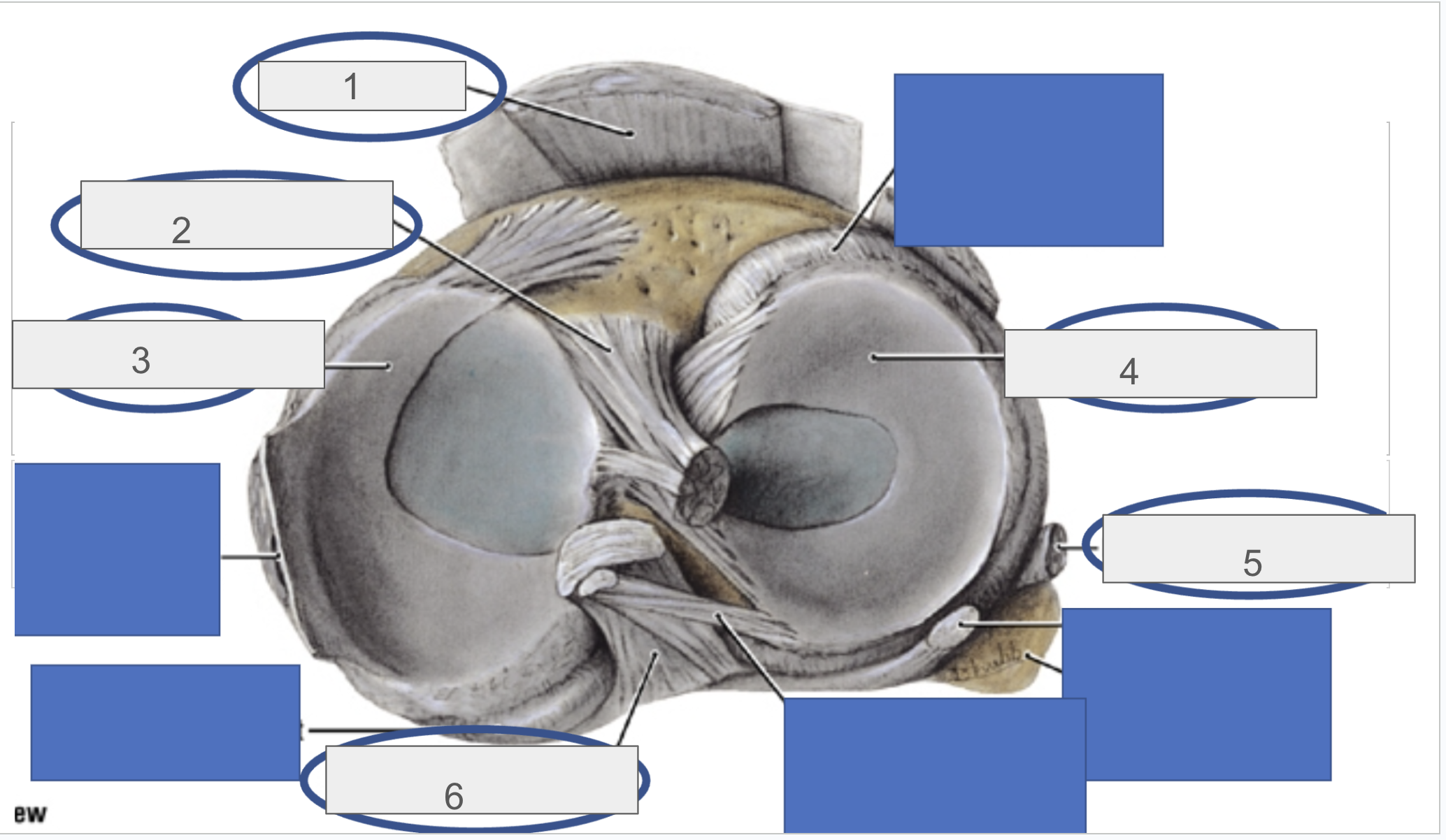
what does bubble #5 represent?
fibular collateral ligament

what does bubble #6 represent?
posterior cruciate ligament
Medial Meniscus
Shock Absorber
Attachment site MCL
More often injured than lateral
what are the hamstring muscles ?
Semitendinosus
Semimembranosus
Biceps Femoris
what are the quadricep muscles?
Rectus Femoris
Vastus Lateralis
Vastus Intermedius
Vastus Medialis
Semitendinosus Origin
Ischial tuberosity
Semitendinosus Insertion
Medial Surface of the Superior Tibia
Semitendinosus Action
Knee Flexion, Hip Extension
Semitendinosus Innervation
Sciatic nerve of the Tibia L5,S1,S2
Semimembranosus Orgin
Ischial Tuberosity
Semimembranosus Insertion
Posterior part of the medial condyle of the tibia
Semimembranosus Action
Flexes Knee, Extends Hip
Semimembranosus Innervation
Sciatic Nerve Tibial Portion (L5, S1, S2)
Biceps Femoris Origin
(Long Head) Ischial Tuberosity
(Short Head) Linea Aspera of Femur
Biceps Femoris Insertion
Fibular Head
Biceps Femoris Action
Both Heads Flex Knee, Long Head Extends Hip
Biceps Femoris Innervation
(Long Head) Tibial Nerve (L5, S1)
(Short Head) Peroneal Nerve (L5, S1)
Rectus Femoris Origin
Anterior Inferior Iliac Spine
Ilium superior to Acetabulum
Rectus Femoris Innervation
Femoral Nerve L2, L3, L4
Rectus Femoris Action
Knee Extension, Hip Flexion
Rectus Femoris Insertion
Tibial Tuberosity
Vastus Lateralis Origin
Posterior Lateral Femur
Vastus Lateralis Insertion
Tibial Tuberosity
Vastus Lateralis Action
Knee Extension
Vastus Lateralis Innervation
Femoral Nerve L2, L3, L4
Vastus Intermedius Origin
Anterior Lateral Femur
Vastus Intermedius Insertion
Tibial Tuberosity
Vastus Intermedius Action
Knee Extension
Vastus Intermedius Innervation
Femoral Nerve L2,L3,L4
Vastus Medialis Origin
Anterior Medial Femur
Vastus Medialis Insertion
Tibial Tuberosity
Vastus Medialis Action
Knee Extension
Vastus Medialis Innervation
Femoral Nerve L2,L3,L4
Sciatic Nerve
Roots: L4-S3
Tibial Nerve
Roots: L4-S3
Muscles Innervated:
Long head of biceps femoris
Semitendinosus
Semimembranosus
Adductor magnus (hamstring portion)
Location: Sciatic Nerve divides into tibial and Peroneal (Fibular)
What does the sciatic nerve divide into ?
Tibial and Peroneal
Peroneal Nerve
Roots: L4-S2
Muscles Innervated:
None (Gives rise to
superficial and deep branches)
Location: Sciatic Nerve divides into tibial and Peroneal (Fibular)
Superficial Peroneal Nerve
Roots: L5,S1,S2
Muscles Innervated: Peroneus Longus (Next week)
Location: Lateral leg
Deep Peroneal Nerve
Roots: L4, L5
Muscles Innervated: Tibialis Anterior, Extensor Hallucis Longus (Next week)
Location: Anterior Leg

what is bubble #1?
aorta
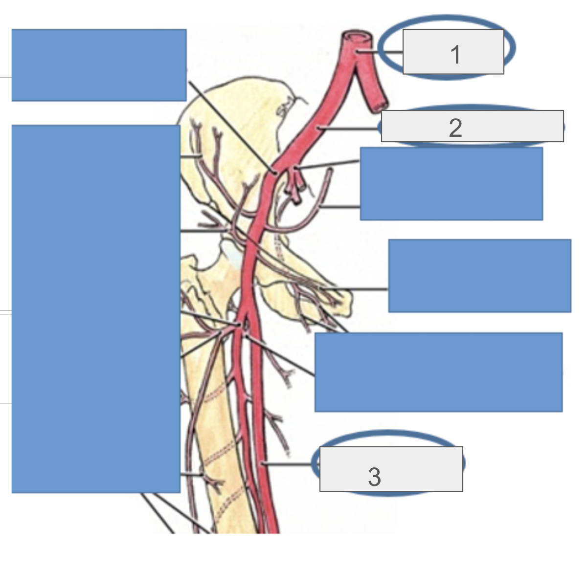
what is bubble #2?
common iliac artery

what is bubble #3?
femoral artery

what is bubble #1?
femoral artery

what is bubble #2?
popliteal artery
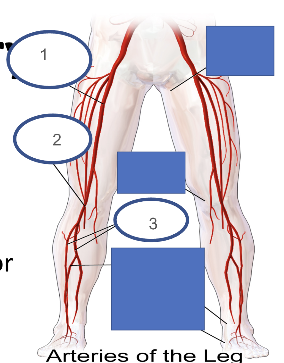
what is bubble #3?
tibial arteries
what are the roots of the sciatic nerve ?
L4-S3
what are the roots of the tibial nerve
L4-S3
What are the roots of the peroneal nerve?
L4-S2
What are the roots of the superficial peroneal nerve ?
L5,S1,S2
what are the roots of the deep peroneal nerve
L4,L5
Popliteal Artery
Continuation Femoral Artery
At the Knee
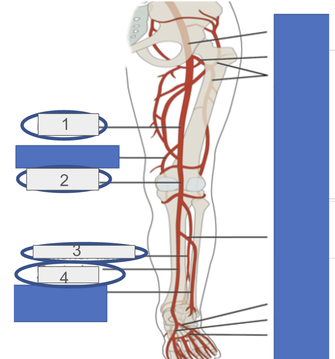
what is bubble #1?
femoral artery

what is bubble #2?
popliteal

what is bubble #3?
anterior tibial