PSYCH 2NF3 midterm
 Call Kai
 Learn
Learn Practice Test
 Spaced Repetition
 Match
Match Flashcards
 Knowt Play
1/191

There's no tags or description
Looks like no tags are added yet.
Name | Mastery | Learn | Test | Matching | Spaced | Call with Kai | Chat |
|---|
No analytics yet
Send a link to your students to track their progress
192 Terms
hypothesis
an educated guess about the way something functions
“educated” because we base hypotheses off our previous experiences and knowledge
once you have a hypothesis the next step is to create an experiment
i.e, engage in hypothesis testing
the blob
Before we had a clear understanding of how things function in the body, we had a 'blob'
People who looked at the body observed a blob of tissue at the top of the head
They hypothesized the purpose of the tissue (not only in the brain, but all over the body)
What was Daniel M Wolpert’s hypothesis?
we have brains to produce adaptable and complex movements
What was the evidence Daniel M Wolpert used to support his hypothesis?
movement is the only way to produce change in the environment
cognitive functions only exist to drive future movements
an underwater creature known as the sea squirt digests it’s own brain and nervous system when it becomes stationary in adulthood
significant historical perspectives on the mind and brain
Aristotle
Galen
Renes Descartes
Phrenologists
significant historical perspectives on the mind and brain → aristotle
his theory was connected to mentalism
proposed that a non-material psyche is responsible for human thoughts, perceptions, and emotions, and this psyche works through the heart to produce action
developed the cardiac hypothesis
mentalism
the idea that we have a mind and it governs our behavior and makes one person different from another
the cardiac hypothesis
the notion that the mind resides in the heart
created by aristotle
came from understandings of feelings and where they can be percieved in the body
significant historical perspectives on the mind and brain → galen
galen (a physician who worked with gladiators) was interested in documenting how different organs function
he observed that we have connections from the bottom of our foot to the top of our head
this inspired him to develop the brain hypothesis
the brain hypothesis
the hypothesis that the entire body is connected to a blob of tissue at the top of the body, so the mind resides in the brain
created by galen
significant historical perspectives on the mind and brain → descartes
proposed the dualism perspective
physicists at the time disputed the dualistic way of thinking and proposed “the mind-body problem”
stated that the mind is located in the brain
Dualism - Descartes
the mind is a non-physical entity within the brain, which is a physical entity
connected to the “mind-body problem,” was people wonder how something non-physical can govern something physical
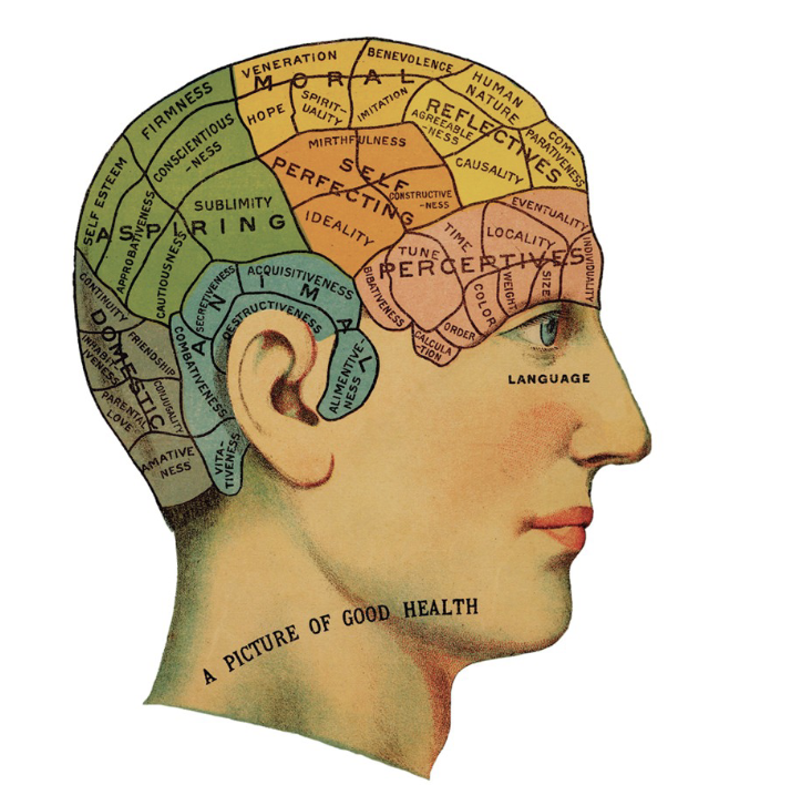
the phrenologists
the phrenologists were a group of individuals interested in mapping out areas of the brain
they proposed that we had unique parts of the brain that mediated specific areas of behavior
believed they could map out brain activity by feeling peoples skulls
Were not taken very seriously because they performed clinical practice using this model
led people to start thinking of the brain as something that has components
What is our current understanding of the relationship between the mind and behavior?
materialism → the idea that all behaviour can be fully explained by the workings of the central nervous system
rooted in evolutionary theories
case studies
the in-depth investigation into a single person or group
responsible for the development of neuropsychology as a feild
became the standard for studying brain function before technology
gyri
the folds of the brain
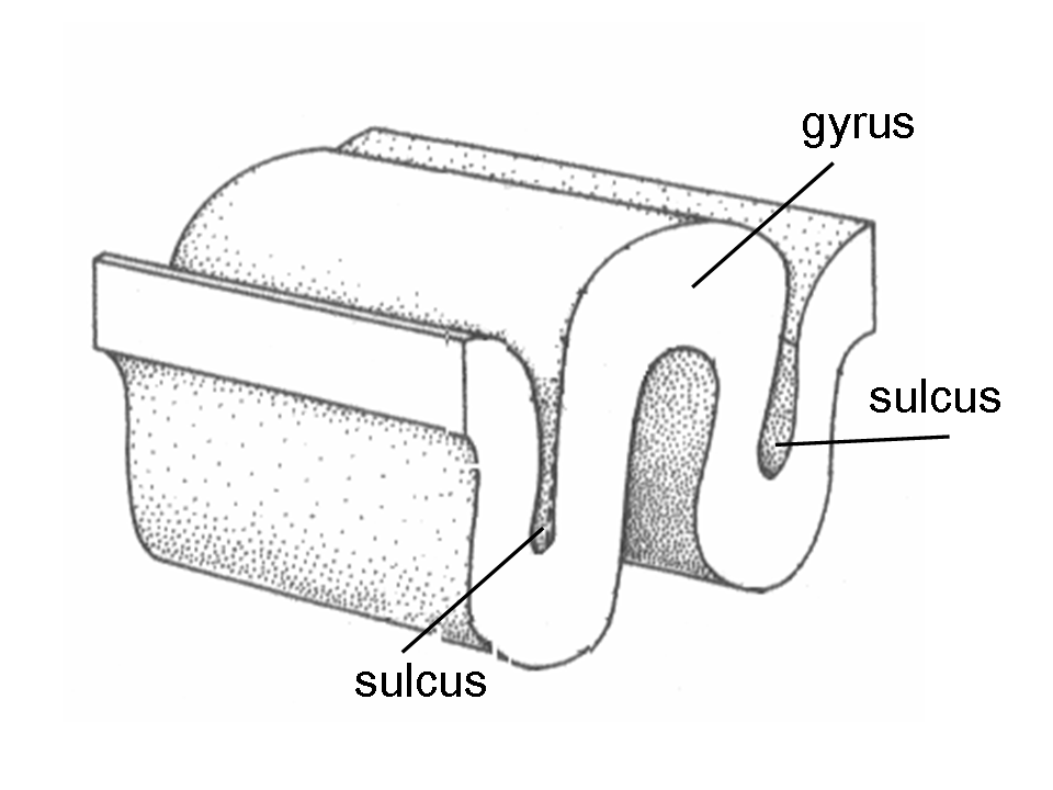
sulci
dips in the brain
big sulci are known as fissures (such as the longitudinal fissure
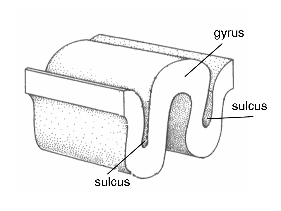
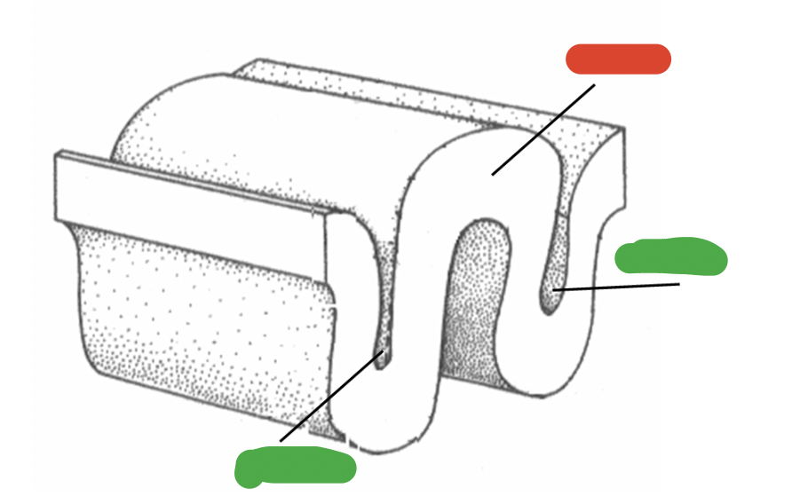
what is the correct name for the green? what is the correct name for the red?
the red = gyri, the green = sulci
Broca’s area
discovered by and named after paul broca
located in the frontal lobe of the left hemisphere
associated with the motor movements used in speech
was interpreted as an example of strict functional localization
Broca’s aphasia
caused by damage to Broca’s area
a language disturbance characterized by motor problems; individuals struggle to get the words out and speak fluently
typically not accompanied by a loss of understanding
speech is limited to short utterances
producing the right sounds/words is a laborious process
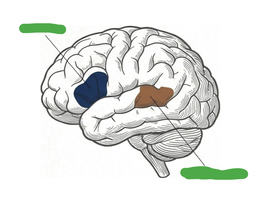
if this image depicts the left hemisphere of the brain, what is the highlighted blue area and what is the highlighted brown area
the blue = broca’s area, located in the left frontal hemisphere
the brown = wernicke’s area, located in the left temporal cortex
Wernicke’s area
discovered and named after Carl Wernicke
located in the left temporal cortex
associated with the organization of words and speech
discovery used as evidence against strict localization
Wernicke’s aphasia
a language disturbance involving the organization of speech
speech production occurs but words often make no sense
patients may include non-existent or irrelevant words in speech
usually results in profound comprehension deficits
are unaware that their own speech doesn’t make sense
Wernicke’s model of speech
Describes the brain as a circuit & states that there's a 5 step sequence in which information moves in the brain
Argues against the idea of strict localization
Implies theres a motor pathway connected to both Wernicke's & Broca's areas
Wernicke's Model Process
1. Sound sensations enter the brain via the auditory pathway
2. Sound representations are stored & processed in Wernicke's area
3. Information is sent to Broca's area through the acuate fasciculus
4. In Broca’s area, sound representations are converted into representations of speech movement
5. Speech movements are sent to the motor pathway for articulation
what is a ‘pathway’?
a bundle of neurons travelling together in the same direction
what is aphasia?
an acquired communication disorder
ACQUIRED because it happens later in life
the brain mappers
a group of individuals who were interested in labelling parts of the brain
using cytoarchitectonics (stamping) korbinian brodmann revealed there were 6 layers of the brain (distinguished by size, shape, and densities of neurons)
the introduction of neuromiaging techniques made his work even more popular
wilder penfield created the sensory homonculus through his works with cortical mapping
the montreal procedure
an epilepsy treatment developed by wilder penfeild
patients are given an anesthesia and are kept awake during open brain surgery
he would stimulate parts of the brain to track different sensations to identify and remove the area causing seizures
he was able to create cortical stimulation maps
neuropsychology
the study of the connection between human behavior and the brain
informed by two hypotheses:
the brain hypothesis
the neuron hypothesis
the brain hypothesis
the idea that the brain is the source of behavior
the neuron hypothesis
a major unit of brain structure and function is the neuron (Nerve cell)
patient H.M case study → background
During the time of brain mapping, patient H.M gained significant attention from neuropsychologists
H.M (or henry molaison) was walking through the park and got knocked down by a bicyclist, injuring his head
after his accident, he began experiencing seizures
his seizures became so bad and frequent in adulthood he was deeply epileptic
epilepsy
an unexpected electrical activity in a specific area of the brain
typically occur in the temporal cortex
patient H.M case study → procedure
to deal with his seizures, H.M decided to have a lobotomy of his temporal lobe to receive the area of his brain causing the frequent seizures
at the time, this was a common treatment for seizures
patient H.M case study → outcome of procedure
As a product of his surgery, he lost the ability to create new memories and had problems with his short term memory
His long-term memory was unaffected and he could access memories from his childhood
His story helped develop “the binding problem”
the binding problem
Memory has multiple different components (short-term, sensory, long-term)
Concerned with how the brain seamlessly creates meaningful comprehension of experiences
the ventral stream
the what pathway
mediates action controlled by conscious visual perception
damage results in visual agnosia
visual form agnosia
the inability to recognize objects visually
with visual form agnosia items can be differentiated by touch
the dorsal stream
the where pathway
mediates action controlled by unconscious visual perception
ex: indivividuals can identify objects but have trouble aiming hands to pick up objects
what are the three portions of a neuron?
The core region of a neuron is is the cell body
The branching extensions are the dendrites
There is many dendrites
The main "root" is the axon
There is only one axon
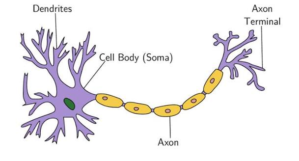
what are the two types of cells found in the nervous system?
the neurons
the glia
localization of function
the idea that specific areas of the brain control each type of behaviour
lateralization of function
the idea that one specific hemisphere of the brain controls a function not shared by the other
given the history of neuropsychology, what are some key insights from brain injury case studies?
neuroplasicity → studies on cats with removed areas of the brain found that the passage of time allowed the brain to rewire and function normally
the brain has a hierarchal organization
our brain can be cut into two brains by cutting the corpus callosum and smaller comissures
we have both conscious and unconscious neural streams
what are the four types of brain damage?
traumatic brain injury
tumours
multiple sclerosis
bacterial infections
traumatic brain injury
damage or injury to the brain caused by an external force our trauma
a disruption in the brain that occurs after birth → it isn’t hereditary, congenital, degenerative, or induced by birth trauma
differentiated in two ways:
open head injuries
close head injuries
traumatic brain injury → open head injuries
involves penetration of the skull
traumatic brain injury → close head injuries
concussions
concussions
movement of the brain in the skull
can’t be seen on routine x-rays, CT scans, or MRI’s
affects the way a person may think
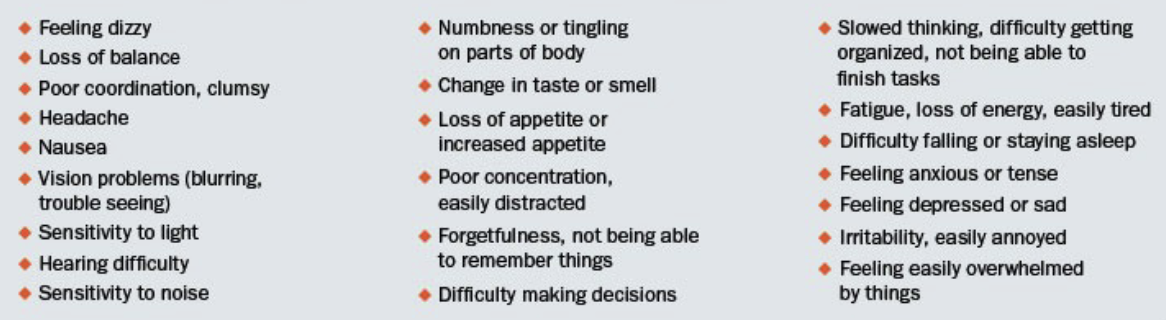
can cause a variety of symptoms
concussions → coups vs countercoups
a coup is the site of the blow
a countercoup is the possible brain damage caused by compression of the side of the skull opposite to the coup
a severe coup or countercoup may be accompanied by bleeding or a subdural hematoma
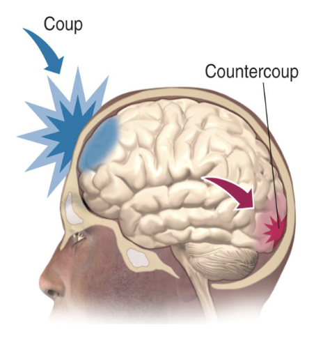
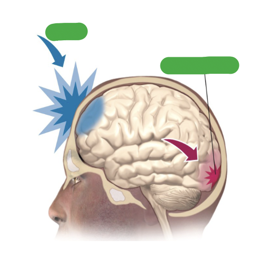
in a concussion, what is the blue arrow pointing to and what is the red arrow pointing to?
the blue arrow is pointing to the coup, the point of impact on the skull
the red arrow is pointing to the countercoup, an area where brain damage can occur opposite side to the site of a blow
Dementia Puglistica (Boxer’s Syndrome)
another name for chronic traumatic encephalopathy (CTE)
a degenerative brain disease thought to be caused by repetitive head trauma
commonly experienced by athletes ** but can occur in anyone
the repeated head trauma is thought to scar brain tissues
considered a “dementia” because there is memory and motor related brain disturbances
Dementia Puglistica → symptoms
Progressively declining cognitive ability
Short-term memory loss
Tremors
Loss of coordination
Difficulty in speech
Changes in gait
Emotional disturbances
Dementia Puglistica → case study on p-tau
a patient who died very suddenly was a retired professional boxer
his brain scan revealed an accumulation of p-tau proteins in the neurons and glial cells
Tau proteins
Help stabilize microtubules in the brain
Super important for the transportation of nutrients & other molecules within nerve cells
They lose their ability to stabilize microtubules when phosphorylated & instead clump together
P-tau
phosphorylated tau proteins
important markers of brain injury & disease
found in abnormal deposits in the brains of individuals with Boxer's syndrome
believed to contribute to the degeneration of brain cells & the development of cognitive & behavioural symptoms associated with the condition
what E4 variant puts athletes at a higher risk of developing brain injuries?
APOE; it helps the body repair from injury
concussions and prolonged rest
studies have shown that prolonged rest is not necessary after experiencing a concussions
after an initial day or two of rest, a concussed person should try to go about daily life until symptoms become too difficult
sitting around thinking about symptoms can make them seem worse and people adopt a sick role psychologically
why are some concussions more difficult to recover from?
axonal health
axons are insulated by fat and if the axons are damaged, it can impact how fast electrical signals are sent leading to delayed reaction times
mental health
ex: post-injury anxiety
concussions → stats canada data and GBA+
concussions in sport are recognized as a public health issue because of their frequency and potential for long-term consequences
previous dats suggests that concussions happen more often in young people; specifically males
however, after a gender based analysis (GBA+), children of both sexes had similar concussion rates until adolescence, when females increasingly experienced more concussions
females have been found to experience more symptoms, worse reaction times, and greater cognitive decline than males from concussions
tumours
an abnormal growth of cells
diagnosed through imaging (which is crucial for establishing a correct diagnosis)
there are two different categories of tumours:
malignant tumours
benging tumours
which can further be classified as gliomas or meningiomas
symptoms → pressure in skull and disruptions related to location
categories of tumours
malignant
benign
categories of tumours → malignant
Lack distinct boundaries; can move around areas of the brain
Likely to recur following surgical removal
Travel through metastasis
Metastasis
the process in which malignant tumours shed their cells and travel to other cites
categories of tumours → benign
tumours contained within their own membrane
types of tumours → gliomas
tumours that develop in the glial cells
types of tumours → meningiomas
tumours that grow out of the meninges
close to the surface of the brain so they are easy to remove
typically benign
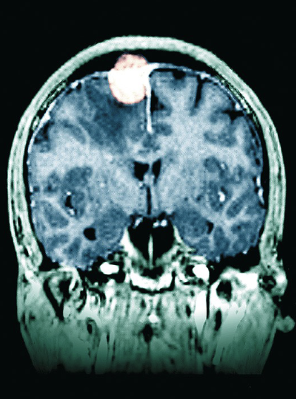
grading of tumours as outlined by the WHO
I: benign, slow-growing, responds well to surgery
II: malignant, slow-growing, increased likelihood of recurrence
III: malignant, increased likelihood of recurrence and requires aggressive treatment
IV: rapidly fatal
factors influencing treatment options for tumours
The type, size, and grade of the tumor
Whether the brain tumor is putting pressure on vital parts of the brain
If the tumor has spread to other parts of the body
The possible side effects that have emerged
The patient's preferences and overall health
types of treatment for tumours
surgery
radiation therapy:
high radiation to target and destroy cancer cells
can be delivered internally or externally
chemotherapy:
medications that can be given orally
works by targeting and destroying rapidly dividing ccells
targeted therapy:
involves using drugs specifically designed to interfere with specific molecules that contribute to the growth and survival of cancer cells
minimizes damage to normal cells (which are affected in other treatment methods)
multiple sclerosis
a chronic autoimmune disease that creates scarring in the white matter of the brain and spinal cord (CNS)
caused by the immune system mistakenly attacking the myelin on neurons
leads to inflammation, damage, & disruption of nerve signals
specific complications with multiple sclerosis
Damages oligodendrocytes (the glial cells that form the myelin on CNS axons)
Leads to the demyelination of axons
Affects white matter in different locations
population distribution of multiple sclerosis
first appears in young adults between the ages of 20-40
there is modest heritability and environmental factors
it is 2x more common in those assigned female at birth compared to males
theories explaining this finding include hormonal factors, genetic factors, immune system differences between men and women, and lower vitamin D (which is more common in women)
canada is among contries with the highest rate of multiple sclerosis in the world
symptoms associated with multiple sclerosis
Numbness, tingling
Fatigue
Muscle spasms
Walking difficulties
Pain
what is an example of a viral infection that can cause brain injury?
human immunodeficiency virus or HIV; the virus that causes AIDS
AIDS can develop into AIDS Dementia Complex
how does HIV turn into AIDS?
Once you have HIV in your system, if it is detected early enough, individuals can take medications to keep the viral load low (and be HIV negative)
However, if it goes untreated, you move from HIV negative to HIV positive
If you have a low enough white blood cell content and are HIV positive, you can develop Acquired Immune Deficiency Syndrome (AIDS)
Acquired Immune Deficiency Syndrome (AIDS) Dementia Complex
AIDS ca lead to a collection of neurological symptoms
initial symptoms include difficulty concentrating, forgetfulness, decreased work productivity, low sex drive, social withdrawl, general apathy, and muscle weakness
it is commonly misdiagnosed as depression
advanced symptoms include severe motor and cognitive disturbances
Acquired Immune Deficiency Syndrome (AIDS) Dementia Complex - Canadian Perspective
Had a 90-90-90 goal
Aimed to train healthcare providers to be really vigilant & know what questions to ask to accurately diagnose, treat, & suppress HIV
In 2016 the process was close but not at the goal yet; the COVID pandemic decreased progress as well
How does HIV work indirectly?
HIV invades macrophages, microglia, astrocytes, and vascular endothelial cells
Infected cells release cytokines which results in tissue damage and apoptosis (cell death)
HIV can bind to special protein structures in the brain resulting the excessive intake of calcium and later neuronal death
Microphages
a type of white blood cell impacted by HIV that destroys microorganisms and dead/dying cells
Microglia
the macrophages of the CNS; impacted by HIV
Astrocytes
the glila cells that provide structural supports to neurons by holding them in place; impacted by HIV
vascular endothelial cells
specific cells impacted by HIV that layer the blood vessels
Important factors in case studies
past medical history
family medical history
general physical exam
mental health status
blood pressure
the amount of pressure that your blood is exerting on your artery walls
measurements of 120/80 are considered normal
how to read blood pressure ratio
the top number shows how much pressure is exerted when the heart is beating
the bottom number is a measurement of the pressure between beats
cranial nerves
the nerves that emerge indirectly from the brain in contrast to the spinal nerves
numbered in roman numerals from 1-12 from the front of the brain to the back
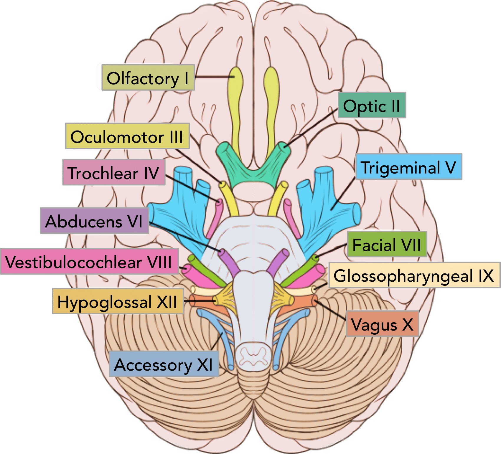
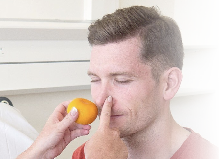
cranial nerves → olfactory nerve CN1
Used in smell
Damage results in the loss of ones sense of smell
Tested by offering something familiar to the patient and asking them to smell and identify it
cranial nerves → the optic nerve CN2
used in vision
damage results in the loss of vision (anopsia)
Colour blindness is tested with Ishihara plates
Visual fields are tested by asking the patient to look straight ahead & identify movement in different quadrants of the visual field
Visual acuity is tested using Snellen charts
Using snellen charts, what does 20/20 vision mean?
The patient sees the same line of letters at 20 feet that a healthy person sees at 20 feet
Using snellen charts, what does 20/40 vision mean?
20/40 vision means that the patient sees at 20 feet what a healthy person sees at 40 feet
fundoscopy
a test that allows diagnosticians to see right into the fundus of the eye
the fundus
the interior surface of the eye
includes the retina, optic disc, macula, fovea, and posterior pole
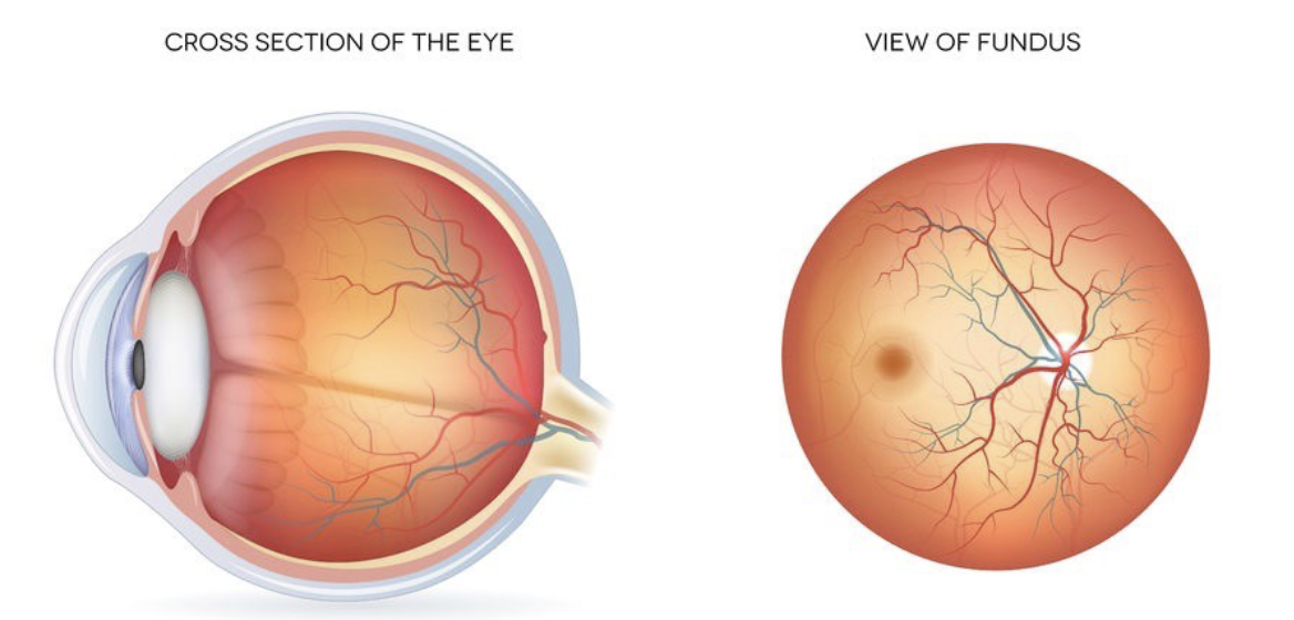
the retina
Contains specialized cells called photoreceptors
Responsible for detecting light & converting it into action potentials that the brain can interpret as visual information
the optic disc
the specific point where the optic nerve exits the eye and connects to the brain
creates a blind spot because it lacks visual receptors
the macula
a small, highly specialized area located on the center of the retina
it is essential for activities that require fine details like reading
the fovea
a region of the highest visual acuity in the eye that is responsible for our sharpest and most detailed vision
the posterior pole
the back portion of the eye; specifically the region encompassing the optic nerve head, macula, and surrounding structures
cranial nerves → the oculomotor nerve CN3
Used in eye movements
Tested by examining ones reaction to light, eyelid movement, and deviation of eye outwards
Damage results in double vision, large pupils, uneven dilation of pupils, and drooping eyelid