Pathophysiology of Osteoarthritis and Gout
 Call Kai
 Learn
Learn Practice Test
 Spaced Repetition
 Match
Match Flashcards
 Knowt Play
1/71

There's no tags or description
Looks like no tags are added yet.
Name | Mastery | Learn | Test | Matching | Spaced | Call with Kai | Chat |
|---|
No analytics yet
Send a link to your students to track their progress
72 Terms
what is Osteoarthritis?
Osteoarthritis (OA) is a chronic joint disease that leads to destruction and immobility of joints
how does Osteoarthritis occur?
It occurs when the protective cartilage that cushions the ends of bones wears down over time
What are the 2 types of OA
Primary (idiopathic)
Secondary
characteristics of Primary
- most common form of disease
- no predisposing factor is apparent
chaarcteristics of Secondary
- pathologically is not distinguishable from primary
- has a known underlying cause
what is the most common joint disease?
OA
Joint distribution of OA in males and females <55 years is similar
WHo is most at risk of OA In patients >55 years
- hip OA is more common in males
- OA of hands, thumb base, and knee are more common in females
Risk factors for OA

a combination of what can cause the disease or progress it further?
A combination of loading and susceptibility (local, systemic) risk factors is required to cause disease or its progression.
Joints most often affected by OA?
OA usually affects the joints in a non-symmetric manner
OA most often affects hands, knees, hips, and spine
what does a healthy joint look like (4) what structures does it contain
The ends of bones are encased in smooth cartilage that are protected by joint capsule lined with synovial membrane and fluid
Synovial membrane (synovium) = a thin layer of tissue that lines the inside of the joint capsule. Its main job is to produce synovial fluid.
Synovial fluid = the slippery liquid inside the joint cavity. It lubricates the joint, reduces friction, and helps nourish the cartilage.
Joint capsule = the outer “bag” that surrounds and stabilizes the joint
Articular cartilage = covers the ends of the bones so they move smoothly
what does a joint affected by OA look like?
The cartilage becomes worn away, spurs grow out from the edge of bone, synovial fluid increases, and joint becomes stiff and sore
who is most affected by Osteoarthritis of knees and what are its effects?
- more common in females than males
- may affect one or both knees
Effects
Increased subchondral bone density osteophytes, narrowing of joint space
who is affected by OA of hips and what are its effects?
- affects both males and females (males: more common)
- may affect one or both hips
Cartilage wears away → joint space narrows → underlying bone becomes denser (sclerosis) + cysts can develop + bone spurs (osteophytes) form around the edges.
who does OA of the hands effectr the most
- mainly affects females
- most often affects the base of thumb and joints at the end of fingers
forms Bouchard’s nodes and Heberden’s nodes
what is Heberden’s nodes
formation of hard knobs at farthest finger joints
what is Bouchard’s nodes
formation of hard knobs at middle finger joints
what is spondylosis?
OA of the neck and back
most common type of arthritis in spine
- can occur anywhere in spine but is more common in
lower back and neck
- may cause no problem
- pain and stiffness are most common symptoms
where does OA of the foot affect the most?
generally affects the joint at the base of big toe
Pathologic changes of joint in OA include?
surface of joint is damaged
surrounding bone grows thicker
leading to joint damage and immobility
what happens to cartillage when OA starts to develop?
progressive loss of articular cartilage but it also involves other tissues
what other tissues are involved (4)
- subchondral bone
- synovium
- ligaments
- neuromuscular tissues
what is early stage of OA look like?
cartilage is thicker than normal
what does Progression of disease look like
- joint surface is breached, vertical clefts (fibrillation)
- cartilage is metabolically active
- chondrocytes replicate and form clusters
chrondrocytes are cells that maintain cartillage break down old cart and make new cart via enzyme
Fibrillation = small vertical cracks/clefts develop in the cartilage.
cartilage becomes metabolically active because the chondrocytes are working hard to respond to the damage.
what does Late Stage look like
cartilage becomes hypocellular
In cartilage, the main cells are chondrocytes, which maintain and produce the cartilage matrix.
So:
Cartilage becomes hypocellular = ↓ number of chondrocytes in the cartilage
what is Bone remodeling and hypertrophy
As cartilage disappears, the bone underneath it experiences more mechanical stress.
That underlying bone is called subchondral bone:
sub = below + chondral = cartilage
The bone responds by adding new bone to its surface (appositional bone growth).
This causes the subchondral bone to become:
thicker + denser = subchondral sclerosis
what is the Extracellular matrix of a normal cartilage is composed of
Proteoglycans (PGs)
Collagen
Matrix metalloproteinases (MMPs)
what are Proteoglycans (PGs
which are responsible for the compressive stiffness of the tissue and its ability to withstand load
functon of Collagen
provides tensile strength and resistance to shear
what does Matrix metalloproteinases (MMPs) do?
are enzymes that break down proteins in the extracellular matrix (ECM).
MMPs appear to have an important role in the loss of cartilage matrix in OA
The extracellular matrix contains structural proteins such as collagen and elastin, so increased MMP activity can cause tissue destruction.
where do primary changes in OA begin?
begin in the cartilage
what stimulayes Synthesis and secretion of MMPs
might be stimulated by IL-1β or by other factors (e.g. mechanical stimuli)
what are Signs and symptoms of OA (7)
pain
stiffness of involved joint (mainly after a period of inactivity) last around 20 min
Localized tenderness bone or soft tissue swelling (arthritis)
Bone crepitus (sensation of bone rubbing against bone)
Warmth
Deformation of involved joints
Marked loss of joint motion
where is the pain located and what is the cause (3)
deep pain localized to the involved joint
- usually aggravated by joint use and relieved by rest
- nocturnal pain interferes with sleep and is observed
particularly in advanced OA of hip
does joint pain happen in Articular cartilage?
Articular cartilage is aneural so joint pain in OA patients must arise from other structures

what is diagnosis of OA based on?
Diagnosis of OA is usually based on clinical symptoms
and radiographic features
• There is NO correlation between radiographic findings
and severity of disease
Main findings of OA in X-Ray include:? (4)
narrowing of joint space
subchondral bone sclerosis (thickening of bone)
subchondral cysts
osteophytosis (bone spurs)
is their specific lab test for OA?
nuh uh
Lab findings may help to identify the underlying cause of secondary OA
why do lab test not work?
OA is usually not systemic so the lab tests such as ESR CBC, urine analysis are usually normal
what about synovial fluid?
Synovial fluid reveals mild leukocytosis (mainly mono-nuclear cells)
Synovial fluid analysis is particularly important for excluding other conditions (gout, septic arthritis, RA)
what are treatment goals of OA?
Treatment of OA is aimed at reducing pain, maintaining mobility, and minimizing disability
what Therapeutic strategies are used?
• weight loss
• mild exercise
• NSAIDS
• analgesics
• glucocorticoids (intra- and periarticular injections
not systemic)
• surgery (replacement of joint
what is GOUT?
• Gout is a metabolic disease caused by precipitation of monosodium urate within and around joints
• Gout is a common crystal-induced arthritis
Where does uric acid come from?
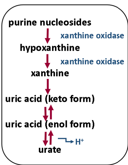
Uric acid is the final product of purine breakdown in humans.
how does uric acid form gouts?
↑ uric acid in blood (hyperuricemia)
→ uric acid has low solubility
→ at high enough concentrations it can form urate crystals
→ crystals deposit in joints
→ immune system reacts to crystals
→ inflammation → gout attack
What happens to uric acid in the kidneys?
UA is filtered from blood by glomerulus and almost completely resorbed by proximal tubules of kidney
A small fraction of resorbed UA is secreted by distal nephron and excreted in urine
who is most at risk of gout?
Gout is more often in men ( : ratio is about 5:1)
It is more common in middle age and older men (peak age is 30-50 years)
Women generally develop signs and symptoms after menopause
Both genetic and environmental factors contribute to development of gout
which joint does gout mostly affect
The most common affected joints in gout are fingers, first toes, ankles, knees, and hips
what forms does gout have?
primary
secondary
what is primary form?
In primary form (90% of cases), gout is the major manifestation of disease and the cause is usually unknown
what is secondary form?
In secondary gout (10% of cases), uric acid level is increased because of a known underlying disease that usually dominates the clinical picture
what causes elevated uric acid results? (3)
Increased uric acid production
Decreased uric acid secretion
Increased reabsorption
what can cause increased uric acid production (3)
- inherited enzyme deficiencies
- myeloproliferative disorders (chemotherapy in leukemia)
- disorders causing ↑ATP degradation (e.g. alcohol abuse)
what can cause Decreased uric acid secretion (2)
- renal problems
- competitive anions (e.g. ketoacidosis)
what can cause Increased reabsorption
drugs (e.g. diuretics)
- dehydration
- idiopathic causes
What causes urate crystals to form? (3)
Decreased solubility - low temp, low ph
Trauma / disturbance to joint or soft tissue
Dehydration of the joint resulting in super-saturation in the joint
what happens to joints when Precipitation and accumulation of urate crystals inside the joint?
leads to joint damage and deformation
how does the immune system react to gout?

why does the immune system work against you when fighting against gout. what does the condition devlop into?

what happens if the immune system keeps attacking gout?

what are the morphologic changes in gout? (4)
Acute arthritis
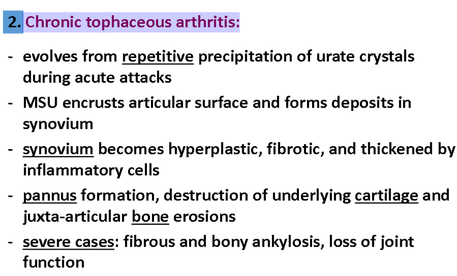
Chronic tophaceous arthritis
Tophi in various sites
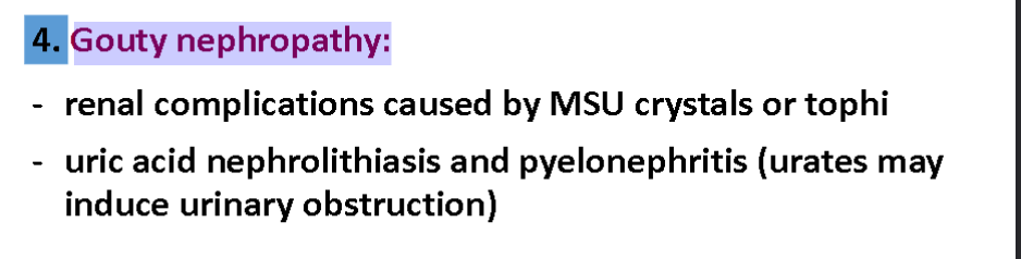
Gouty nephropathy
what is Acute arthritis

what is Chronic tophaceous arthritis:
what is Tophi in various sites:

what is Gouty nephropathy:
what are the 4 stages of quite and their symptoms

what are symptoms of acute gout

what does Chronic gout look like
after several acute mono- or oligoarticular attacks, some patients develop chronic non-symmetric synovitis
Hyperuricemia (plasma urate >6.8 mg/dl) is necessary, but not sufficient, for development of gout.
what are Major risk factors for gout

how can you Diagnostic tests for gout (2)
Laboratory tests
Radiography

Treatment goals for gout
Treatment goal is to control inflammation and pain
what are the Two approaches used to accomplish the last flashcards goals
control of inflammation: NSAIDS and Glucocorticoids
decrease deposition of urate in joints: hyperuricemia therapy (↓ production, ↑ excretion, ↑breakdown of uric acid)