6 - outer, middle and inner ear
1/61
There's no tags or description
Looks like no tags are added yet.
Name | Mastery | Learn | Test | Matching | Spaced | Call with Kai |
|---|
No analytics yet
Send a link to your students to track their progress
62 Terms
what is auricular haematoma
collection of blood underneath perichondrium of ear
what causes auricular haematoma
trauma
Hair cell depolarisation is mediated by…
mechanically gated-potassium channel leading to depolarising influx of potassium ions
treatment of auricular haematoma
incision and drainage
pressure dressing to prevent reaccumulation
antibiotics to prevent infection of cartilage
common complication of auricular haematoma
cauliflower ear - necrosis of cartilage leading to deformity
foreign body removal times
- button batteries
within HOURS - can cause permanent damage through corrosion
foreign body removal times
- organic
within DAYS - will cause infection
foreign body removal times
- inorganic
within DAYS
how are foreign bodies removed from ear
appropriate instrument removal
sometimes under general anaesthetic if cannot get it out
define otitis externa
inflammation of the external ear canal
presentation of otitis externa
ear pain
discharge
itching
hearing loss
management of otitis externa
antibiotic/steroid ear drops
suction for discharge
antibiotics for infection - amoxicillin
steroids for inflammation - prednisolone
prevention of otitis externa
no water or cotton buds
- OE is often called swimmers ear and results from improper drying of ears
define malignant otitis externa
osteomyelitis of temporal bone
-not malignant in cancer sense but malignant in the fact it can spread through skull base and cause serious morbidity or mortality
who is most at risk for malignant otitis externa
elderly diabetics
presentation of malignant otitis externa
severe pain in elderly diabetic
granulations in external auditory meatus
+/- cranial nerve palsies
management of malignant otitis externa
ciprofloxacin long term
what is the causative agent of malignant otitis externa
Pseudomonas aeruginosa
what is otitis media
infection of the middle ear
what is a complication of otitis media
untreated and recurring otitis media may result in a perforated eardrum
presentation of otitis media
redness, swelling, bulging, of tympanic membrane
fluid and or pus trapped under eardrum - glue ear
management of otitis media
observation for 3 months
if not resolving or recurring
- otovent
- grommet
what is acute supparative OM
pus in middle ear
presentation of acute supparative OM
otalgia +/- ottorhoea
- increasing ear pain followed by discharge when eardrum bursts and resolution of pain
management of acute supparative OM
conservative - observation
- antibiotics ONLY IF INFECTED
what is tympanosclerosis
calcification in tympanic membrane +/- middle ear
what causes tympanosclerosis
-chronic ear infections
-trauma to tympanic membrane
otoscope findings in tympanosclerosis
white patches on tympanic membrane
management of tympanosclerosis
observation
symptoms of tympanosclerosis
asymptomatic but can sometimes affect hearing
what are the 2 types of chronic supparative OM
perforated tympanic membrane
cholesteatoma
- both lead to long term discharge from ear
what is cholesteatoma
benign cyst in the mastoid cavity
complications of chronic supparative OM
complete hearing loss
CN VII palsy
meningitis
brain abscess
causes of perforated tympanic membrane
infection
trauma
grommet
presentation of perforated tympanic membrane
recurrent infections +/- hearing loss
management of perforated tympanic membrane
water precautions +/- myringoplasty
pathophysiology of cholesteatoma
2 types of
- eustachian tube dysfunction
-impaired skin migration
presentation of cholesteatoma
1. Drainage with foul odor
2. Fullness or pressure in ear
3. Hearing loss
4. Ache behind ear at night
5. Dizziness
6. Muscle weakness on ipsilateral face
management of cholesteatoma
refer to ENT
mastoidectomy - drill into mastoid bone to remove
what is otosclerosis
conductive hearing loss + normal tympanic membrane
pathology of otosclerosis
fixation of stapes by extra bone
management of otosclerosis
hearing aid
stapedectomy
what is the external ear made of and it’s overall function
auricle (pinna)
external acoustic meatus (ear canal)
tympanic membrane (eardrum)
to collect, amplify and transmit sound waves to the middle ear
what is the auricle made of and what’s it’s function?
elastic cartilage
collects and directs sound into the ear canal
external acoustic meatus
ear canal - 2.5cm long tube leading to the eardrum
outer 1/3 cartilaginous with glands (produces earwax)
inner 2/3 bony
conducts sound to tympanic membrane
tympanic membrane
eardrum - thin membrane separating external and middle ear
vibrates in response to sound waves
epithelial lining of external ear canal and middle ear
external ear canal:
outer 1/3 keratinised stratified squamous epithelium
inner 2/3 also lined with keratinized stratified squamous epithelium, but thinner and more tightly attached to bone.
middle ear:
simple cuboidal to columnar epithelium, becomes ciliated pseudostratified columnar with goblet cells near the eustachian tube
structure and function of tympanic membrane
layers:
outer layer : keratinised stratified squamous epithelium
middle layer: fibrous connective tissue (provides strength)
inner layer: simple cuboidal or columnar epithelium
internal auditory meatus
A short canal in the petrous part of the temporal bone.
Transmits cranial nerves VII (facial) and VIII (vestibulocochlear), and labyrinthine vessels.
Leads from the posterior cranial fossa to the inner ear.
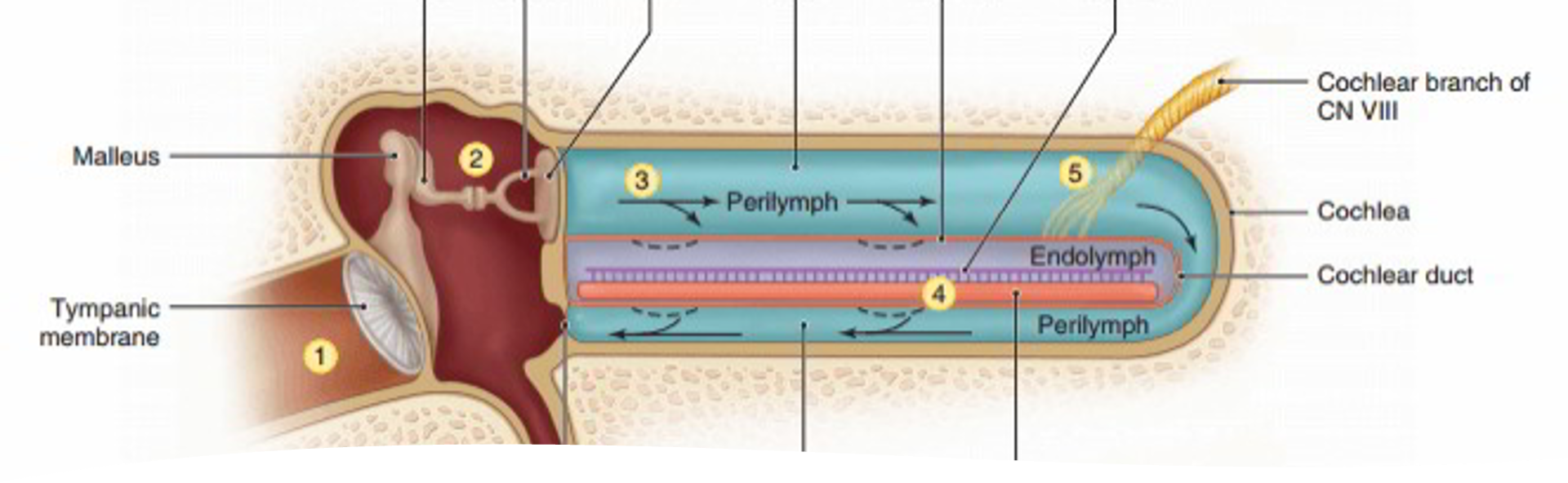
ossicular chain (middle ear bones)
Three small bones that transmit sound from the tympanic membrane to the inner ear:
Malleus (hammer) – Attached to the tympanic membrane.
Incus (anvil) – Connects malleus to stapes.
Stapes (stirrup) – Base (footplate) fits into the oval window of the cochlea.
transmission of sound across middle ear
Sound waves hit the tympanic membrane, causing it to vibrate.
Vibrations are passed through the ossicles (malleus → incus → stapes).
Stapes footplate pushes on the oval window, transmitting vibrations into the fluid-filled cochlea (inner ear), where they are converted into nerve impulses.
physiology of hearing - inner ear 1
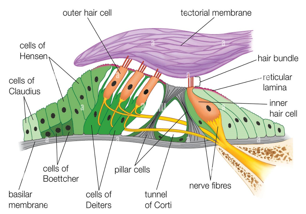
footplate of stapes moves in and out of oval window creating a travelling wave in the scala vestibuli and scala tympani of the cochlea
causes movement of the basilar membrane and movement of inner and outer hair cells in the organ of corti in relation to the tectorial membrane
physiology of hearing - inner ear 2
cilia of the hair cells are deflected and ion channels open
cations flow from the endolymph into the hair cells
depolarisation takes place and an impulse is sent up the cochlear nerve
inner hair cells activate the afferent nerves
outer hair cells modify the response of the inner hair cells
physiology of the inner ear - 3
For every frequency there is a specific place on the basilar membrane where the hair cells are maximally sensitive to that frequency. This is known as a tonotopic arrangement.
importance of eustachian tube in middle ear function
equalises air pressure - during swallowing or yawning, allows pressure to be equal on both sides preventing damage to the eardrrum ensuring it can vibrate properly for hearing
drains fluid
protects ear from pathogens
inter connection of middle ear, mastoid air cell system and post nasal space (nasopharynx)
interconnected systems that rely on each other for air pressure regulation and the transmission of air
The postnasal space (nasopharynx) connects to the middle ear via the Eustachian tube which allows for pressure equalisation. The middle ear is connected posteriorly to the mastoid air cells, a network of air-filled cavities, through a bony passage called the aditus. The mastoid air cells are thought to act as a gas reserve for the middle ear, further helping to stabilize its pressure
the course of facial nerve in the middle ear cavity and its importance to the surgeon
The facial nerve runs through the middle ear, traveling posterosuperior to the oval window, then inferiorly along the medial and posterior walls of the tympanic cavity before exiting downward in the mastoid.
Its importance to surgeons is that this nerve is vital for facial expression and other function
bony anatomy of the inner ear and function of the parts
Labryinth - the complex, fluid-filled system of channels in the inner ear responsible for hearing and balance
Cochlea – controls hearing
Vestibular system – controls balance
bony labrynth, membranous labrynth, perilymph
The bony labyrinth encloses the membranous labyrinth, which is a system of connected fluid-filled sacs and tubes.
The space between the bony and membranous labyrinths is filled with a fluid called perilymph.
The membranous labyrinth itself contains another fluid, endolymph, which plays a crucial role in transmitting sound and balance information.
intra-cranial relations of the middle ear and mastoid system
This connection means that infections can spread from the middle ear to the mastoid air cells and, in severe cases, to the cranial cavity itself, leading to serious complications like meningitis or brain abscesses. The system is also adjacent to important nerves, including the facial nerve (CN VII) and the vestibulocochlear nerve (CN VIII), which are at risk during an infection.
basic principles of pure tone audiometry and tympanometry
pure tone audiometry measures hearing sensitivity by presenting pure tones of varying frequencies and volumes to determine the softest sound a person can hear at each frequency
tympanometry measures middle ear function by testing the movement of the eardrum and its compliance with changes in air pressure
neural pathway of hearing
The neural pathway of hearing begins with sound waves causing vibrations that travel from the outer ear to the inner ear's cochlea, where they are converted into electrical signals by hair cells.
These signals are sent via the cochlear nerve to the brainstem, which processes them in the cochlear nucleus.
The pathway continues through the midbrain (inferior colliculus) and thalamus (medial geniculate nucleus) before finally reaching the auditory cortex in the temporal lobe, where sound is consciously perceived.