Lab 10: The Digestive System
 Call Kai
 Learn
Learn Practice Test
 Spaced Repetition
 Match
Match Flashcards
 Knowt Play
1/103

There's no tags or description
Looks like no tags are added yet.
Name | Mastery | Learn | Test | Matching | Spaced | Call with Kai | Chat |
|---|
No analytics yet
Send a link to your students to track their progress
104 Terms
order of structures of alimentary canal
mouth
pharynx
esophagus
gastroesophageal sphincter (GES)
stomach
pyloric sphincter
small intestine
ileocecal valve
large intestine
anus
order of structures in the intestines
pyloric sphincter
duodenum
jejunum
ileum
ileocecal valve
cecum
ascending colon
transverse colon
descending colon
sigmoid colon
rectum
anal canal
parietal peritoneum
lines the outer abdominal wall
visceral peritoneum
lines the digestive organs themselves
mesentery
supports the small intestine
greater omentum
hangs between stomach and transverse colon like a fatty apron
lesser omentum
hangs between liver and lesser curvature of stomach
mesocolon
supports large intestine
retroperitoneal organs in digestive system
esophagus
kidney
portions of large intestine: cecum, appendix, ascending + descending colon, rectum
esophagus
function: peristalic contractions to move food from pharynx to stomach
stratified squamous
longitudinal + circular muscle
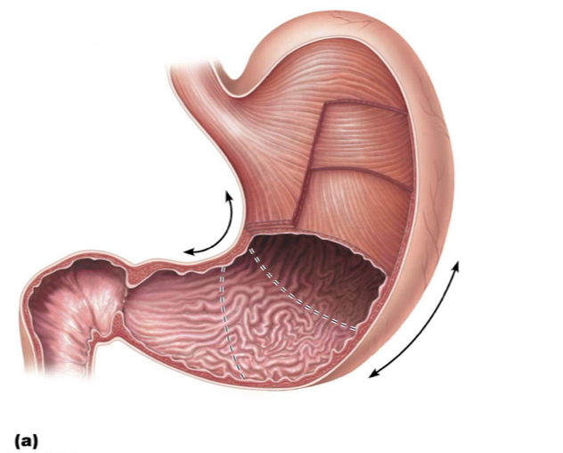
stomach
mechanical + chemical breakdown
simple columnar
gastric pits
longitudinal, circular, oblique
rugal folds
small intestine
absorption of nutrients into bloodstream and chemical digestion
simple columnar
peyer’s patches, villi, microvilli
lacteals
longitudinal, circular
circular folds
large intestine
contraction + storage of stool; nutrient + water resorption
simple columnar
longitudinal + circular
haustra, epipolic appendages, teniae coli
3 types of folds that maximize the SA of small intestine
circular folds
villi
microvilli
vitamins that are produced and absorbed in the large intestine
vitamin K
vitamin B
amount of baby teeth on maxilla + mandible
10 on each
how many adult teeth on the maxilla + mandible
16 on each
Incisors
shearing, cutting, biting
Canines
gripping, tearing, ripping
Pre-molars
crushing, tearing, grinding
molars
crushing, grinding, breaking down food into smaller pieces
3 sets of salivary glands
parotid
submandibular
sublingual
which set of salivary glands empties saliva by the upper molars
parotid
how does saliva aid in digestion
chemical breakdown (amylase + lipase)
dissolves food particles, allowing them to interact with taste buds
organ that makes bile
liver
organ that stores and secretes bile
gallbladder
alkalinity of pancreatic enzymes serves what purpose
neutralizes acidic chyme
pancreatic fluids are secreted into which part of the small intestine
duodenum
if a gallstone gets stuck in the opening where the bile duct enters the small intestine, which other organ will have the backup of its secretion?
pancreas—condition is gallstone pancreatitis
are the same organs retroperitoneal in cats as in humans
no organs are retro in cats
main functions of the digestive system
take in food
break it down into nutrient molecules
absorb molecules into bloodstream
rid body of any indigestible remains
alimentary canal
continuous muscular tube that runs from mouth to anus
functions of the AC
digests food: breaks down into smaller fragments
absorbs fragments through lining into blood
organs of the AC
mouth
pharynx
esophagus
stomach
small intestine
large intestine
anus
where does the AC constrict
sphincter
where does the AC dilate
stomach
accessory glands of the AC
salivary glands
tongue
teeth
pancreas
liver
gallbladder
teeth + tongue function
mechanical breakdown
digestive glands
produce secretions that help break down foodstuffs
salivary glands function
moisten food
liver function
produces bile
filters blood
stores glycogen
pancreas function
endocrine: secretes insulin and glucagon
exocrine: secretes H
why is it beneficial to have non-keratinized stratified squamous epithelium in the mouth, esophagus, and anus
many layers
resistance to abrasions and friction
why is it beneficial to have simple columnar epithelium in the stomach, small intestine, and large intestine
apical side faces lumen of AC
basal side faces bloodstream
one-way absorption of molecules
membrane proteins =
transporters
functions of saliva
cleanses mouth
dissolves food chemicals for taste
moistens food; compacts into bolus
begins breakdown of starch with enzyme amylase
major salivary glands include
parotid
submandibular
sublingual
parotid
anterior to ear and external to masseter muscle
Parotid duct opens into oral vestibule next to second upper molar
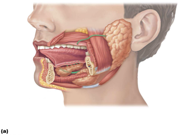
submandibular
medial to body of mandible
Duct opens at base of lingual frenulum

sublingual
anterior to submandibular gland under tongue
Opens via 10–12 ducts into floor of mouth

teeth location
lie in sockets in gum-covered margins of mandible and maxilla
mastication
process of chewing that tears and grinds food into smaller fragments
dentition and the dental formula
all but 3rd molars (wisdom teeth) are in by end of adolescence
3rd molars may or may not emerge around 17-25 years of age
incisors
rip/tear flesh of apple
canines
pierce flesh
premolars (bicupsids)
grind flesh
molars
best at grinding flesh due to “fit”
impacted tooth
a tooth that remains trapped in the jawbone
Can cause a good deal of pressure and pain
Wisdom teeth are most commonly involved.
Treatment: surgical removal
propulsion
involuntary, smooth muscle contractions propel and breakdown ingested food through GIT via peristalsis and segmentation
peristalsis
adjacent segments of the AC alternately contract and relax
food is moved along the tract
primarily propulsive
segmentation
nonadjacent segments of the AC contract and relax
back-and-forth sloshing mixes food and breaks it down mechanically
4 layers of AC
mucosa
submucosa
muscularis externa
serosa
mucosa functions
secretion
absorption
protection
submucosa
structural and elastic functions
contains blood and lymph vessel and nerve fibers
muscularis externa
deep circular layer (can form sphincters)
superifical longitudinal layer of smooth muscle for peristalsis and segmentation
serosa
made up of the visceral peritoneum
formed from alveolar CT covered with mesothelium (single layer of squamous epithelium) in most organs
peritoneum
serous membranes of abdominal cavity that consists of:
Visceral peritoneum: membrane on external surface of most digestive organs
Parietal peritoneum: membrane that lines body wall
Peritoneal cavity (the abdominopelvic cavity)
Fluid-filled space within the parietal peritoneum
Fluid lubricates mobile organs
mesentery functions
double layer of peritoneum; layers are fused back to back
Extends from posterior of abdominopelvic cavity wall to digestive organs
Provides pathway for blood vessels, lymphatics, and nerves
Holds organs in place and also stores fat
intraperitoneal organs
located within the peritoneal cavity
retroperitoneal organs
located outside, or posterior to, the peritoneum
Includes most of pancreas, duodenum, and parts of large intestine
Food passes from mouth to
oropharynx and then into laryngopharynx
pharynx function
Allows passage of food, fluids, and air
Stratified squamous epithelium lining with mucus-producing glands
External muscle layers consists of two voluntary skeletal muscle layers
inner layer of muscles runs longitudinally
Outer pharyngeal constrictors encircle wall of pharynx
esophagus
flat muscular tube that runs from laryngopharynx to stomach
esophagus is … when not involved in food propulsion
collapsed
Gastroesophageal sphincter GES
Keeps orifice closed when food is not being swallowed
Mucus cells on both sides of sphincter help protect esophagus from acid reflux
digestive processes in the stomach
1.Serves as holding area for food
2.Carries out mechanical breakdown of food (churns)
3.Denatures proteins using acidity (HCl)
•Denature means breaks bonds so unfolds proteins
4.Goblet cells secrete mucous to protect lining from acid (HCl)
5.Pepsin carries out enzymatic digestion of proteins
•Milk protein (casein) is broken down by rennin in infants
•Results in curdy substance
6.Delivers chyme to small intestine
anatomy of stomach
microscopic structure of esophagus

accessory organs are associated with small intestine
liver
gallbladder
pancreas
bile
fat emulsifier
pancreas function
supplies most of enzymes needed to digest chyme, as well as bicarbonate to neutralize stomach acid
small intestine
located between pyloric sphincter+ ileocecal valve
20 feet long when relaxed
3 divisions: duodenum, jejunum, ileum
circular folds
Permanent folds that force chyme to slowly spiral through lumen, allowing more time for nutrient absorption
villi
Fingerlike projections of mucosa with a core that contains dense capillary bed and lymphatic capillary called a lacteal for absorption
microvilli
Cytoplasmic extensions of mucosal cell that give fuzzy appearance called the brush border that contains membrane-bound enzymes brush border enzymes, used for final carbohydrate and protein digestion
Small intestine modifications and lymphoid nodules pattern
the abundance of small intestine modifications decrease in number over the small intestine, the number of Peyer’s Patches increase
teniae coli
three bands of longitudinal smooth muscle in muscularis
haustra
pocket-like sacs caused by tension of teniae coli
epiploic appendages
fat-filled pouches of visceral peritoneum
subdivisions of large intestine
cecum
appendix
colon
rectum
anal canal
cecum
first part of large intestine (retroperitoneal)
appendix
masses of lymphoid tissue
retroperitoneal
MALT
Bacterial storehouse capable of recolonizing gut when necessary
Twisted shape of appendix makes it susceptible
4 portions of colon
ascending—retro
transverse
descending—retro
sigmoid
ascending colon
travels up right side of abdominal cavity to level of right kidney
transverse colon
travels across abdominal cavity
descending colon
travels down left side of abdominal cavity
sigmoid colon
S-shaped portion that travels through pelvis
rectum
retro
3 rectal valves stop feces from being passed with gas
anal canal
last segment of large intestine that ends at anus
2 sphincters: internal and external