Module 3
 Call Kai
 Learn
Learn Practice Test
 Spaced Repetition
 Match
Match Flashcards
 Knowt Play
1/41

There's no tags or description
Looks like no tags are added yet.
Name | Mastery | Learn | Test | Matching | Spaced | Call with Kai | Chat |
|---|
No analytics yet
Send a link to your students to track their progress
42 Terms
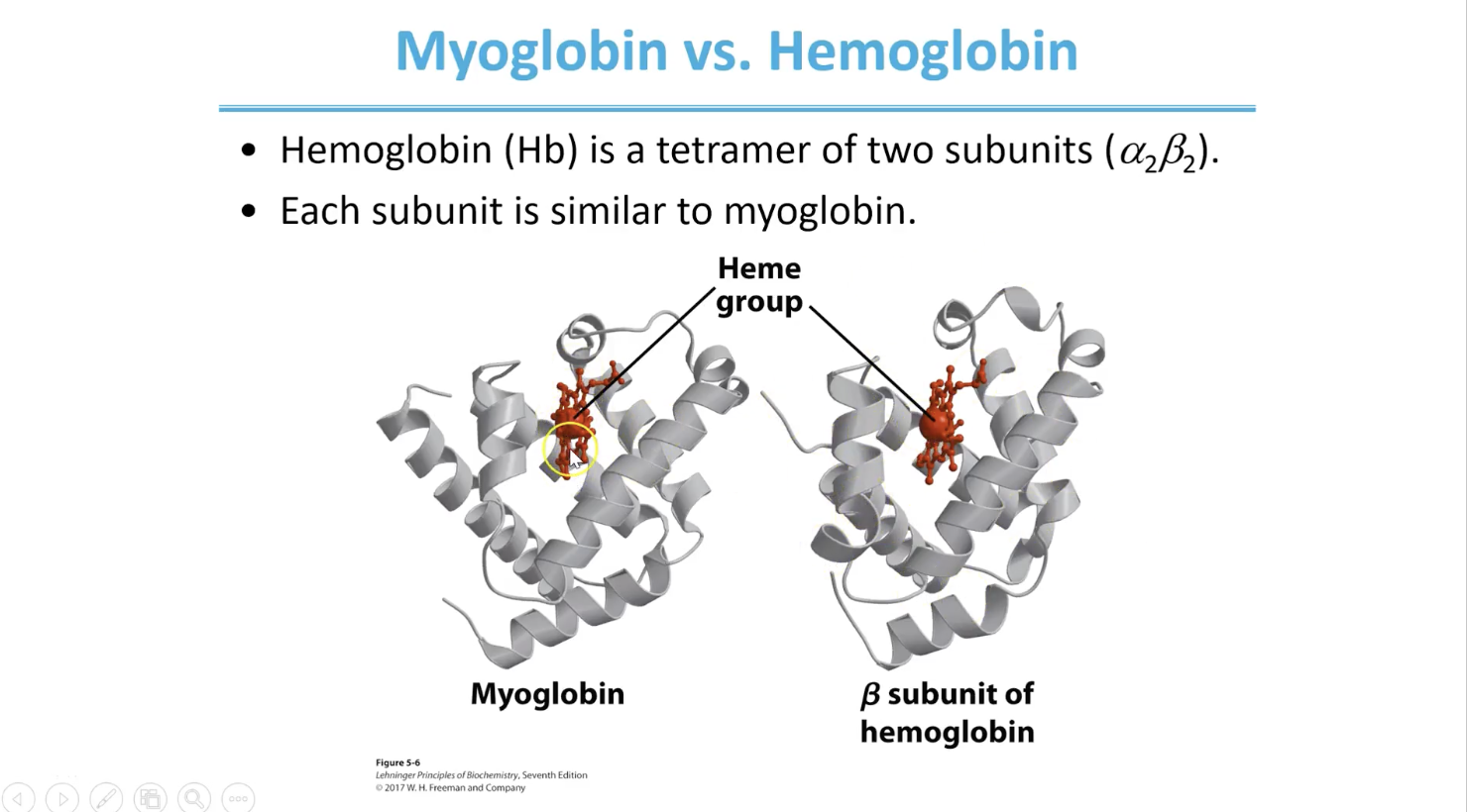
Myoglobin vs Hemoglobin
Myoglobin: monomer, singular polypeptide creating single domain
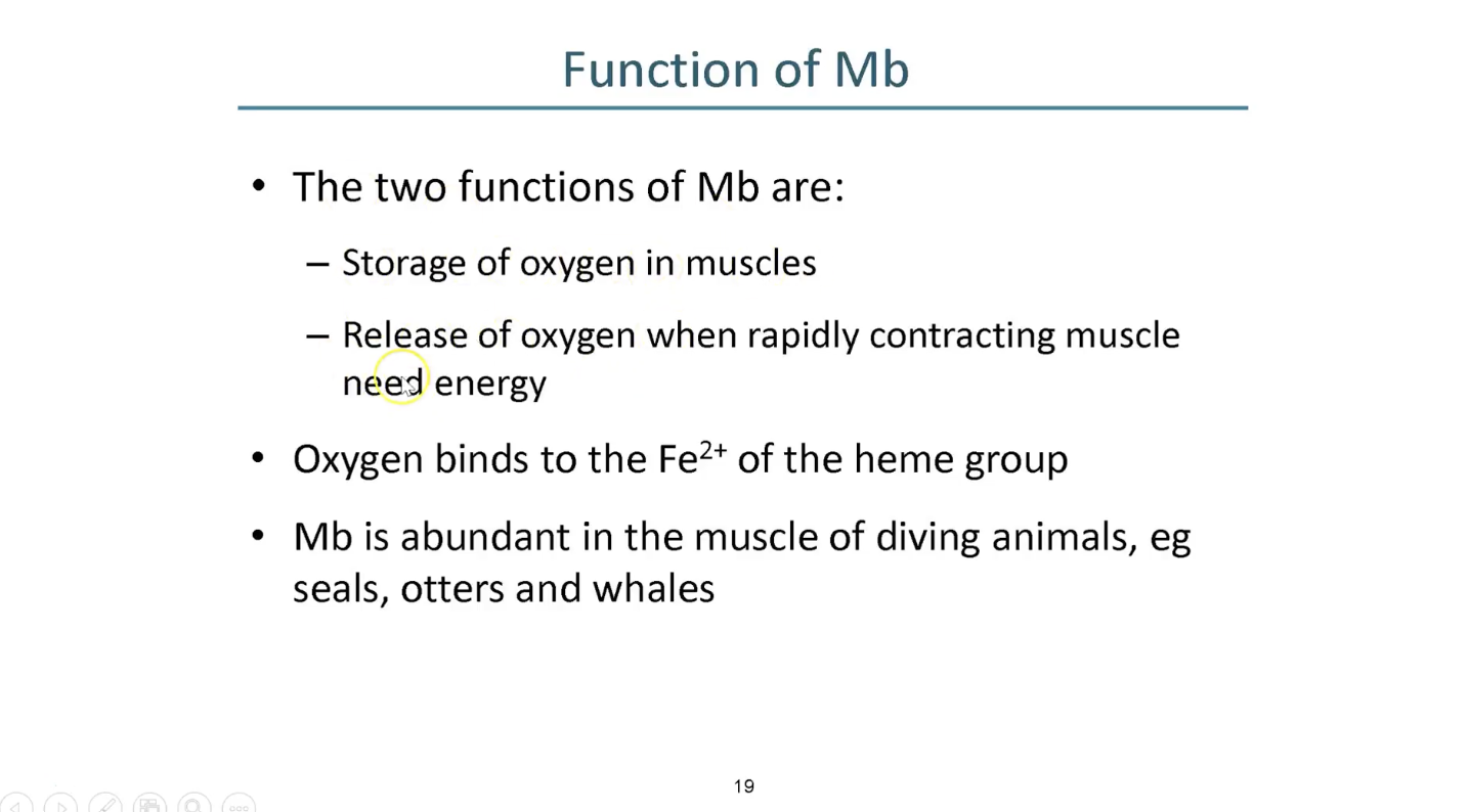
stores oxygen
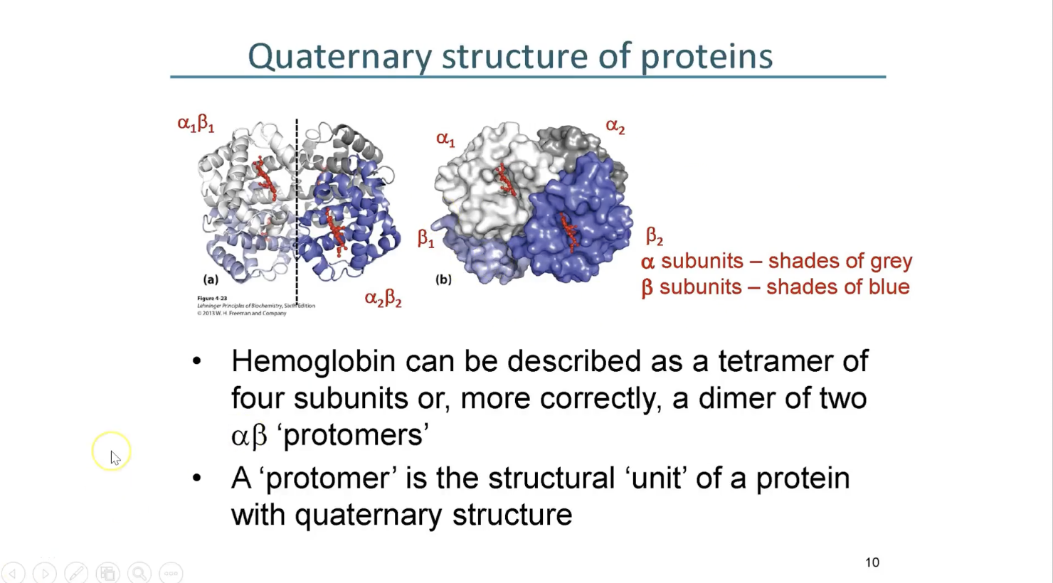
Hemoglobin: tetramer with quart structure - similar fold but 4 subunits
transports oxygen
Heme group coordinates the oxygen that binds
Other ways to describe hemoglobin
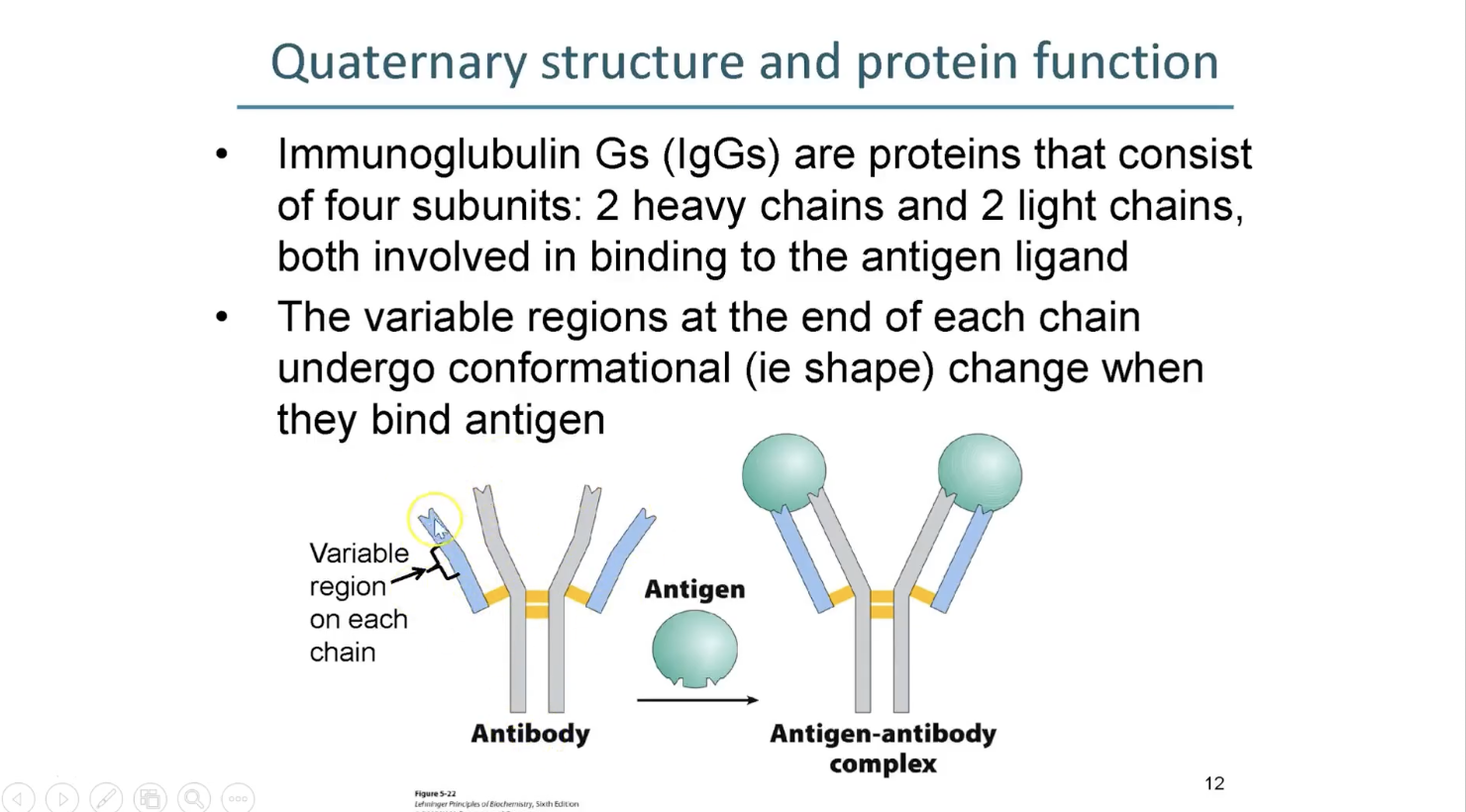
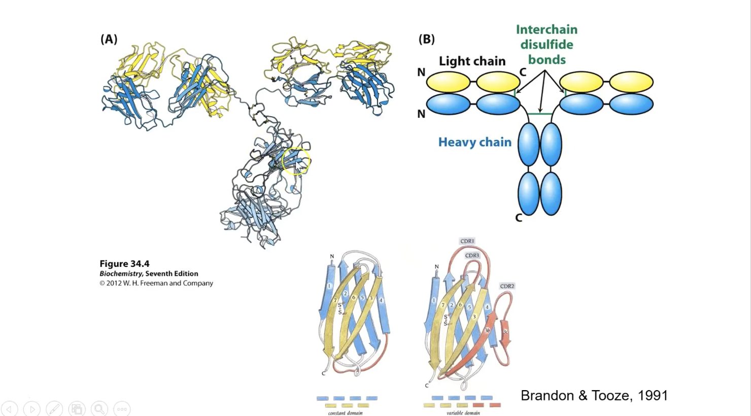
Immunoglubulin Gs (antibody)
Tetramer bc composed of 4 chains
but symmetry indicates that its a dimer of light chain - heavy chain pairs
Different representations of antibodies (Immunoglubulin G)
4 domains in left corner 4 ovals in schematic (two yellows and 2 blues)
4 domains hanging at bottome (all blues) form the constant region
right in bottom is a variable version of Immunoglubulin G fold
similar to left original one but with some adjustments and additions
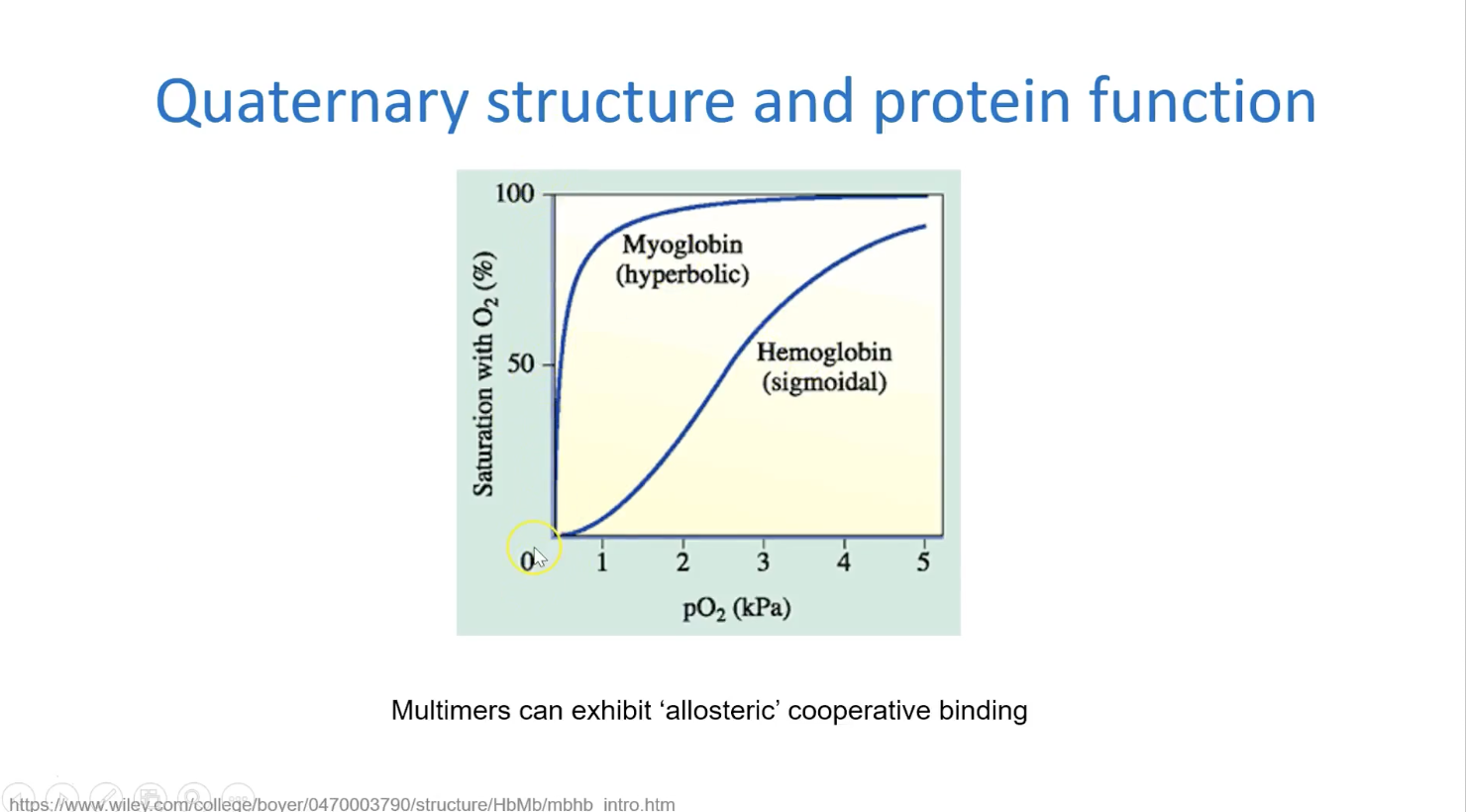
Why is hemoglobin multimeric? - why does it have multiple domains/binding. sites
Binding properties comparison
Myoglobin Function
Release of O2 is when oxygen conc in tissue drops
Mb and diving mammals
Proteins and ligand explanation with Mb
O2 is the ligand
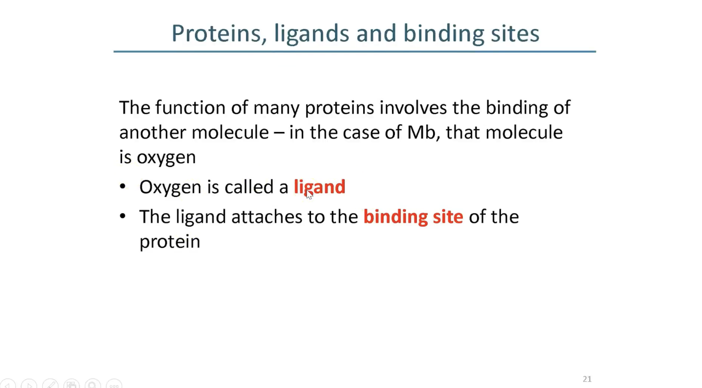
Mb and oxygen binding
prosthetic: bc its seperate to the protein
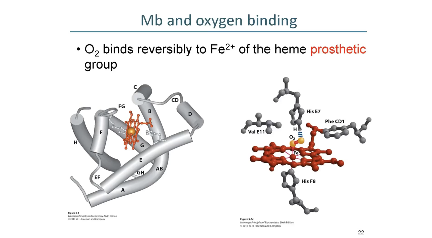
Heme prosthetic group
1 ligand above and below heme ring
on other sides is the his residue and then O2
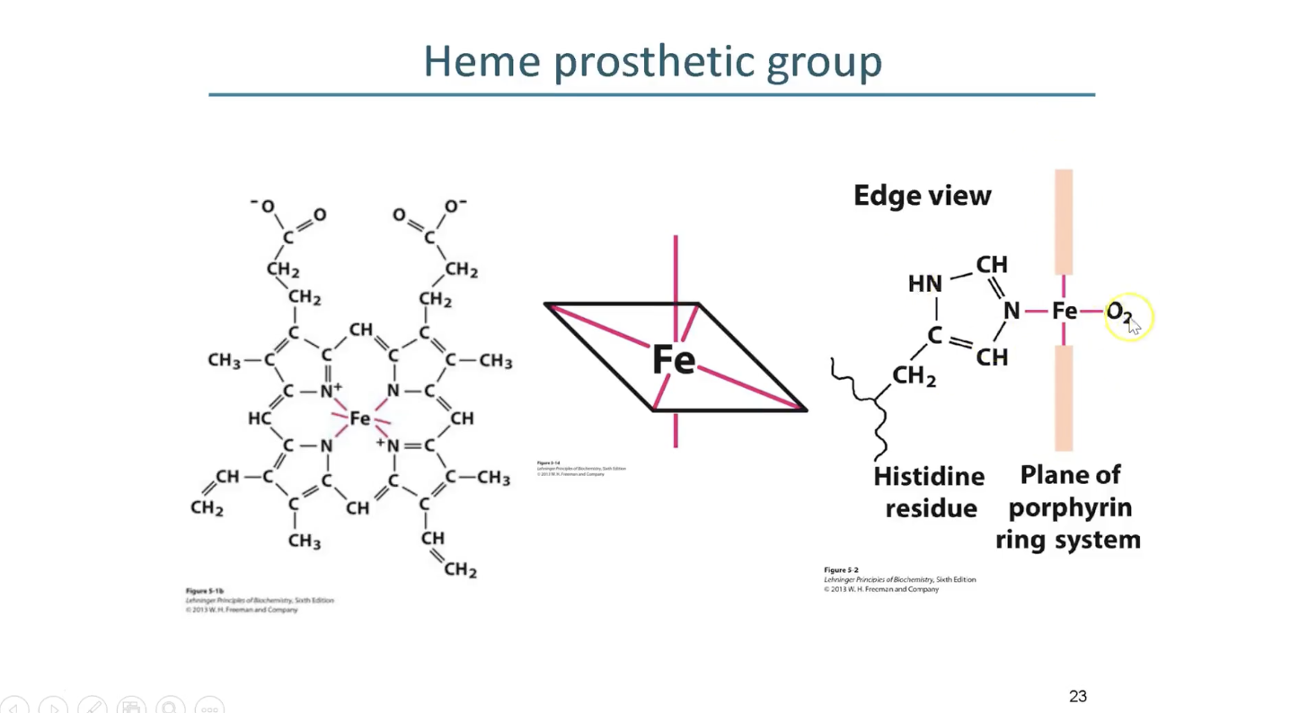
Equilibrium Association and Dissociation Constants
Why Kd is preferred over Ka
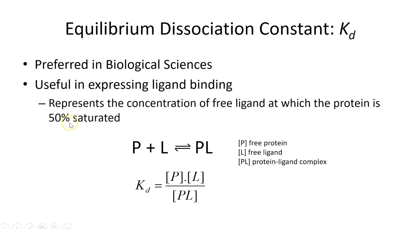
How Kd looks on the graph + important eq to remember
x-axis: theta = shows how many binding sites occupied
hyperbolic in shape
kd is also indicated on the graph - it is the y-axis value when 50% of binding sites are occupied
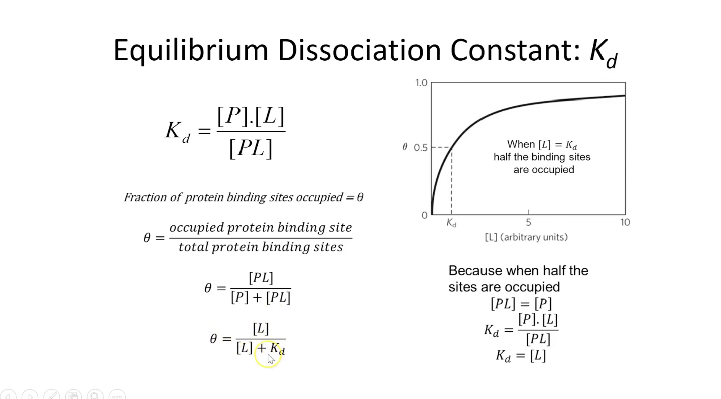
What Kd value tells us about how tight interactions are
low affinity: weak binding
high affinity: tight binding receptor
avidin-biotin is so tight that its practically irreversible
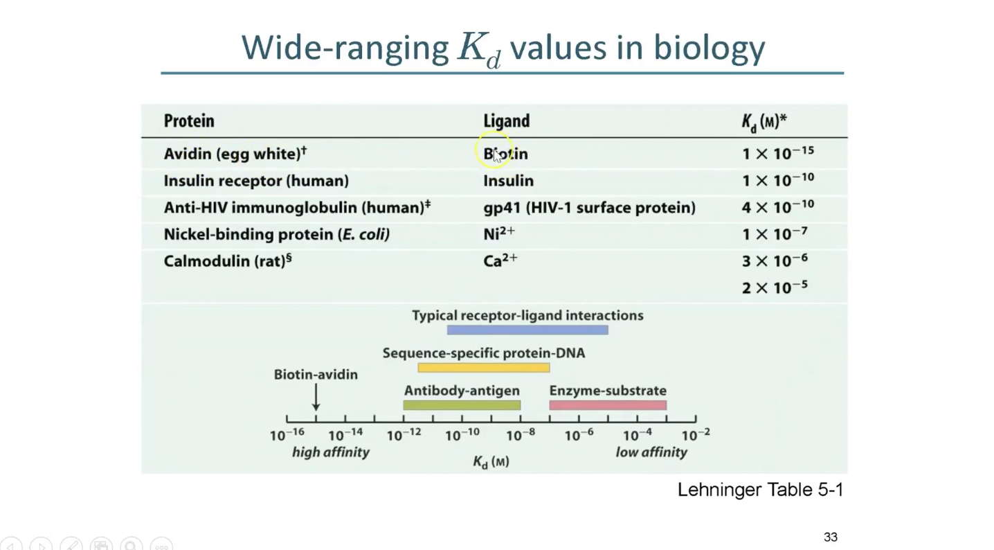
Biotin
vitamin that can only be provided by the diet
it binds to protein avidin in raw egg white - eggs’ defence mechanism against bacteria
avidin in egg white will extract the biotin out of cell and kill them
Mb and oxygen binding curve
y-axis: conc
x-axis: saturation of binding sites
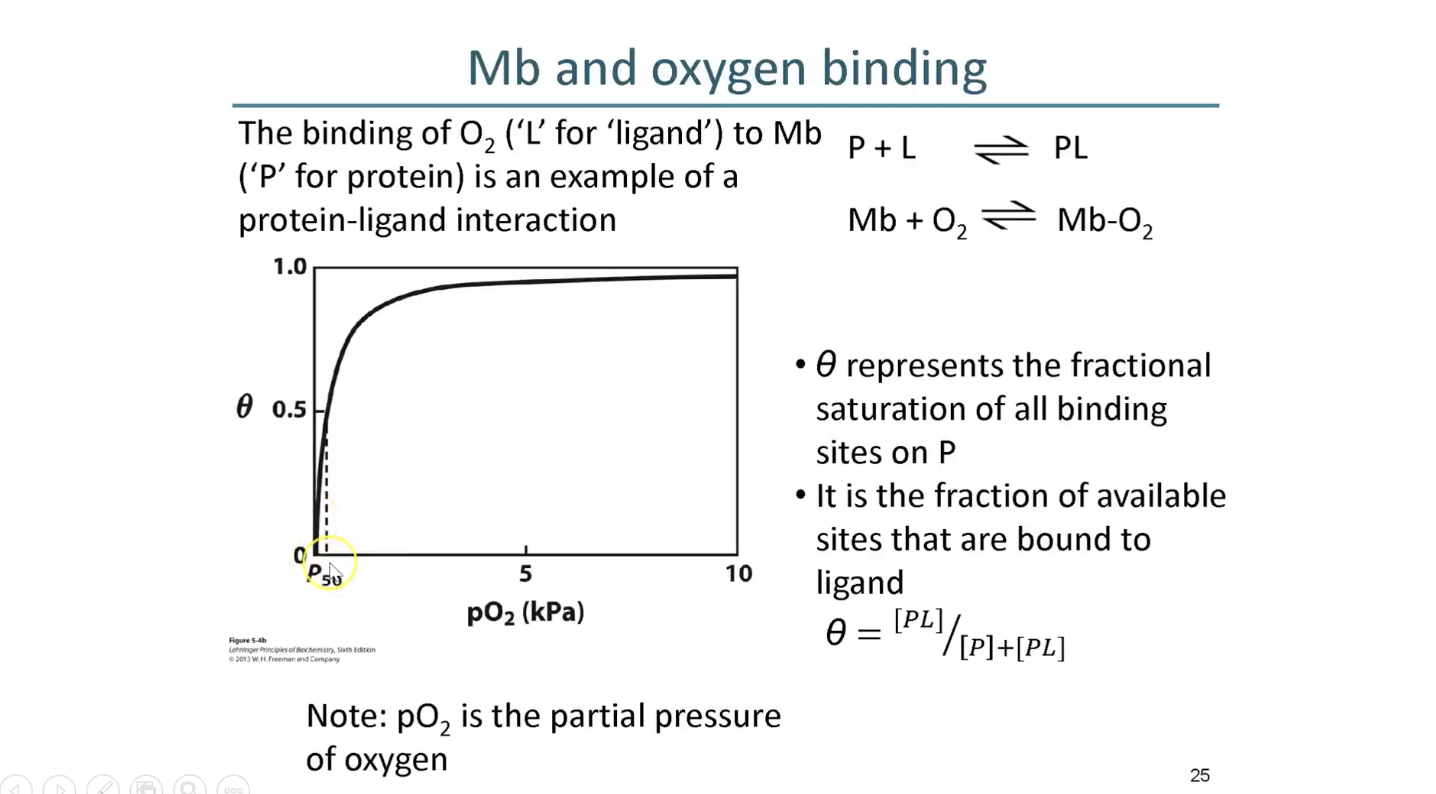
Could myoglobin transport O2?
Myoglobin binds O2 very tightly and O2
Myoglobin would pick up O2 very tightly in the lungs and become almost saturated but when moved to the tissues, it will deliver very little
List of traits that would be necessary for an oxygen transporting molecule on the side
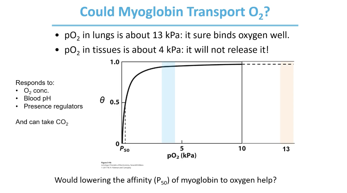
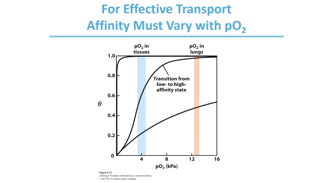
Affinity varying with pO2 (hemoglobin)
How it can change it’s affinity to binding O2 like this is bc it changes between conformations that have a high affinity (at lung partial pressure) and then low affinity at tissues partial pressure
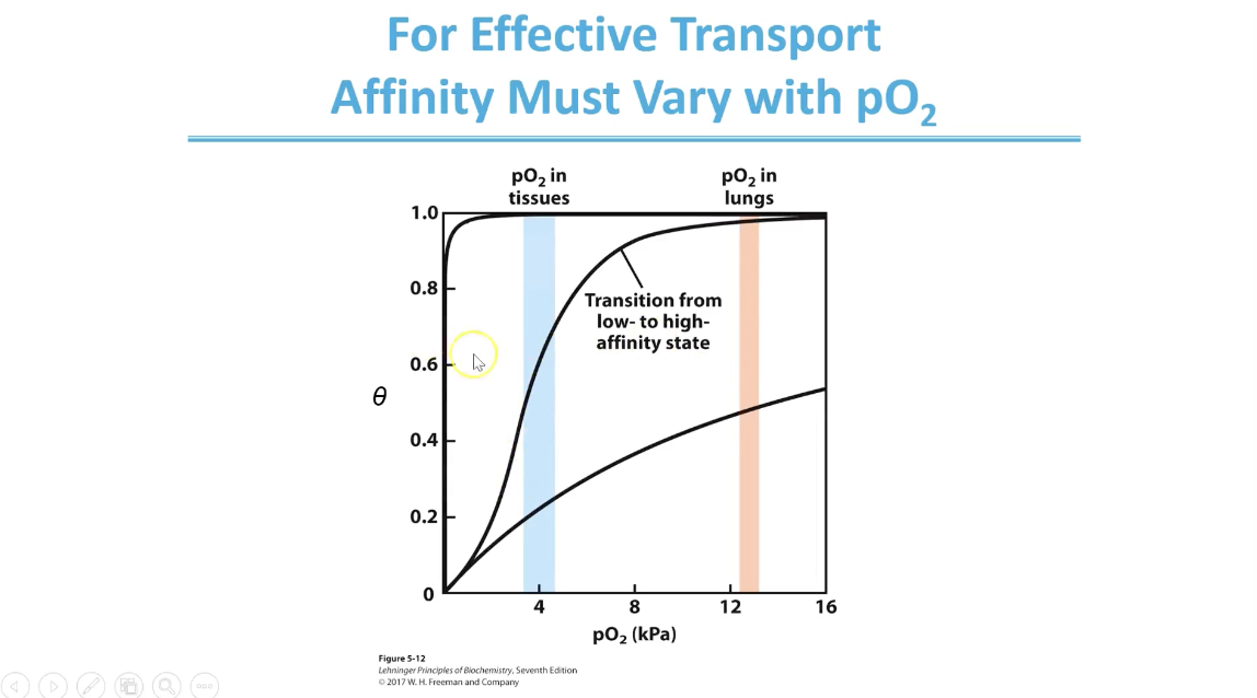
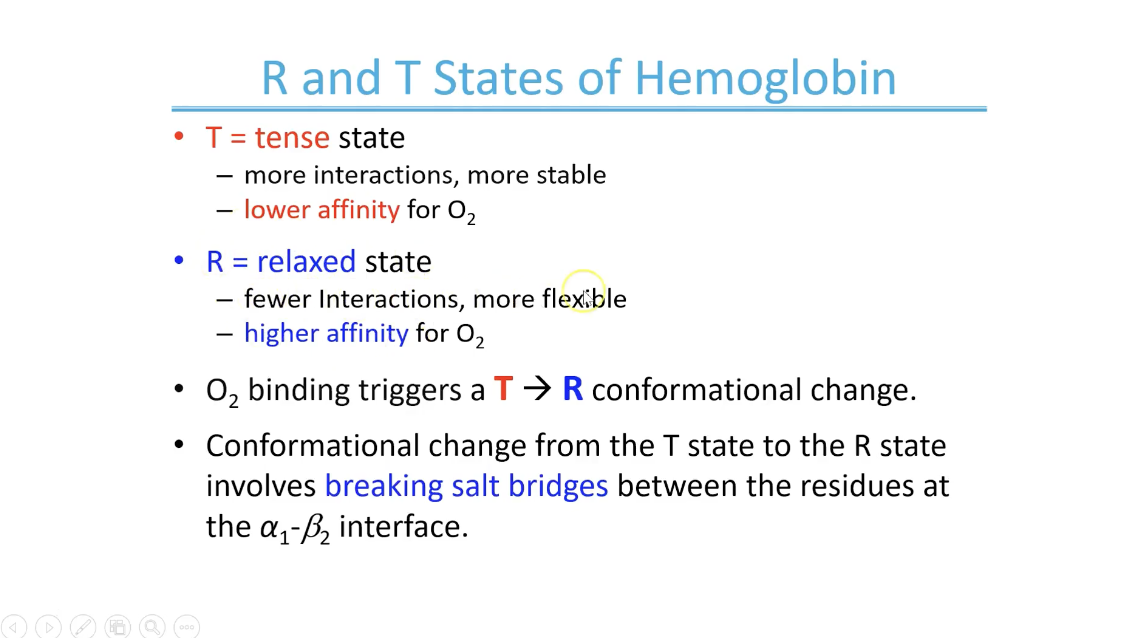
Mechanism that helps switch bw high affinity and low affinity states based on O2 conc
R and T States of Hemoglobin
O2 binding in one subunit can trigger the conformational change in both that subunit and others
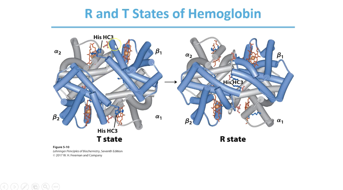
R and T States crystal structure
salt bridge interactions stabilise the T state
O2 binding destabilises these salt bridge interactions and allows transition to R state
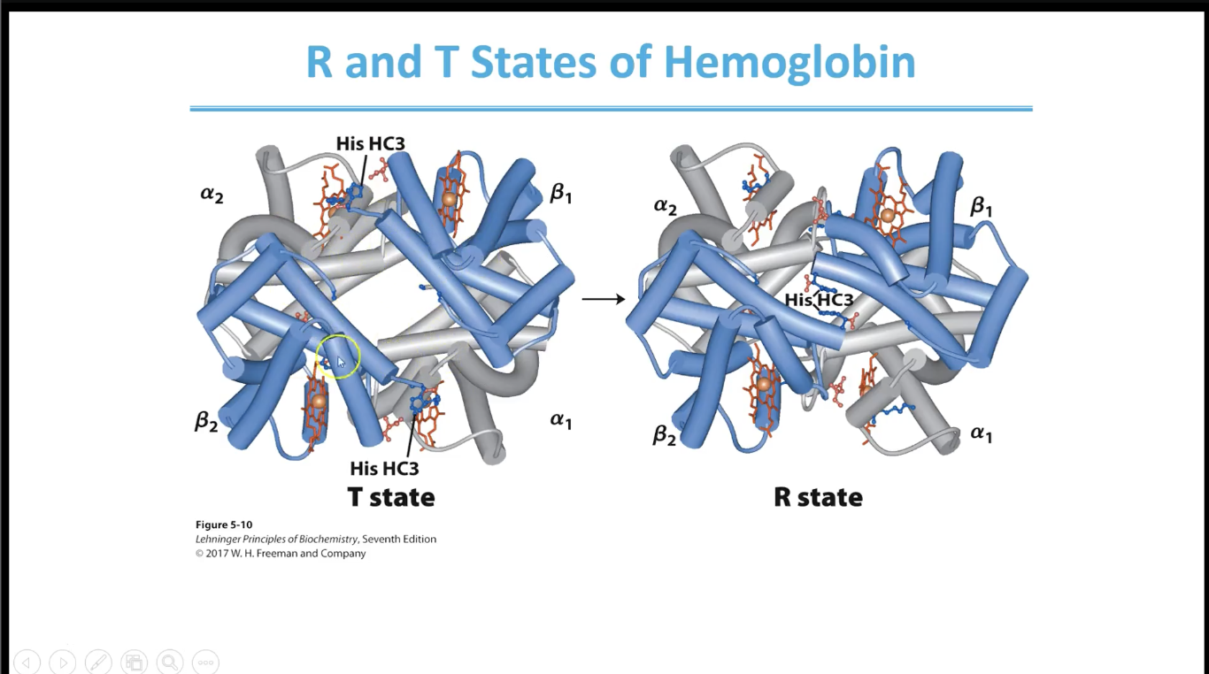
Important specific subunit interactions
Salt bridge bw positive charge on histadine HC3 side chain and Aspartic acid FG1 carboxylate group
Lys C5 and H HC3 also have salt bridge interactions
Examples of subunit interactions
if oxygen binds to any one of the heme groups, it destabilise and changes to R group
Conformation change triggered y O2 bonding
O2 pulls Iron 2 through the heme ring to other side and flattens the heme ring which stabilises the transition to the R state
Ka changes for cooperative binding - Hill Equation
Ka is the reciprocal of Kd
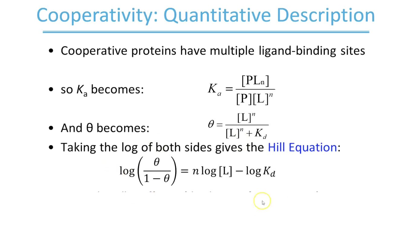
Hill equation breakdown
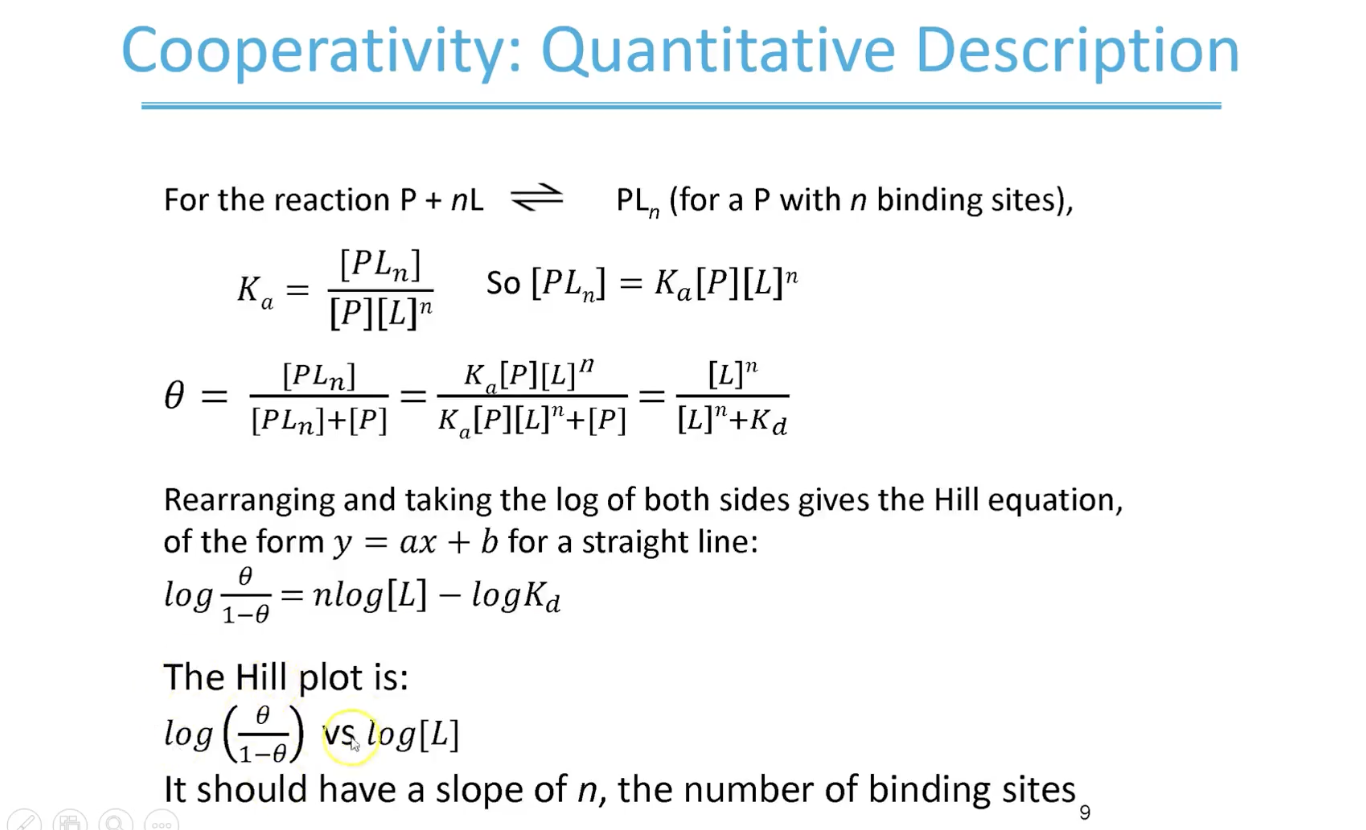
What the slope of Hill plot gives us
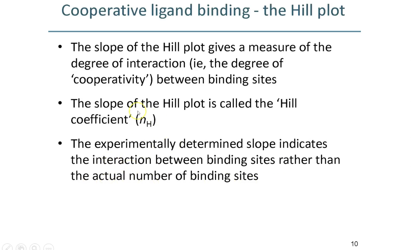
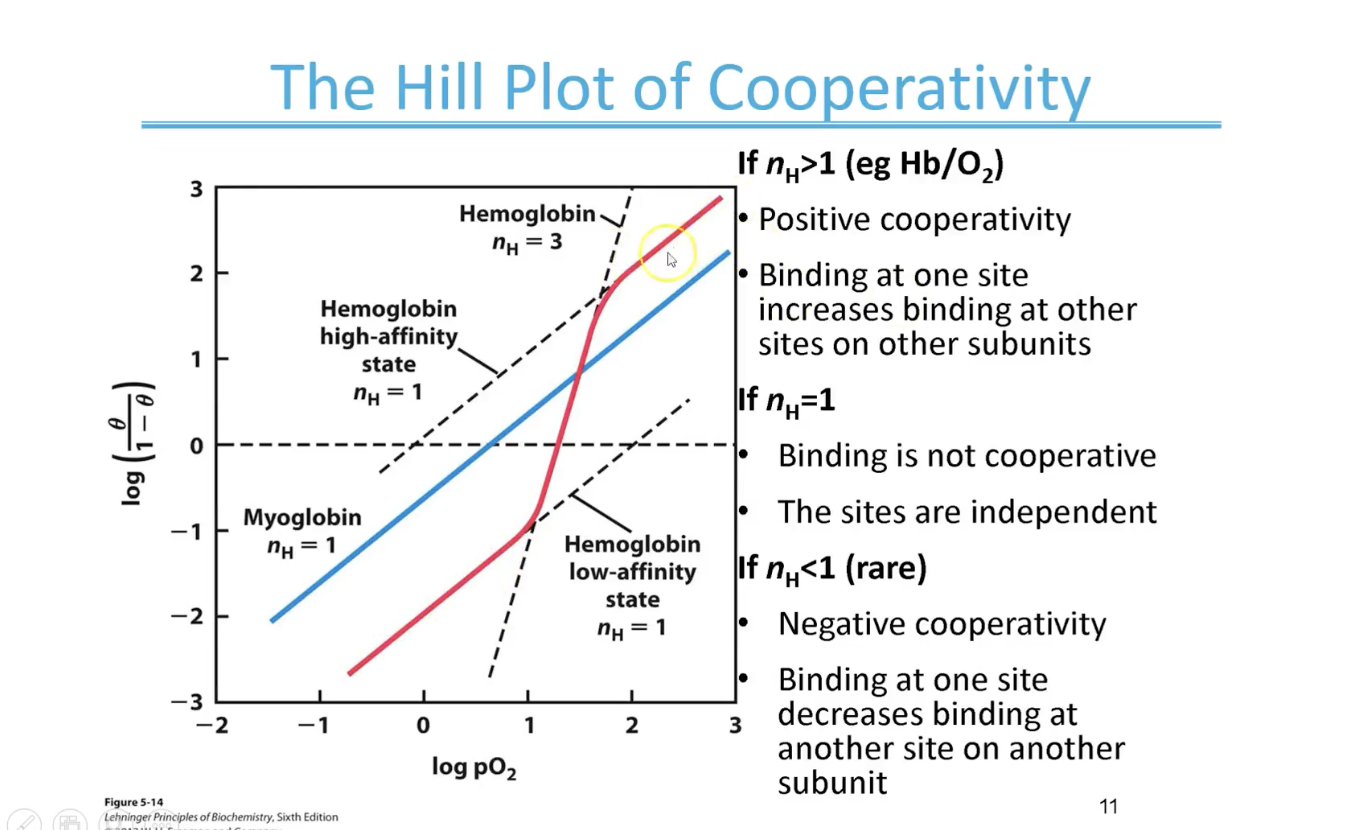
Hill Plot cooperativity interpretation for hemoglobin
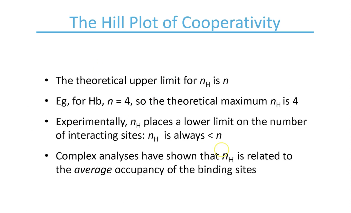
Upper limit of Hill plot - max
What are the two models of cooperativity
concerted or sequential
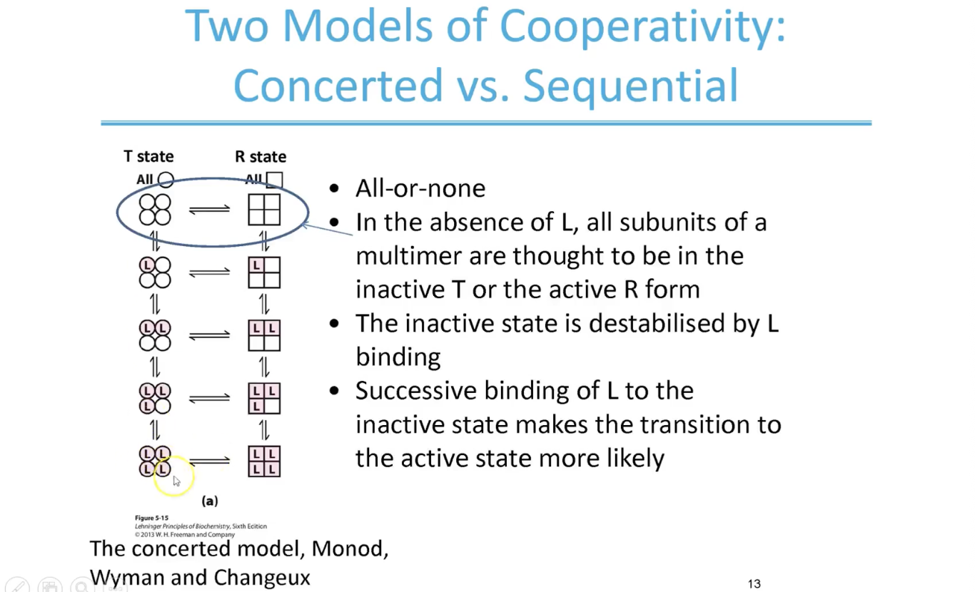
Concerted model
ALL OR NONE
circles represent subunits in the low affinity T states and squares represent subunits in high affinity R states
shaded L means there’s a ligand bound to that subunit
In the absence of L, all subunits are in the inactive T or active R form but bc T form is more stable → more would be in that state
successive binding of ligand to subunits increases the likelihood of that transition from T state to R state
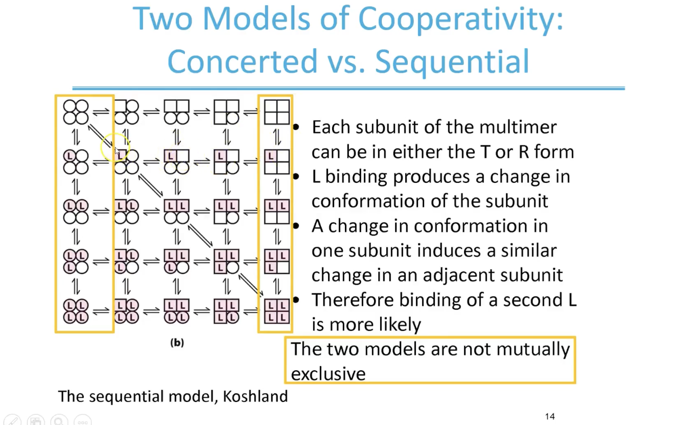
Sequential model
The highlighted in yellow shows the elements of concerted model - physical reality is probably better represented by a composite model somewhere in the middle
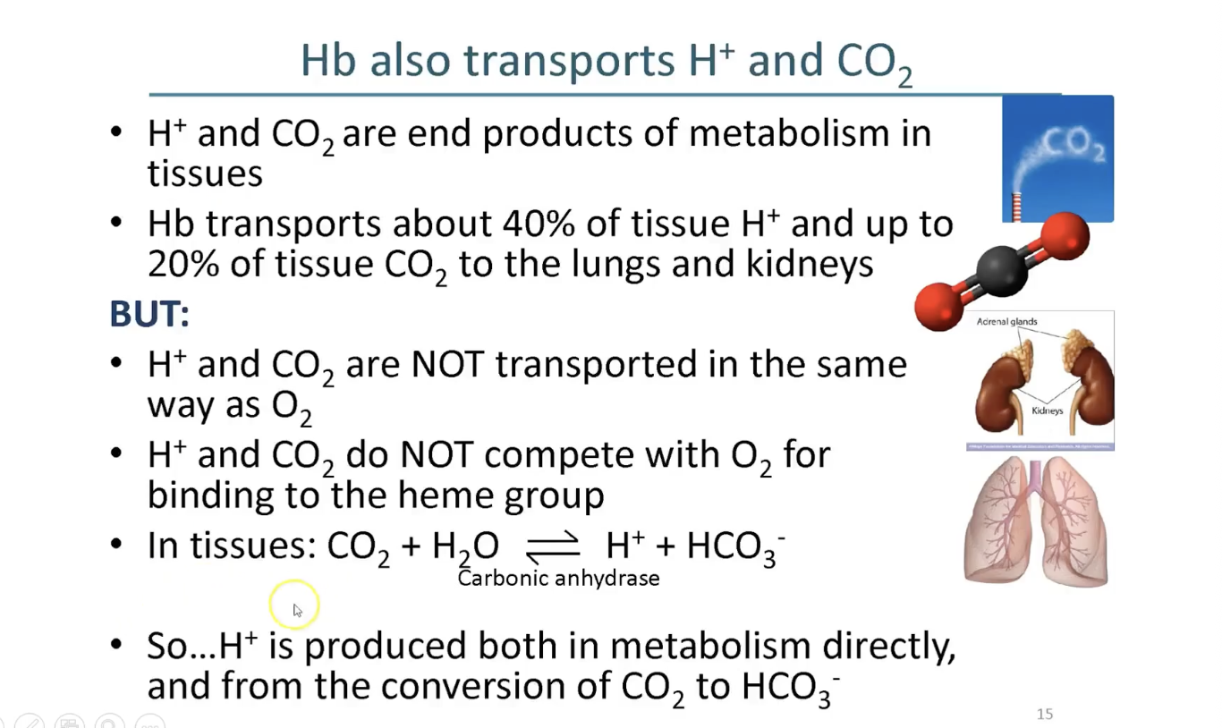
Hb also transports H+ and CO2 differently
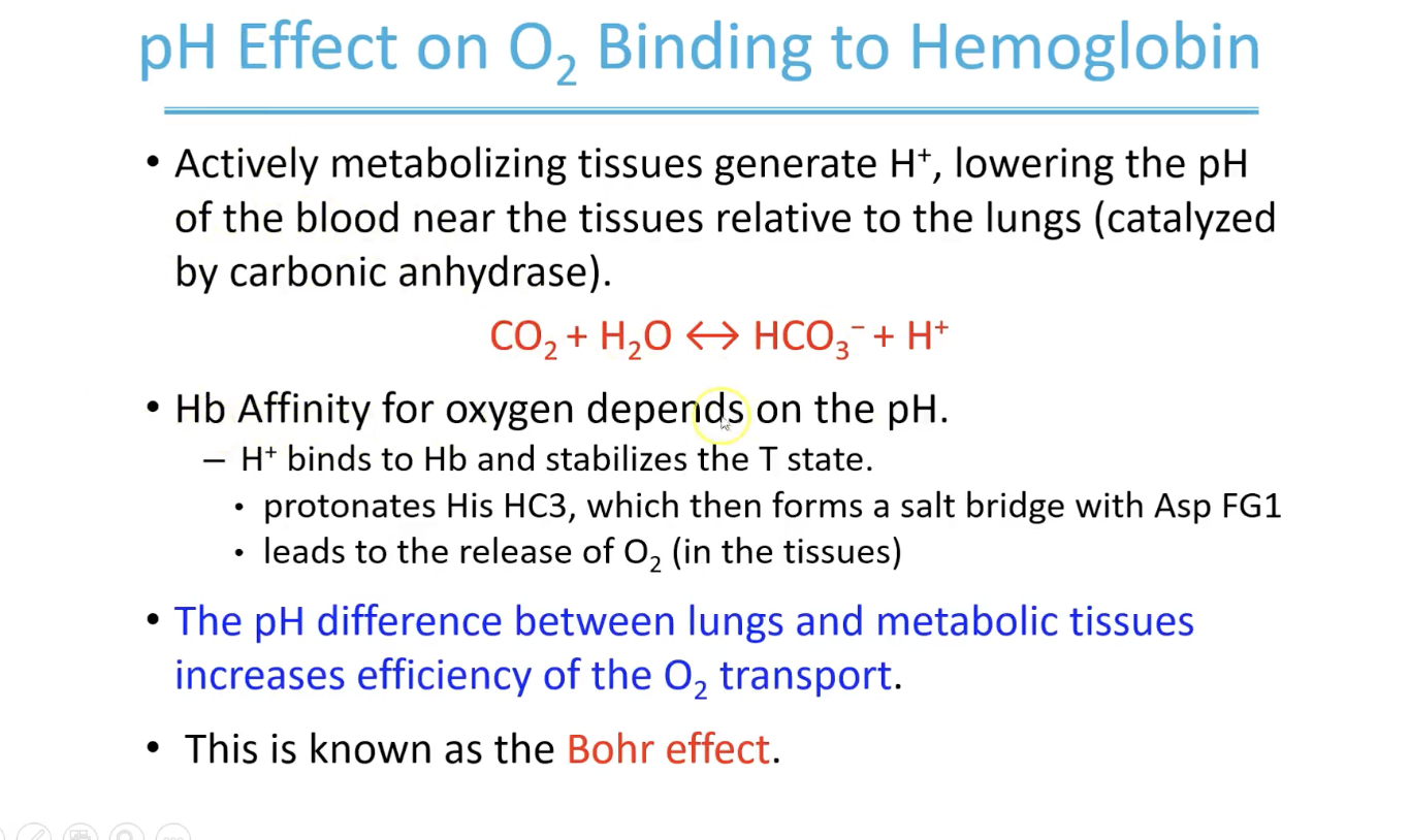
pH effect on O2 binding to hemoglobin
right shift indicates weaker binding and left shift indicates tighter binding
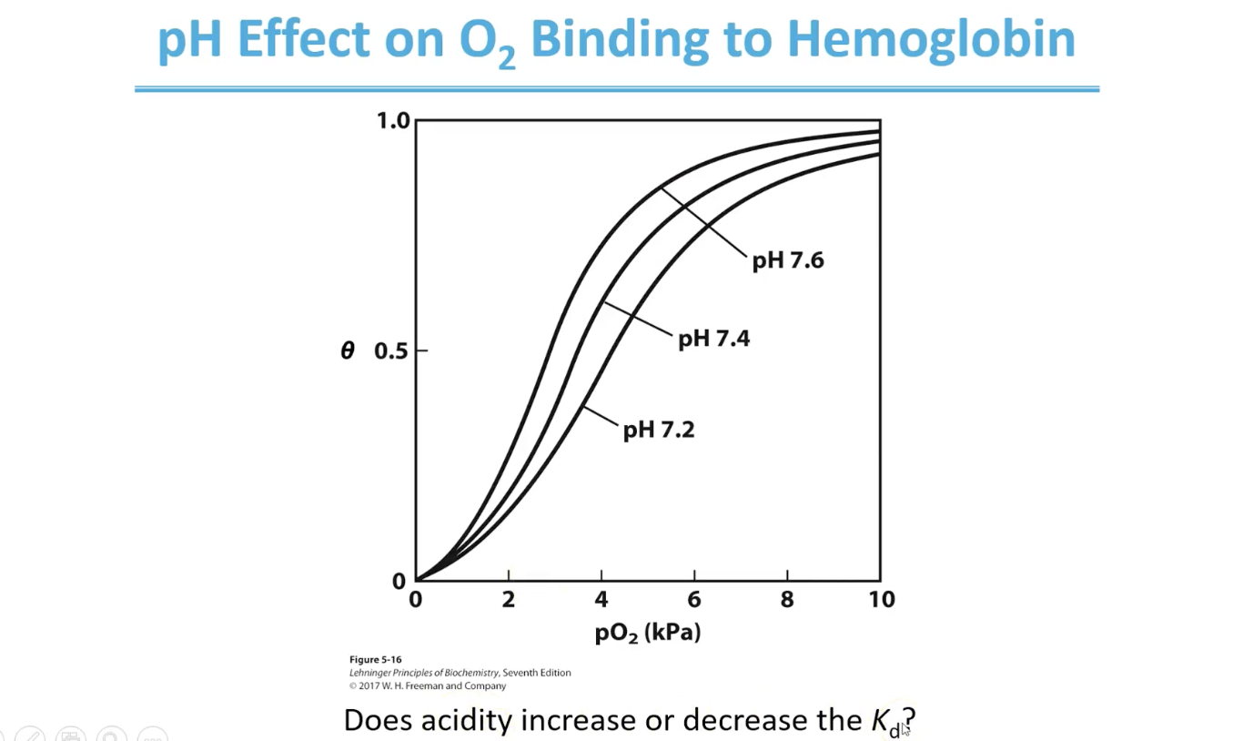
Effect of H+ on binding of O2 to Hb (lower pH)
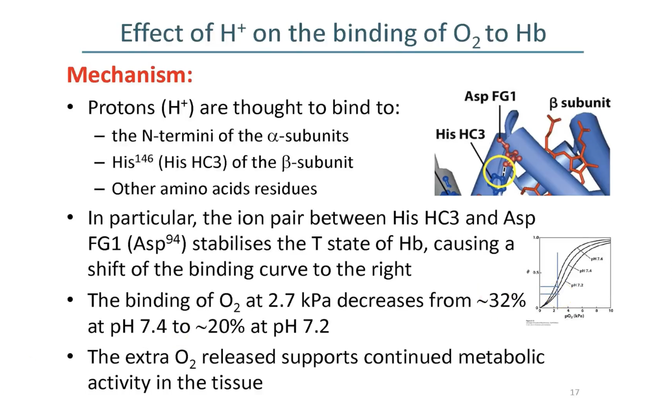
Lower pH = lower affinity of Hb for O2 = T state stabilised
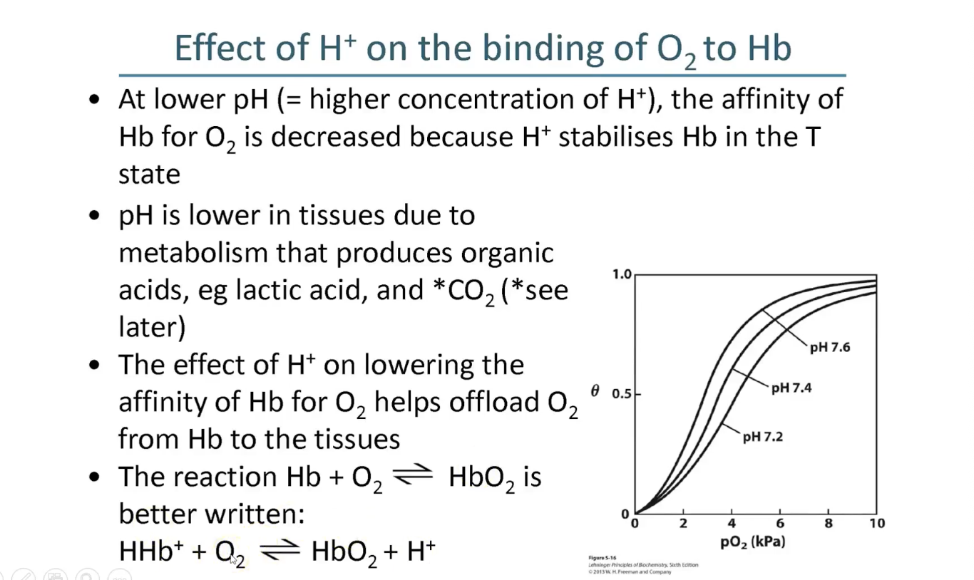
Mechanism by which Hemoglobin picks up protons
His HC3 forms that salt bridge with Asp FG1
Effect of H+ on bindings curve mechanism
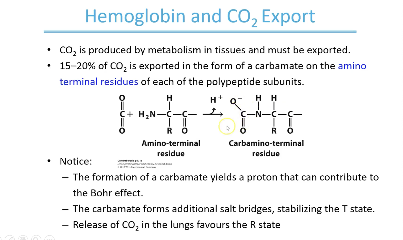
Hemoglobin and CO2 export
The negative carbonate group also helps form additional salt bridges stabilising the T state but when CO2 released - these salt bridges are disrupted and R state is favoured
Overview of BPG
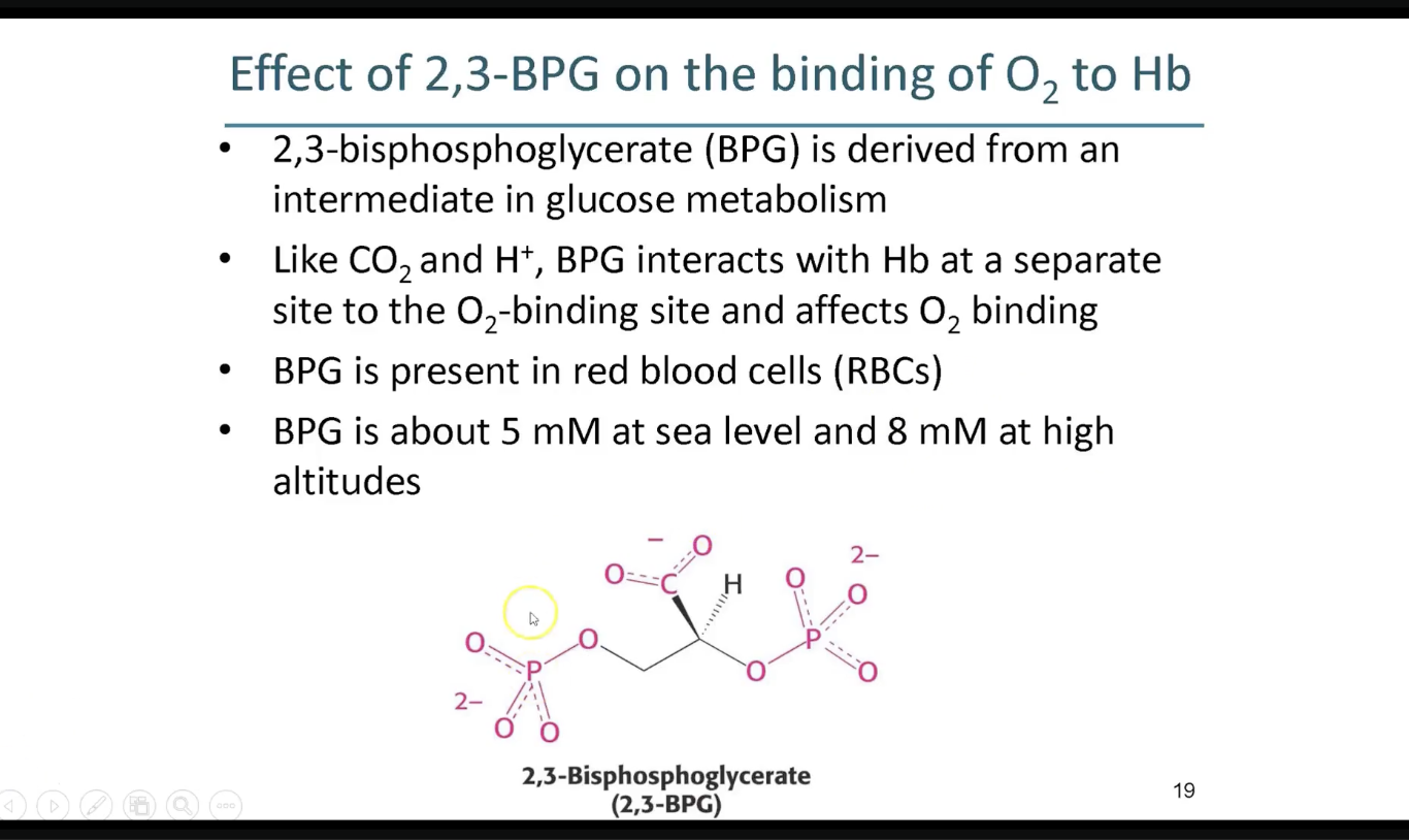
2,3-BPG binding function - T state
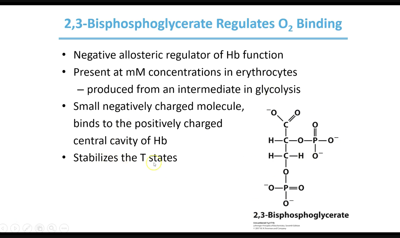
How BPG effects binding of O2 to Hb
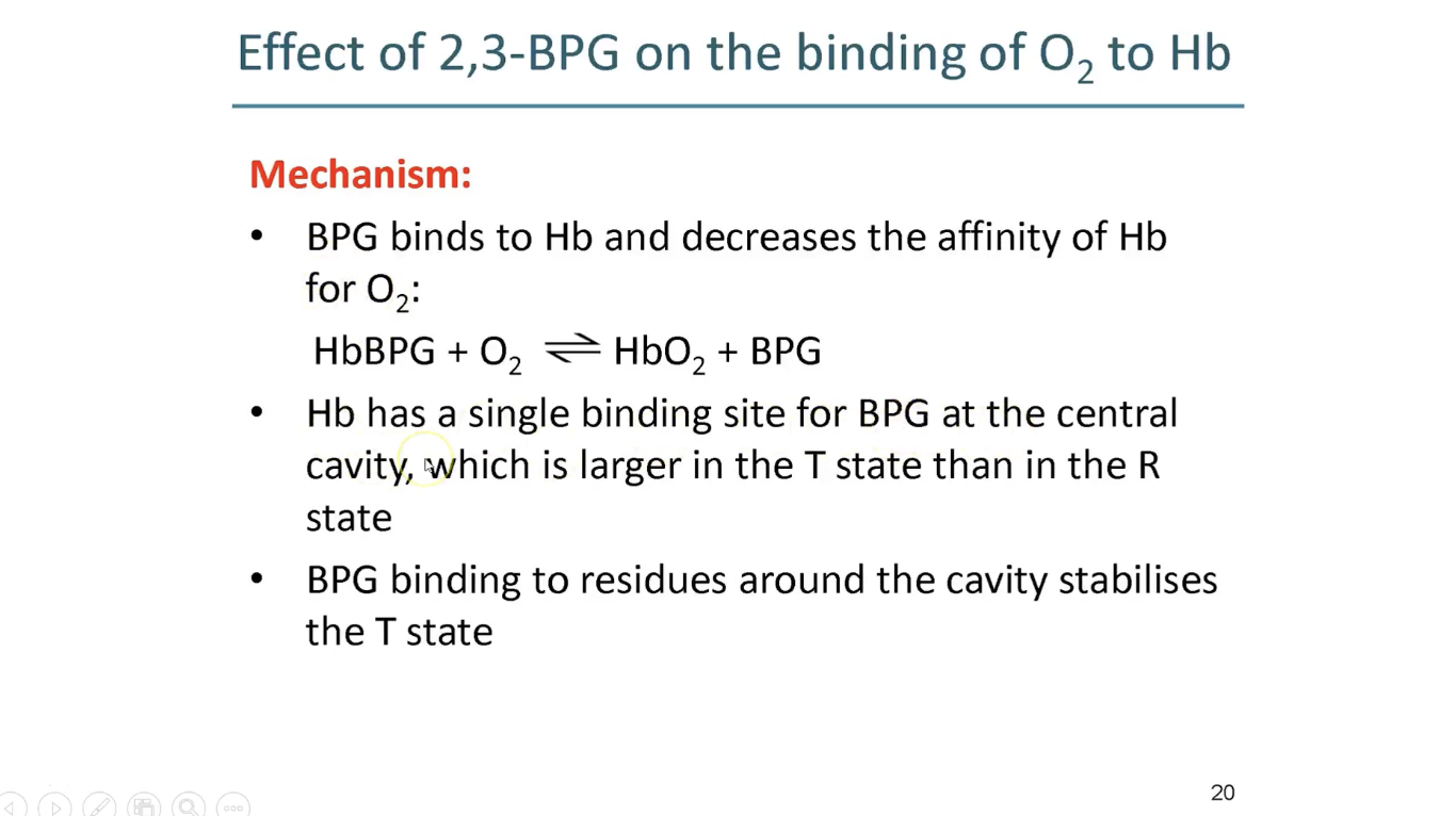
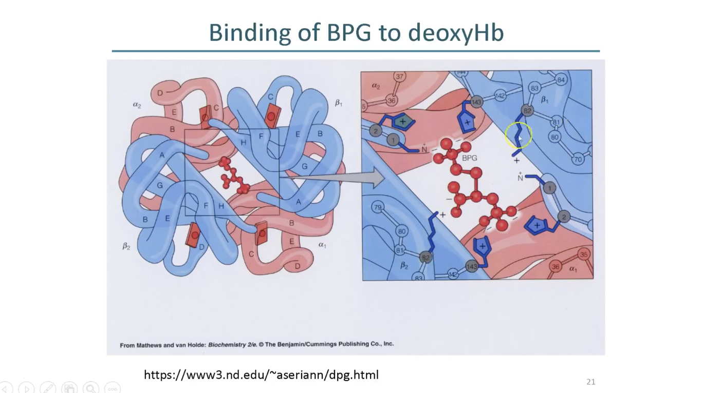
BPG binding to deoxyHb
BPG is binding into the cavity of T state of hemoglobin
it interacts with positively charged residues (Lys)
Bohr effect and precense of BPG stabilise T state