HUMB C9
 Call Kai
 Learn
Learn Practice Test
 Spaced Repetition
 Match
Match Flashcards
 Knowt Play
1/40

There's no tags or description
Looks like no tags are added yet.
Name | Mastery | Learn | Test | Matching | Spaced | Call with Kai | Chat |
|---|
No analytics yet
Send a link to your students to track their progress
41 Terms
Spinal cord Regions (superior to inferior)
Cervical, Thoracic, Lumbar, Sacral, Coccygeal
Spinal nerves
31 pairs:
8 cervical
12 thoracic
5 lumbar
5 sacral
1 coccygeal
vetebral bones
30 bones:
7 cervical because C8 nerve exits superior to T1 vetebra
12 thoracic
5 lumbar
5 sacral
1 coccygeal
Spinal nerve and vertebra relative position
7 cervical nerves exit superior to their corresponding vertebra
C8 nerve exits superior to T1 vetebra
All nerves inferior to C8 nerve exit inferior to their corresponding vertebra
enlargements
Thicker areas of spinal cord, located where plexuses are
2 - Cervical and lumbosacral
Conus medullaris
Cone-shaped area inferior to lumbosacral enlargement where spinal cord tapers off and ends
Lumbar and sacral nerves exit below
cauda equina
bundle of lumbar, sacral, and coccygeal nerve roots that descend inferior to conus medullaris, through vetebral canal providing dual innervation to lower limbs and pelvic area
Meninges - Def and functions
Connective tissue surrounding brain and spinal cord
Protection, contains CSF, form partitions inside skull
meninges layers
Outer: epidural space - adipose tissue
Dura mater - thick, strong
subdural space
Arachnoid mater
Subarachnoid space – CSF and blood vessels
Inner: Pia mater - thin covering with many small blood vessels
Nerve coverings
3 layers of CT
Endoneurium– surrounds each axon
Perineurium– surrounds a fascicle (bunch of axons)
Epineurium – surrounds several fascicles
Brain regions
Forebrain: Cerebrum and Diencephalon
Midbrain
Hindbrain: Pons, Medulla oblongata, and Cerebellum
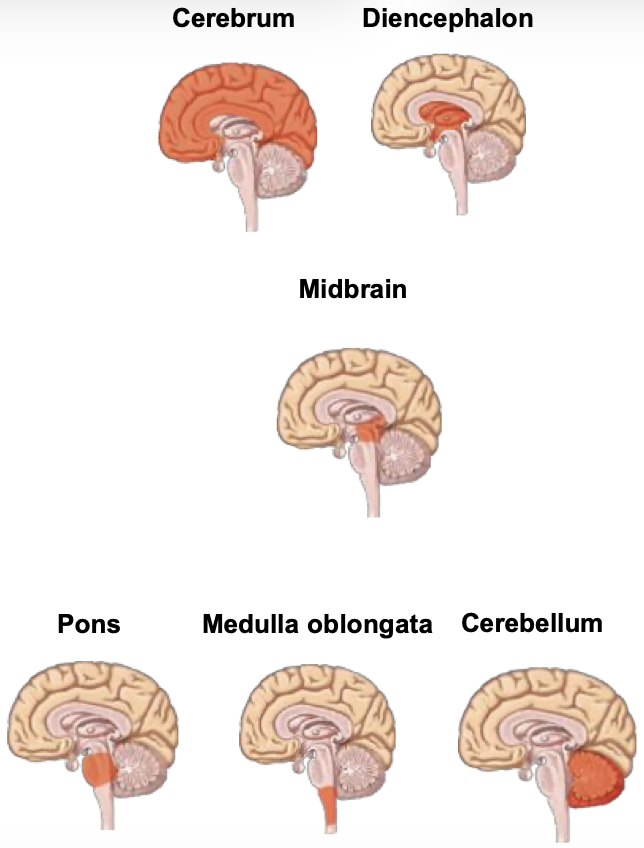
Which regions make up brainstem?
midbrain, pons, and medulla oblongata
medulla oblongata
3cm long
Connects to spinal cord
White matter forms motor and sensory tracts
autonomic reflexes (coughing, vomiting, swallowing), cardiovascular, and respiratory centre
Pons
white matter forms longitudinal (sends info b/w brain and spinal cord) and transverse (sends info b/w cerebrum and cerebellum) tracts
sleep and respiratory centre
Midbrain
Receives and integrates visual, auditory, and tactile sensory input
Responds by orienting head and eyes
Cerebellum
communicates with cerebrum (via transverse tracts) to control locomotion (movement)
Fine motor, posture, balance control
Diencephalon - Thalamus
Paired structure
Cluster of nuclei (grey matter)
Sensory relay centre
Influences mood and strong emotion (fear, rage)
Diencephalon - Hypothalamus
Inferior to thalamus
Cluster of nuclei (grey matter)
Controls endocrine system (Infundibulum connects to pituitary gland)
behaviour, autonomic muscle control, body temp, food and water intake, sexual development
Gyri, Sulci, and Fissures
Gyri (singular - gyrus): elevated tissue or fold that increases surface area, allowing more neurons to fit within brain
In between gyri are sulci (singlular - sulcus): folds
Fissure is a deep sulcus
Cerebrum landmarks
longitudinal fissure seperates right and left hemispheres
lateral fissure seperates frontal and parietal lobes from temporal lobe, underneath is insular lobe
central sulcus seperates frontal lobe from parietal lobe
Cerebrum lobes
Frontal, parietal, occipital, temporal, insular
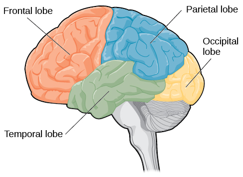
Cerebrum - pre and post central gyri
Pre-central gyrus: In frontal lobe, anterior to central sulcus, primary somatic motor cortex
Post-central gyrus: In parietal lobe, posterior to central sulcus, primary somatic sensory cortex
Cerebrum - Frontal lobe
pre-central gyrus
motor function, motivation, planning, mood regulation, sense of smell
Cerebrum - Parietal lobe
post-central gyrus
receives and evaluates sensory info (pressure, temp), not special senses (smell, hearing, vision, taste)
Cerebrum - Occipital Lobe
receives and integrates visual input
Cerebrum - Temporal Lobe
receives and evaluates input for smell and hearing
memory
Cerebrum - Insular Lobe/Insula
receives and evaluates taste information
Cerebral Cortex
consciousness, memory, reasoning
Corpus callosum
Biggest commisural fibers that transmits information between hemispheres
Located in limbic system
Brain Nucleus/Nuclei
Clusters of neuron somas
Limbic system
Deep area running through medial parts of both hemispheres and parts of diencephalon
Memory, motivation, emotion, learning
Influences autonomic and endocrine systems
Meninges 1
Skull
Dura mater: periosteal dura (superficial), meningeal dura continuous with dura mater of spinal cord (deep)
Dural venous sinus: space between periosteal and meningeal dura, contain veins
*In some areas periosteal and meningeal dura are fused
falx cerebri
Main dural fold extending into a fissure
limits excessive movement of brain in cranium
Meninges 2
Subdural potential space: very small, filled serous fluid
Arachnoid mater
Subarchnoid space with blood vessels and CSF
Pia mater directly attached to cerebrum
Ventricles
4 continuous ventricles - 2 lateral (1 in each hemisphere) continuous with 3rd then 4th ventricles
4th ventricle continuous with spinal cord
All have a choroid plexus that produces CSF
Lined with ependymal cells - circulate CSF
Cerebrospinal fluid (CSF)
Most produced by choroid plexuses, some by blood
Protects and cushions brain and spinal cord
Allows brain to float to reduce weight
Similar to blood serum, without most of proteins
Cranial nerves
12 pairs
9 pairs connect to brainstem
can have single or multi function (parasympathetic, somatic motor, sensory)
Autonomic NS Divisions
sympathetic (fight and flight) and parasympathetic (rest and digest)
both maintain homeostasis by adjusting body functions
Sympathetic has greater influence during stress and physical activity
Parasympathetic has greater influence during resting
Location of ANS (S + PS) somas in spinal cord
Sympathetic preganglionic neuron somas in thoracolumbar division in lateral horn of grey matter
Parasympathetic preganglion neuron somas in craniosacral division in lateral horn of grey matter
Both Sympathetic and Parasympathetic postganglionic neuron somas in autonomic ganglia (outside grey matter)
Dual ANS Innervation
Structure/organ has both sympathetic and parasymathetic neurons
Not all organs have dual innervation
ANS Regulation
Sensory receptors
Spinal cord reflex centres - defecation, urination
Brainstem reflex centres - pupil dilation, coughing, digestive activities, heart rate, respiration
Hypothalamus - influences hormonal release from pituitary galnd, body temp
Cerebrum and limbic system - thoughts and emotions influence ANS through hypothalamus