Chapter 4 - Protein Structure
 Call Kai
 Learn
Learn Practice Test
 Spaced Repetition
 Match
Match Flashcards
 Knowt Play
1/49

There's no tags or description
Looks like no tags are added yet.
Name | Mastery | Learn | Test | Matching | Spaced | Call with Kai | Chat |
|---|
No analytics yet
Send a link to your students to track their progress
50 Terms
proteins are made of…
≥1 polypeptides (chains of amino acids)
structure and charges of amino acids at physiological pH
isolated amino acid has both a positive and negative charge
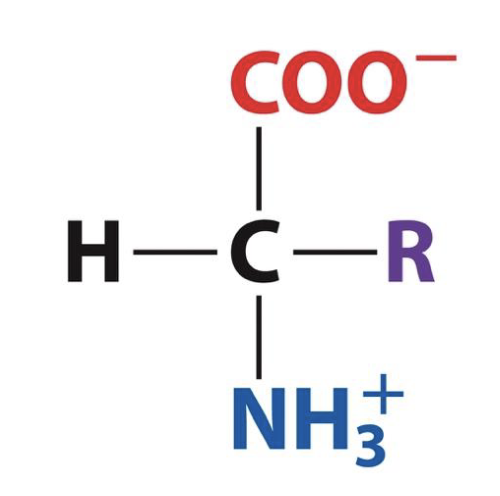
formal name of isolated amino acide
alpha amino acids, because central carbon is alpha carbon
number of amino acids, how they’re grouped
20, grouped by properties of R groups → hydrophobic, polar, charged (and one nonpolar)
hydrophobic amino acids
alanine, phenylalanine, leucine, isoleucine, methionine, valine, tryptophan, proline
polar amino acids
serine, threonine, tyrosine, cysteine, asparagine, glutamine, histidine
charged amino acids
aspartate, glutamate, lysine, arginine
weird nonpolar amino acid
glycine → R group is hydrogen, so it’s not polar or hydrophobic or charged
chirality of amino acids
all but glycine are chiral (19/20)
chiral form of amino acids found in proteins
form on the left
amino acid with no chirality
glycine
formation of peptide bond, what it looks like
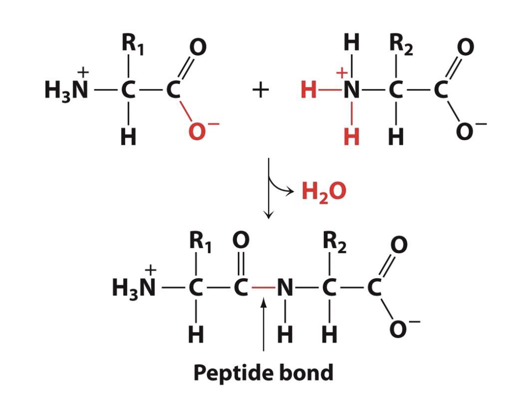
reaction in which amino acids are linked
condensation reaction, where water is removed (from amino and carboxylic acid groups)
two ways peptide bonds can be broken
hydrolysis via exopeptidases or endopeptidases (enzymes that cut peptides)
exopeptidases act upon/cut peptides…
at the end of polypeptide chain
endopeptidases act upon/cut peptides…
from the middle of the polypeptide chain
which is the N-terminus of polypeptide
the end with amino group; beginning of the chain (first formed)
which is the C-terminus of polypeptide
the end with carboxylic acid; end of the chain (last formed, last to leave ribosome)
how to tell charge for a specific amino acid or N/C terminus given pH?
If pK of aa/terminus < pH, will be deprotonated from. If pK of aa/terminus > pH, will be protonated form. If you want the charge of the entire polypeptide at a certain pH, find the charge for each aa, N terminus, and C terminus, and add them together.
how many of the 20 amino acids are found in each protein, usually?
all 20
primary structure of protein
sequence of amino acids
secondary structure of proteins
conformation of polypeptide backbone, not taking side chains into account (how backbone folds - 2D structure)
tertiary structure of proteins
3D conformation of folded polypeptide - how the backbone and side chains fold/interact
quaternary structure of proteins
spatial arrangement and interaction of multiple polypeptides in proteins containing more than one polypeptide chain
conformation/rotation of peptide bonds
there is no rotation around the C-N bond!
Regular secondary structures
alpha helices & beta sheets → repeating conformations of polypeptide backbone
why are alpha helices and beta sheets so common?
they are stabilized by hydrogen bonds
typical length of alpha helices
~10 amino acid residues
parallel beta sheets
all N-terminuses and C-terminuses are aligned; polypeptide chains are aligned in the same direction
antiparallel beta sheets
N-terminuses and C-terminuses alternate on each side of the beta sheet; polypeptide chains are aligned in opposite directions
typical structure of globular proteins
hydrophobic core (often called domain) and hydrophilic surface
largest force governing protein structure
hydrophobic effect
other common forces holding together protein structure (not hydrophobic effect)
ion pairs, interactions with zinc ions, disulfide bonds, thioester bonds, isopeptide bonds
relative chronology of protein structure development
hydrophobic collapse is first → hydrophobic residues to center, hydrophilic residues to surface
next is development of secondary structures like alpha helices and beta sheets → they DO NOT form immediately
molecules helping with protein folding
chaperone proteins
traits of all/most proteins
inherently flexible, often containing some level of disorder
monomorphic proteins
only have a single stable tertiary structure
metamorphic proteins
have two or more possible tertiary conformations
intrinsically disordered proteins
have no fixed structure → primary structure remains the same, but secondary/tertiary are variable
relative energy/stability of monomorphic, metamorphic, and intrinsically disordered proteins
IDPs least stable/highest amount of free energy, then metamorphic, and monomorphic proteins have the lowest free energy/most stability
intrinsically disordered regions
regions with no particular secondary/tertiary structure in a protein; the protein may have both stable regions and IDRs (different than IDP, where entire protein’s structure is variable)
common functions of IDRs
as linkers/spacers, like ‘hinge’ regions connecting antibody proteins’ domains
perform like molecular springs
may wrap around and solubilize calcium phosphate → essential for biomineralization
aggregate in tangled network undergoing liquid-liquid phase separation
liquid-liquid phase separation
IDRs aggregate and separate from liquid environment, though remaining in a liquid state (more gel-like) → this aggregate is protein-rich and sometimes called a membraneless organelle
subunits in a protein
each individual polypeptide chain (since proteins are typically made of multiple)
general name of proteins if same subunits
homodimer, homotrimer, homotetramer… homo prefix with suffix of number of subunits
general name of proteins if different subunits (not all the same, even if like 2/4 are the same)
heterodimer, heterotrimer, heterotetramet… hetero prefix with suffix of number of subunits
polypeptides’ spatial arrangement is known as…
protein quaternary structure
some diseases characterized by misfolded protein aggregates
Alzheimer’s, Parkinson’s, TSEs (transmissable spongiform encephalopathies) (these are neurodegenerative disease)
amyloid deposits
aggregates of misfolded proteins → different types for each disease
name comes from amyloids’ starch-like appearance
most toxic form of amyloid-beta
the dimer