all cells arise from other cells
1/24
There's no tags or description
Looks like no tags are added yet.
Name | Mastery | Learn | Test | Matching | Spaced | Call with Kai |
|---|
No analytics yet
Send a link to your students to track their progress
25 Terms
importance of mitosis
genetically identical daughter cells for:
1. growth: gametes fuse to form a diploid with parental DNA. all cells that grow from this original zygote must be genetically identical
2. repair: when cells are damaged or die, new cells must have identical structure and function
3. asexual reproduction
purpose of meisosis
production of gametes
DNA in eukaryotic cells
- associated with histones (protein) to form chromatin
- chromatin arranged into chromosomes (threadlike structure of chromatin, usually found in the nucleus, carrying genes)
- chromatids: one half of two identical copies of a replicated chromosome
mitotic index
how to make a slide:
- thin layer of cells → so light can pass through
- cells from root tip → that’s where there’s highest rate of mitosis in plants → it’s where stem cells are
- add hydrochloric acid → helps break down cells walls and nuclear membranes → helps separate the cells so you can see them more clearly
- add stain → to increase contrast + view DNA
- push coverslip down (not side to side → could break the chromosomes)
how to calculate mitotic index:
number of cells in mitosis / total number of cells
when does DNA replication occur?
during the S phase of interphase of the cell cycle
the cell cycle
all actively dividing cells pass through a series of stages:
1. interphase:
- G1: cell growth and replication of organelles
- S: DNA replication
- G2: DNA checked for damage/errors
2. mitosis → nuclear division - each new nucleus is identical to the parent nucleus
3. cytokinesis:
- division of cytoplasm
- (mitosis) forms 2 genetically identical diploid daughter cells
mitosis
prophase:
- the chromosomes condense and are now visible
- the nuclear envelope breaks down
- chromosomes free in the cytoplasm
- spindle fibres grow and extend from the poles of the cells to the equator
metaphase:
- chromosomes made up to 2 identical chromatids joined by the centromere
- chromosomes line up along the equator
- spindle fibres attach to the centromere
anaphase:
- spindle fibres contract (uses ATP from mitochondria)
- sister chromatids are pulled apart, to opposite poles of the cell (appear v-shaped)
- the centromere of each chromosome breaks
- these chromatids are now referred to as chromosomes
- the cell starts to elongate
telophase:
- spindle fibres break down
- the chromosomes unwind back into chromatin and are no longer visible under a microscope
- the nuclear membrane starts to reform and divide
regulating the cell cycle
- sometimes cells go through uncontrolled cell division
- the cell cycle is more active than usual
- this can lead to tumours
- genes that regulate the cell cycle: proto-oncogenes and tumour suppressor genes
proto-oncogenes
- normally produces protein products that enhance cell division or inhibit normal cell death
- when altered by mutation it becomes an oncogene
- these can cause a cell to divide in an unregulated manner
- this growth can occur in the absence of normal growth signals such as those provided by growth factors
tumour suppressor genes
- when functioning, the proteins prevent formation of tumours
- when mutated, this leads to uncontrolled cell division
apoptosis
cell death
why are tumours dangerous?
- can put pressure on organs
- can enter bloodstream and spread and form secondary tumours
- can decrease respiration in other cells as it uses up blood supply and glucose for respiration
cancer treatments
1. chemotherapy: some chemical drugs inhibit the synthesis of enzymes needed for DNA replication → so, the cell is unable to enter the S phase
2. some drugs inhibit metaphase: by interfering with spindle formation → so, the cell is unable to divide accurately → the cell undergoes apoptosis → however, it also affects healthy cells
3. radiotherapy: a beam of radiation (gamma rays) is targeted at a mass of cancerous cells. this radiation damages and destroys the DNA in the cells. these cells can no longer divide
malignant tumours
grow rapidly, are less compact and more likely to be life-threatening
how do bacterial cells replicate?
binary fission:
- asexual process (like mitosis)
- quick process → bacterial cells divide approx. every 20mins (under correct conditions → water, glucose, temp)
- does not involve the exchange of genes
steps of binary fission
1. circular DNA replicates, both copies attach to cell membrane
2. plasmids (if present) replicate
3. the cytoplasmic membrane elongates, separating DNA molecules
4. cell membrane pinches inwards between 2 circular DNA molecules dividing cytoplasm
5. new cell wall forms between 2 DNA molecules, dividing the original cell into 2 identical daughter cells, each with a single copy of the circular DNA and a variable number of copies of plasmids
virus
a particle → not a cell → cuz non-living → cuz can’t reproduce on its own
viruses do not belong to any domain or kingdom cuz viruses have no nucleus, no organelles, no cytoplasm or cell membrane - non-cellular
all viruses have: a capsid (protein coat), inside this is the genetic material (DNA or RNA (retrovirus)) that codes for viral proteins, attachment proteins (allow viruses to bind on to other cells)
bacteriophage: a virus that infects bacterial cells
how do virus particles replicate?
1. virus’s surface attachment proteins bind to host cell
2. DNA/RNA injected into host cell which provides “instructions” for host cell’s metabolic processes
3. DNA/RNA is copied
4. host cell begins to produce viral components, nucleic acids, enzymes and structural proteins
5. these are then assembled into new viruses
6. cell bursts (lyses) and releases new viruses
certain viruses can only attack certain cell types. they are said to be specific
receptor sites need to be complementary for the virus to attach and insert its genetic information into the cell
calibrating an eyepiece graticule
- eyepiece graticule placed in the microscope eyepiece (typically 10mm long with 100 divisions)
- stage micrometer slide used to calibrate (usually 2mm long with 0.01mm sub-divisions)
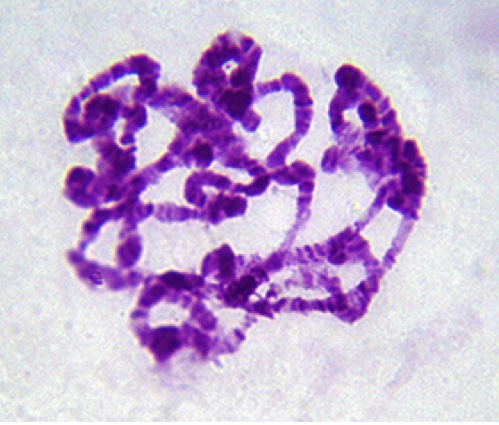
describe and explain the arrangement of the genetic material shown in the image
1. chromosomes are becoming visible
2. because still condensing
name the fixed position occupied by a gene on a DNA molecule
locus
Suggest one way the structure of the chromosome could differ along its length to result in the stain binding more in some areas.
differences in base sequence
what is a homologous pair of chromosomes?
two chromosomes that carry the same genes
Describe two aseptic techniques she would have used when transferring a sample of broth culture on to an agar plate. Explain why each was important.
1. keep lid on Petri dish to prevent unwanted bacteria contaminating the dish
2. wear gloves to prevent contamination from bacteria on hands