Lecture 12 : Retinal Processing and outputs
1/27
There's no tags or description
Looks like no tags are added yet.
Name | Mastery | Learn | Test | Matching | Spaced | Call with Kai |
|---|
No analytics yet
Send a link to your students to track their progress
28 Terms
What is the mechanism for the formation of an antagonistic surround?
Horizontal cell is the key mechanism
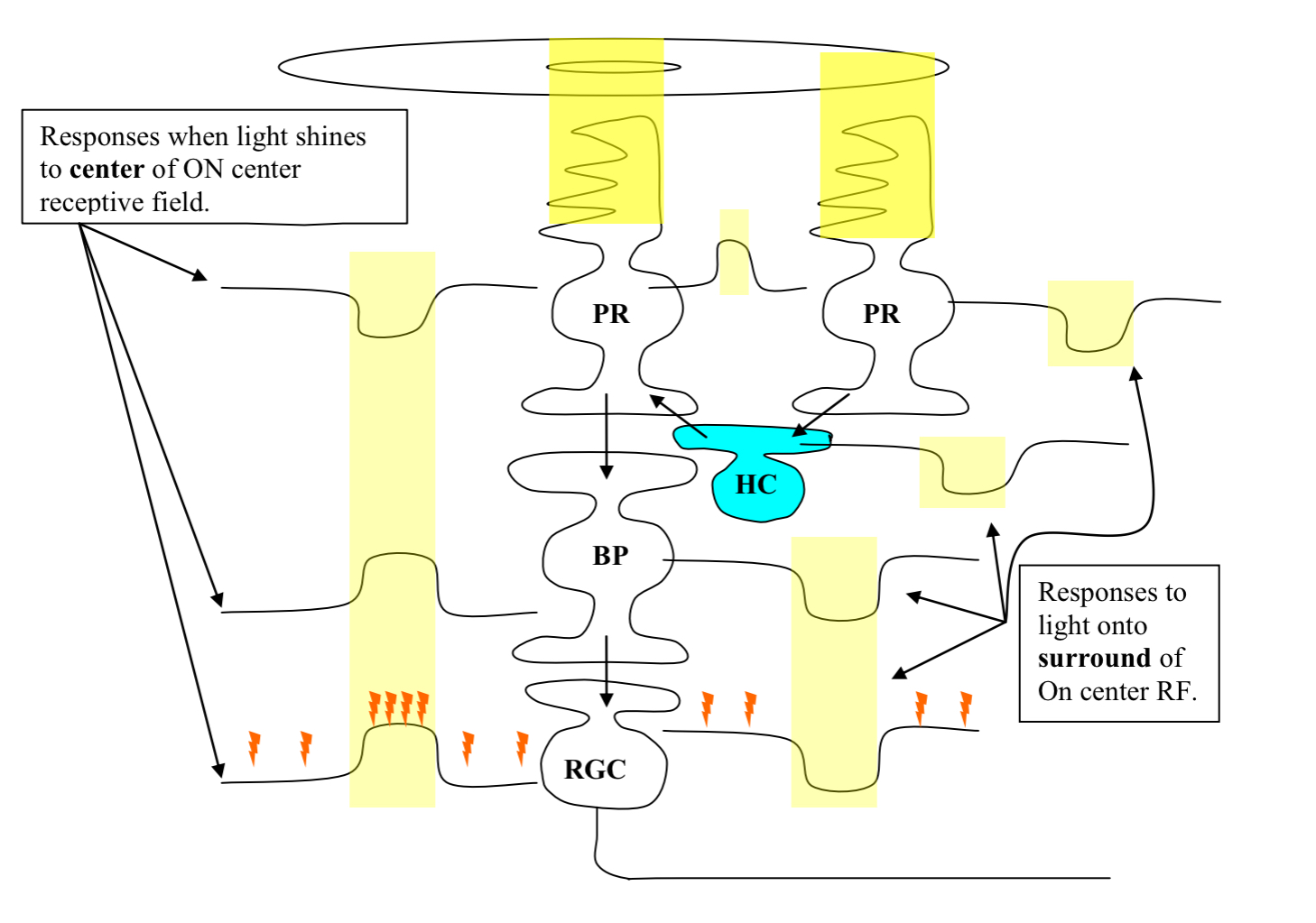
What does this diagram depict?
the role of a horizontal cell (HC) when light falls on a photoreceptor (PR) in a bipolar cell’s (BP) and RGC’s surround

How does light falling on the center and surround of a BP or RGC receptive field produce antagonistic effects?
light in the surround of the bipolar cell’s receptive field produces the opposite effect on the membrane potential of a PR (photoreceptor) than light falling in the center
This is b/c of the horizontal cell’s synaptic connection between PRs in the surround and PRs in the center of a BP’s and RGC’s receptive field
What does this figure show?
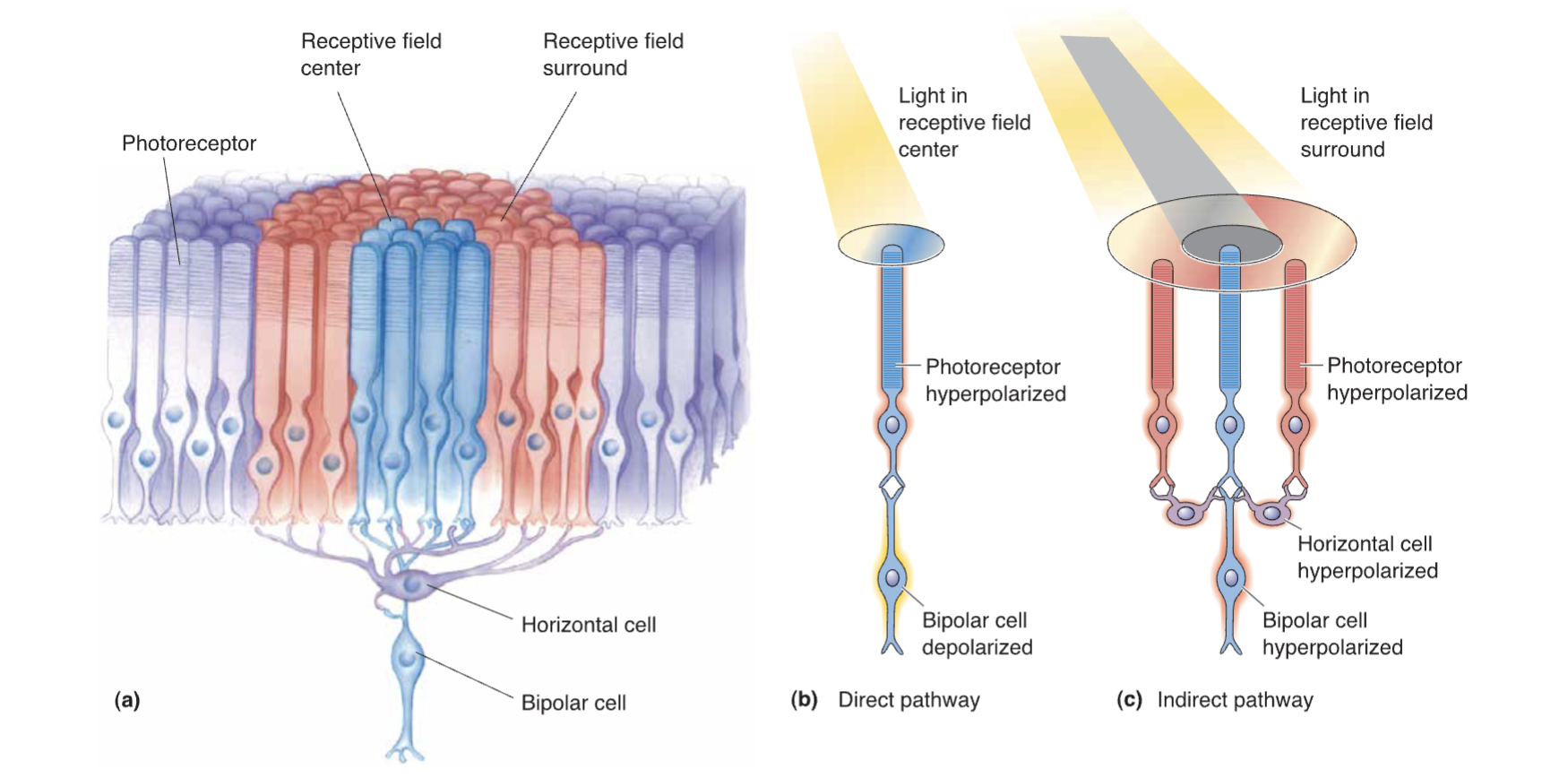
depiction of a single BP cell with synaptic inputs from PRs from its center, surround, and outside its receptive field

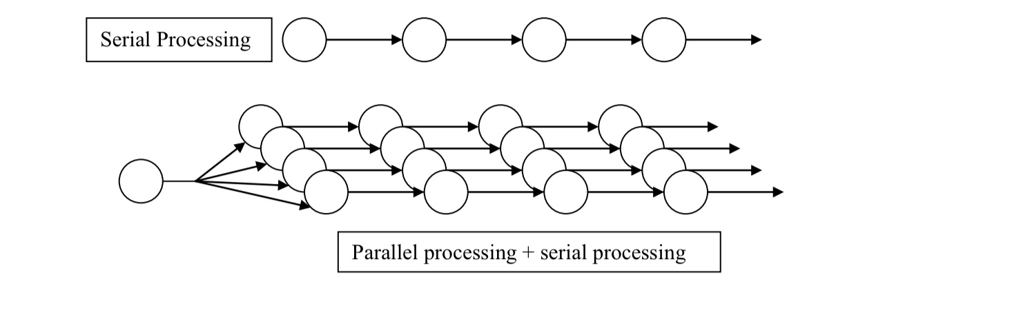
“What happens to the output from the retina” What does this diagram illustrate?
this diagram illustrates general features of serial and parallel processing
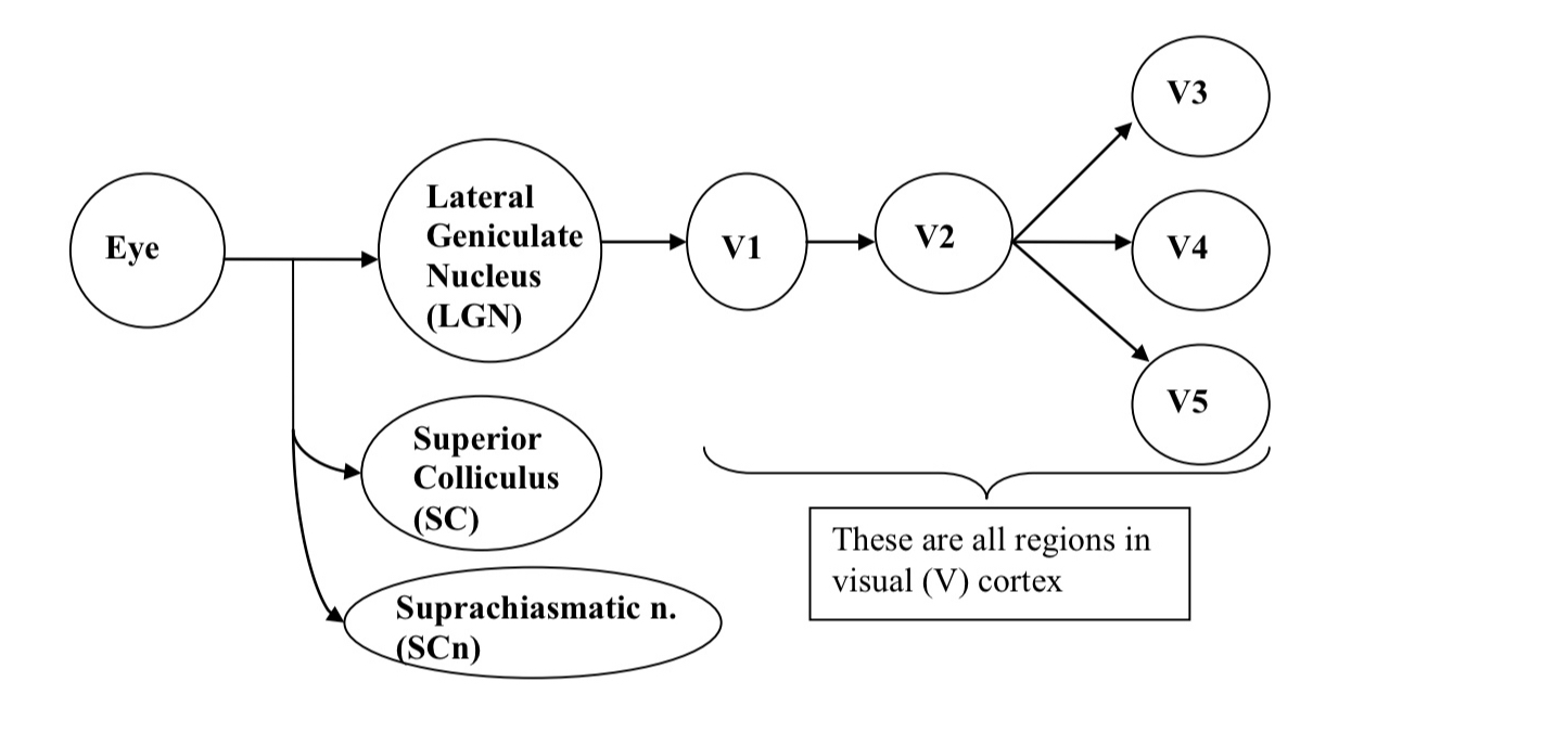
What does this diagram show?
overview conception of the organization of the visual systems
there is massive parallel and serial processing of the retinal ouput

What are some examples of different portions of the visual system having different functions?
Superior colliculus:
tracking system for orienting the eyes towards visual stimuli in the environment
Superchiasmatic nucleus of the hypothalamus (SCn):
like the brain’s clock
neurons in here (SCn) have intrinsic firing frequencies of about 24 hours
controls sleep/wake cycles
Light input to the SCn from the retina resets the SCn clock to the rising and the setting of the sun
How is visual information mapped?
through Visuotopic and Retinotopic maps
What are the characteristics of Visuotopic and Retinotopic maps?
Visuotopic map
every point in the visual world is mapped in a point to point fashion onto various brain areas in the visual system (including the retina)
Retinotopic Map
every point in the retina is mapped in a point to point fashion onto various brain areas in the visual system
What is the difference between Visuotopic and Retinotopic maps?
whether the visual field or the retina is used as the initial point of reference
Visual world is repeatedly represented by Visuotopic maps in different brain regions
How do these orderly maps occur?
orderly location of the RGC in the retina is maintained by orderly point to point neuronal projections from visual brain area to visual brain area
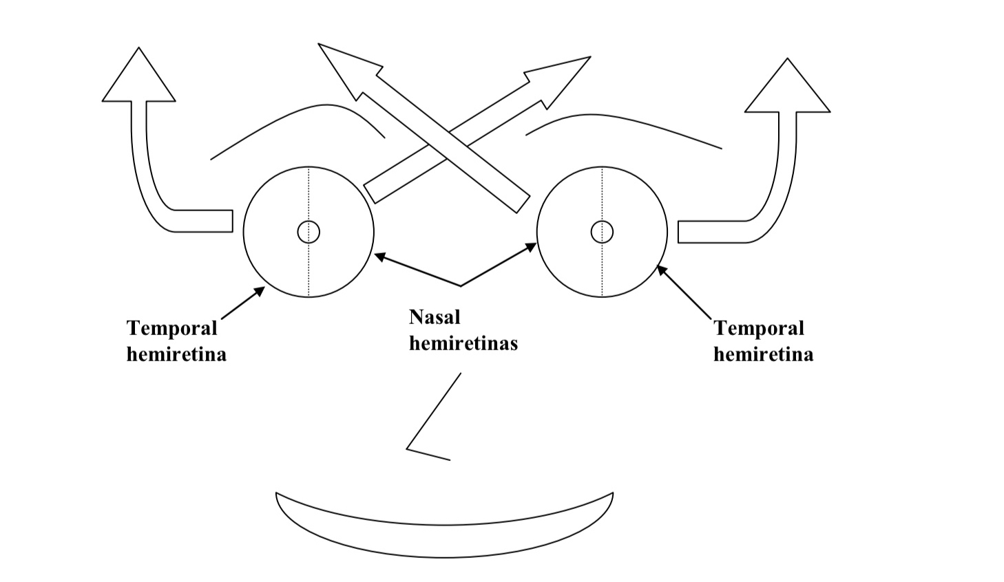
What does this diagram show
How retinas are divided into halves at the fovea
How RGCs from temporal hemiretinas project to the opposite (contralateral) side of the brain

Explain whats occurring in this diagram?
Retinas are vertically divided in half through the fovea
The 2 halves of the retina have different patterns of projections
Nasal Hemiretinas: project to the opposite side of the brain (contralateral projections)
Temporal Hemiretinas: project to the same side of the brain (ipsilateral projections)


What does this diagram depict?
How a visuotopic map is produced
How are orderly representations of the visual field produced?
produced by orderly patterns of anatomical connections

Explain whats happening in this diagram?
the diagram represents a bird’s eye view
Light from objects in the extreme periphery of the visual field (A and F) to the contralateral (opposite) eyes is blocked by the nose so they are detected only in the ipsilateral eye
The Superior colliculus on each side of the brain contains a complete visual representation of the contralateral (opposite) visual field

What is the mechanism for the formation of the visuotopic map?
The nasal hemiretina’s RGCs project in an orderly manner to the contralateral side of the brain
The temporal hemiretina’s RGC project in an orderly manner to the ipsilateral side of the brain

What happens to the axons in this mechanism?
axons from RGCs in either eye that are activated by light from the same object converge on the same neurons in the superior colliculus
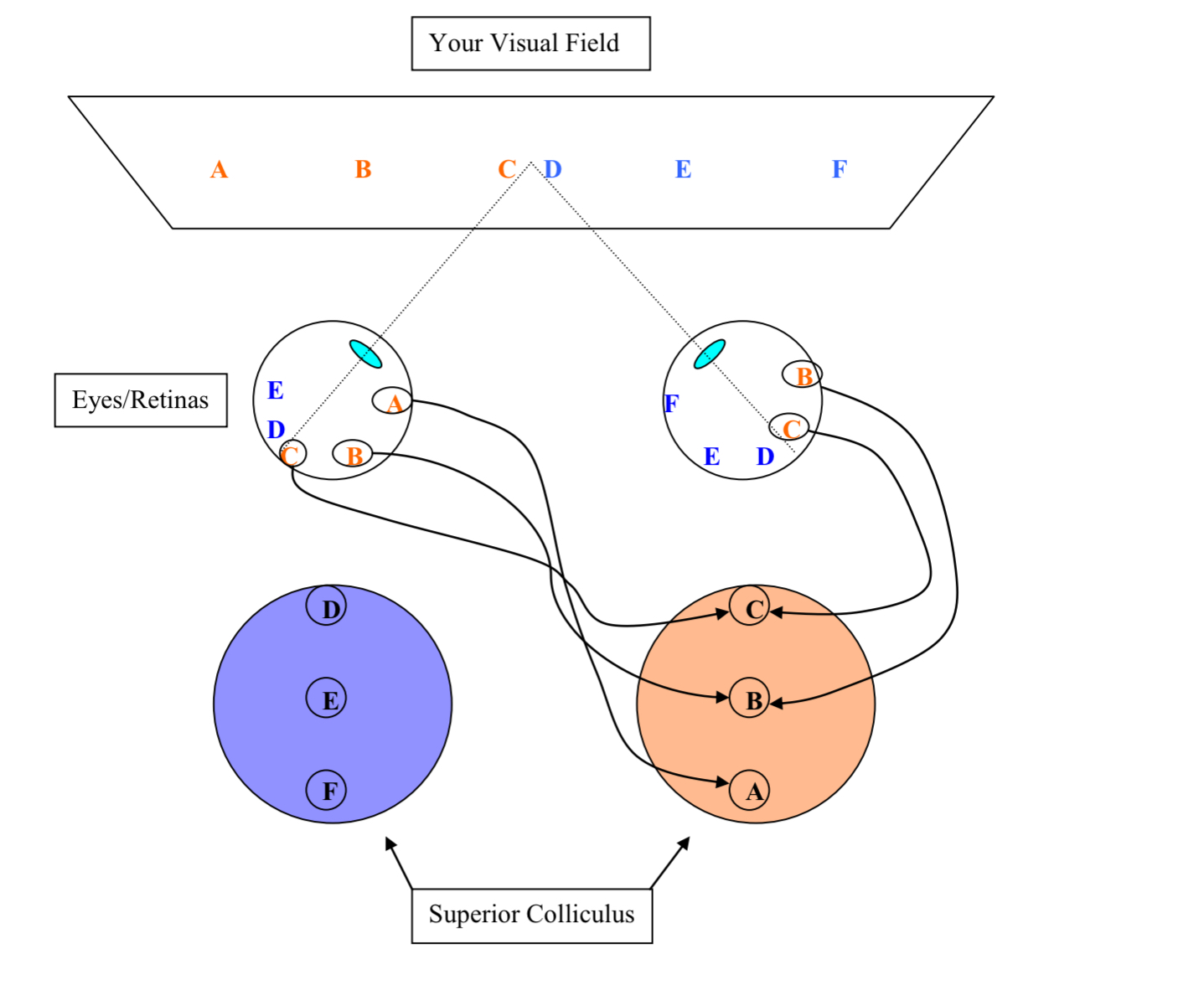
What would happen if you were to record from neurons at point B in the superior colliculus?
the cells there could be driven by inputs from the right eye, the left eye, or both eyes

What happens at A and F locations in the superior colliculus?
cells respond to light input only from the contralateral hemiretina
How do our nervous systems integrate information?
through the same general type of mechanisms even when info comes from different sensory modalities (i.e visual and auditory)
How do we integrate information from both organs (eyes and ears)?
happens because info from the different sources is brought together in the brain in the superior colliculus
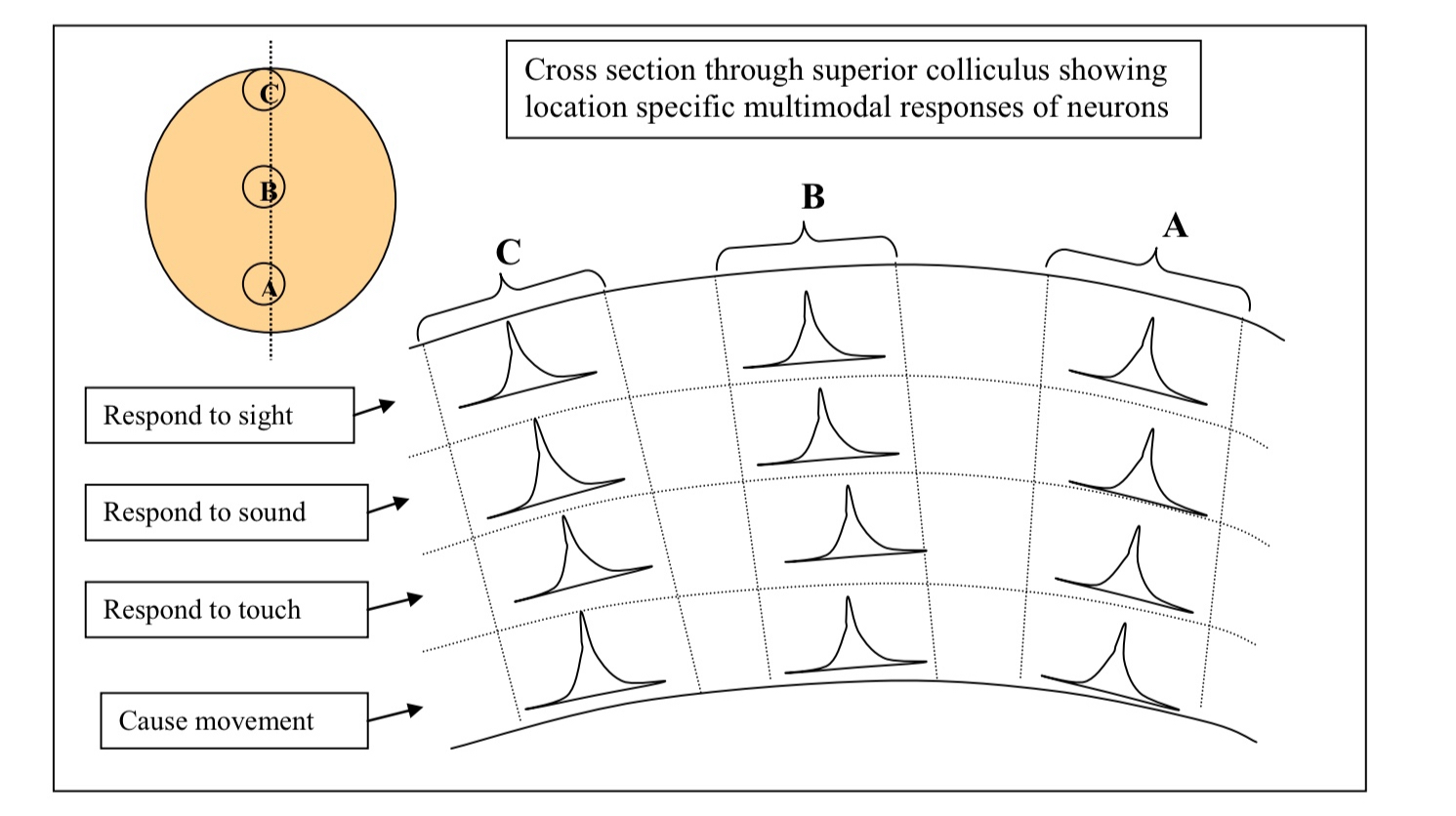
What does this diagram depict?
how info from different sensory modalities is mapped in the superior colliculus
What do cells in each distinct layers in the superior colliculus do?
respond to different modalities

What will a visual stimulus in location B (in the visual field) produce?
causes cells in location B in the superior colliculus to fire
If that visual stimulus makes a noise (like a train whistle), then that will drive neurons at location B (auditory field)
Give some examples on how cells in each functionally distinct layer in the superior colliculus respond to different sensory modalities?
Cells in the Visual Layer: respond to visual info from a single location
Neurons in the Auditory Layer: respond to auditory info from a single location
Cells in the Somatosensory Layer: respond to touch from a single location
What would happen if you stimulate neurons in location B of the motor layer of the superior colliculus?
will cause the eyes and the head to move to look towards location B in the visual field
What is one way to integrate information from different modalities?
bring info together in the brain