red and white lesions and the relationship with oral cancer
 Call Kai
 Learn
Learn Practice Test
 Spaced Repetition
 Match
Match Flashcards
 Knowt Play
1/84

Earn XP
Description and Tags
lecture given 9/11/2026
Name | Mastery | Learn | Test | Matching | Spaced | Call with Kai | Chat |
|---|
No analytics yet
Send a link to your students to track their progress
85 Terms
demarcated hyperkeratosis with epithelial dysplasia

gingival cyst of the adult

focal cemento-osseous dysplasia
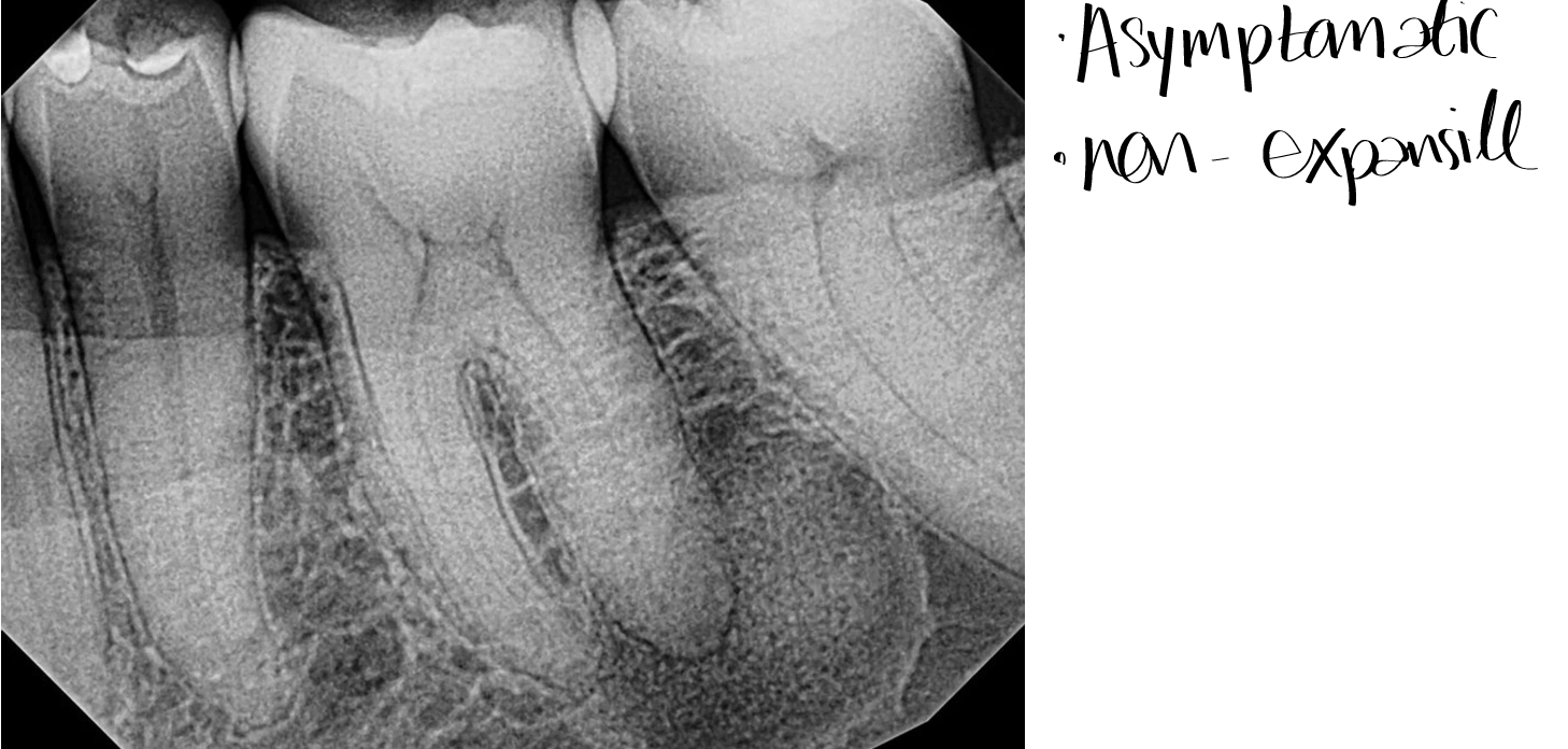
central giant cell granuloma

leukoplakia
white plaque of questionable risk having excluded (other) known diseases or disorders that carry no increased risk for cancer
clinical term, diagnosis is modified after histopathologic evaluation
most common potentially malignant oral lesion with higher risk of malignant transformation over time
DO NOT WRITE THIS TERM ON THE EXAM- IT IS WAY TOO BROAD
leukoedema
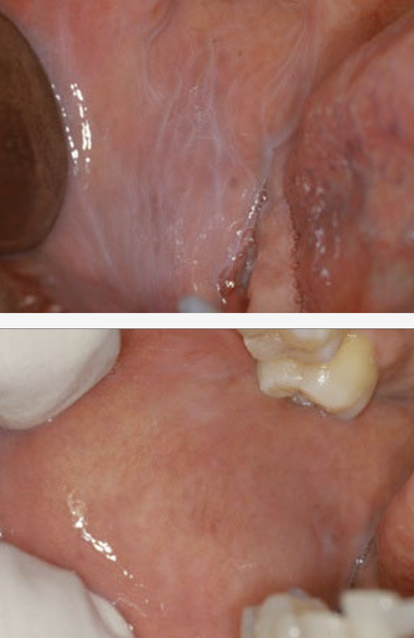
leukoedema
delicate grayish change involving buccal mucosa; disappears on stretching (edema)
may be secondary to mild topical injury
remove inciting agent if identified
no further management
linea alba
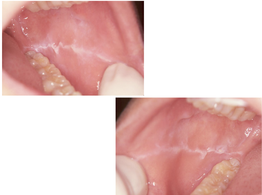
frictional keratosis or morsicatio

frictional keratosis
non keratinized mucosal lesions- linea alba, morsicatio mucosae oris, usually not sharply demarcated, local mild trauma from nibbling
keratinized mucosal lesions- benign alveolar ridge keratosis (BARK), white plaque on the alveolar ridge, painless, may be rough; poorly demarcated, often in the area of previously extracted mandibular third molar (wisdom tooth)
smokeless tobacco keratosis

smokeless tobacco keratosis

smokeless tobacco keratosis
asymptomatic grey-white opalescent area with parallel ridges where tobacco is placed
likely caused by contact irritation and gradient effect
reversible unless leukoplakia develops
manage by discontinuing habit
counsel pt regarding other adverse effects- periodontal disease, staining of dentition
cancer risk is 2x; low compared to cigarettes
follow up
nicotinic stomatitis

nicotinic stomatitis
pebbly, red and white change on palate
caused by heat; typically from pipes, reverse smoking
management via pt education, discontinue habit
reverse smoking may have malignant potential; too few to know
follow up
candidiasis

candidiasis
pseudomembranous type (thrush); white, curdy plaques that may or may not wipe off
hyperplastic candidiasis- do not wipe off easily, often associated with muco-cutaneous disease
treatment- nystatin, clotrimazole, fluconazole, if necessary scraping for cytology, biopsy
leukoplakia

what causes a white lesion to appear white lesion to appear white?
thickened keratin
thickened epithelium
altered epithelial cells- keratinocyte edema, coagulation/degeneration of cells, epithelial dysplasia, squamous cell carcinoma
scar/fibrosis below epithelium (no surface change)
potentially malignant/premalignant
morphologically altered tissue in which cancer is more likely to occur than its apparently normal counterpart
developmental white lesions
white sponge nevus
other congenital genodermatoses
reaction to mild local injury white lesions
leukoedema
contact desquamation
frictional keratoses
autoimmune white lesions
lupus erythematosus
chronic graft vs host disease
tobacco-associated white lesions
smokeless tobacco keratosis
nicotinic stomatitis
infectious white lesions
candidiasis
hairy leukoplakia (EBV- associated)
immune mediated white lesions
lichen planus/lechenoid lesions
migratory glossitis
pre-neoplastic / neoplastic white lesions
dysplasia
squamous cell carcinoma
verrucous carcinoma
leukoplakia
localized- homogenous vs non-homogenous (verrucous/nodular leukoplakia, erytheroleukoplakia (speckled leukoplakia))
proliferative (multi-focal) leukoplakia- homogenous vs non-homogenous (verrcous/nodular leukoplakia, erythroleukoplakia (speckled leukoplakia))
how common is leukoplakia (white), and how often does it become dysplasia or carcinoma?
common
43-47% dysplasia or carcinoma
how common is speckled leukoplakia (red and white), and how often does it become dysplasia or carcinoma?
less common
50-80% dysplasia or carcinoma
how common is erythroplakia (red), and how often does it become dysplasia or carcinoma?
least common
90% dysplasia or carcinoma
what sites are high risk for oral squamous cell carcinoma?
lateral tongue, ventral tongue, floor of mouth, retromolar tongue, soft palate
high risk leukoplakia
lesional presence in high risk areas such as the floor of the mouth, ventral tongue, and soft palate have a higher association with dysplasia or carcinoma than other sites
sharp demarcation at least in parts of the lesion is a worrisome sign
red, raised, or ulcerated component
pt with exoegenous risk factors such as tobacco and alcohol use
non smoker and unknown etiology of lesion
non-reversible type
microscopic atypia
leukoplakia and transformation
when clinical leukoplakia shows histoplathologic dysplasia or carcinoma in situ, the malignant transformation rate is 5-36%
higher risk of malignant transformation in lesions involving lateral-ventral tongue, floor of mouth, and soft palate sites, and non-homogenous lesions
clinical leukoplakia with initial histopathologic findings of hyperkeratosis, unclear if reactive may transform to cancer; not all lesions with histopathologic findings of dysplasia will transform
what level of risk of cancer development is acceptable to watch for a leukoplakic lesion?
overall 10-18% of leukoplakias undergo malignant transformation
in leukoplakic lesions with dysplasia malignant transformation in 5-36% of lesions
squamous cell carcinoma

what are common myths about pre-malignant and malignant oral lesions?
the lesion doesn’t look like cancer
the lesion is not white enough to be cancer
the lesion is too small to be oral cancer
the pt is too young to have oral cancer
epithelial dysplasia
morphologically altered tissue in which cancer is more likely to occur than its apparently normal counterpart
OR
the sum of various disturbances of epithelial proliferation as evidence by histopathologic evaluation
what are morphologic features of dysplasia?
drop shaped rete ridges
basal cell hyperplasia
irregular epithelial stratification
nuclear hyperchromatism
increased nuclear-cytoplasmic ratio
enlarged nucleoli
individual cell keratinization
loss or reduction of cellular cohesion
cellular pleomorphism
loss of basal cell polarity
how are epithelial dysplasias graded and treated?
mild, moderate, or severe / carcinoma in situ
no invasion of the underlying connective tissue has occurred
treat with complete surgical excision
normal
A?
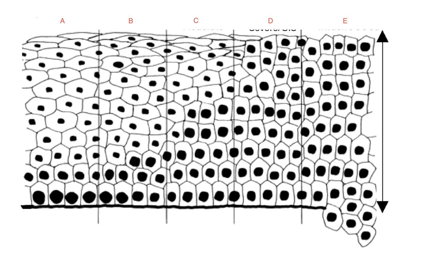
mild
B?

moderate
C?

severe/carcinoma in situ
D?
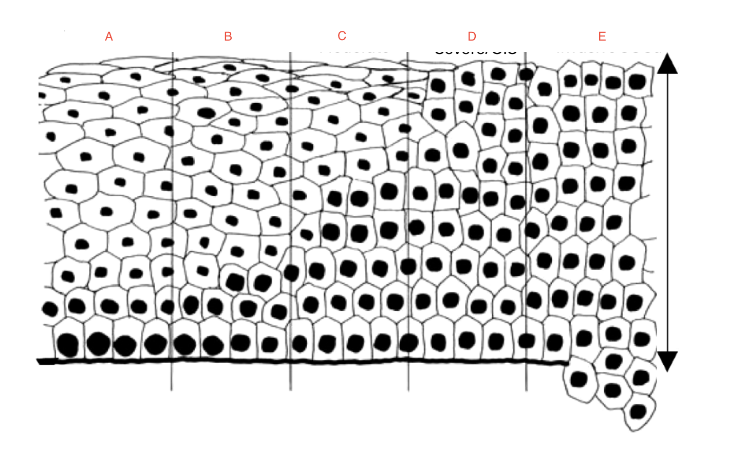
invasive SCCa
E?

normal
what grade is this?

moderate
what grade is this?

squamous cell carcinoma

dysplasia

squamous cell carcinoma, sharply demarcated

how should benign hyperkeratosis be re-evaluated?
is it at least partially well demarcated?
has it ever resolved completely?
has it grown larger?
could it be frictional?
if unlikely to be frictional, what can be causing the hyperkeratosis? early dysplasia?
are there exogenous risk factors?
is this a high risk site?
is it verrucous?
what would you do if this was in your mouth and biopsy was benign hyperkeratosis?
what questions should you ask to weigh no treatment vs treatment?
what is likelihood that this will become invasive?
how long will it take to become invasive?
how morbid, difficult, expensive is removal?
is the pt healthy and what age?
can we safely watch and see what happens?
if we watch with the option to remove it later, have we lost valuable time?
how is localized leukoplakia managed?
all dysplastic lesions should be excised, preferably with clear margins
even if not dysplastic, conservative or narrow excision should be considered, if no clear etiology is identified
wide excision of recurrent lesions
how should you manage an oral leukoplakia?
eliminate the possible cause
if the lesion disappears- definable lesion, manage accordingly
if the lesion does not change- biopsy
squamous cell carcinoma

what are factors that affect squamous cell carcinoma?
extrinsic factors- cigarette smoking, alcohol use, betal quid and areca nut use, immunosuppression, history of previous cancer, family history of cancer, HPV, sunlight, age
exophytic, endophytic, leukoplakic, erythroplakic
squamous cell carcinoma

normal epithelium

dysplasia, bulbous rete ridges
what is this, and what is it demonstrating?

dysplasia

carcinoma

carcinoma

carcinoma

carcinoma

squamous cell carcinoma
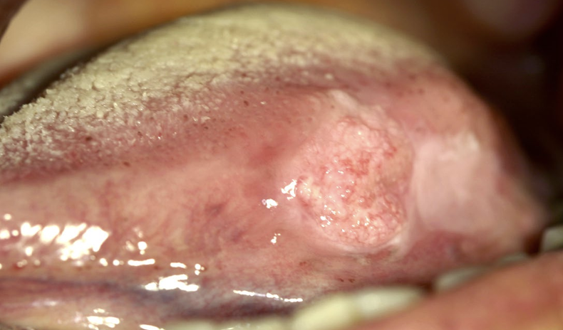
squamous cell carcinoma
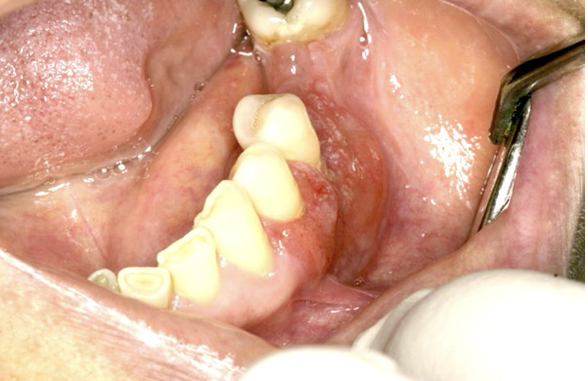
squamous cell carcinoma
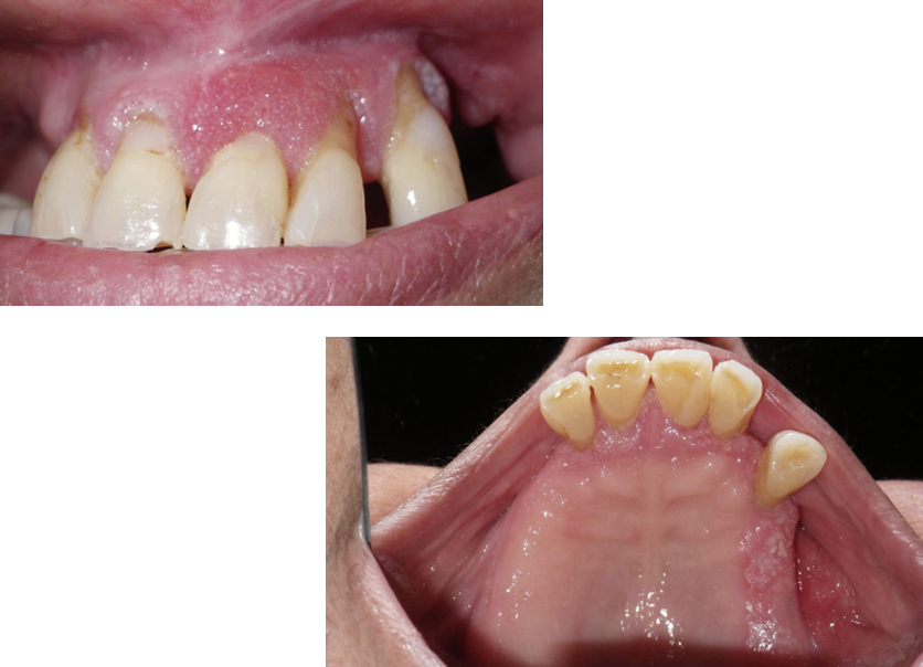
squamous cell carcinoma
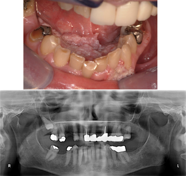
squamous cell carcinoma
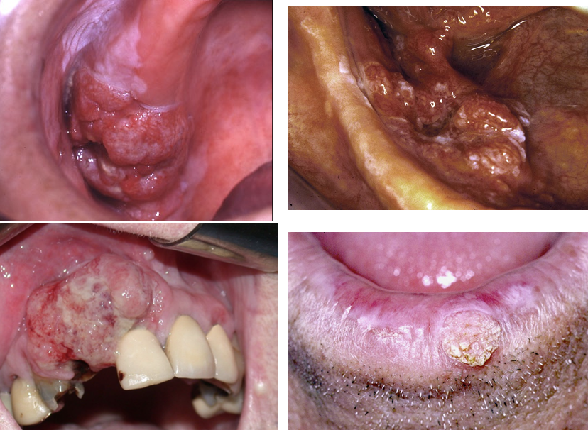
what is the 5 year survival rate for squamous cell carcinoma?
50%
how is squamous cell carcinoma treated?
combination surgery, radiotherapy, and chemotherapy
altered fractionated radiotherapy
chemo-radiotherapy
immunotherapy
proliferative (verrucous) leukoplakia
extensive, irregular white plaques
involves multiple mucosal sites
persistent growth, exophytic, and verrucous
may lead to dysplasia, squamous cell carcinoma
female predilection (4:1 vs males)
<50% with tobacco habit
multifocal lesions, non-contiguous
transform in 50-100% of cases over time
homogenous type is rare
tldr localized leukoplakia
mostly in men
high association with cigarette smoking
single site, usually ventral tongue, floor of mouth
malignant transformation: 10-18%
easy to ablate/excise because localized
tldr proliferative leukoplakia
mostly in women
low association with smoking
multifocal
malignant transformation 50-100%
difficult to treat bc multifocal
what are the common histopathologic findings in proliferative leukoplakia?
typically multiple biopsies
usually early biopsies show benign hyperkeratosis with no evidence of dysplasia
not reliable- should re-evaluate clinically
follow closely
proliferative leukoplakia

proliferative leukoplakia

proliferative leukoplakia

proliferative leukoplakia

erythroplasia
least common and appears as a well demarcated erythematous area with a velvety texture
floor of the mouth, tongue, and soft palate are common locations
90% of changes exhibit carcinoma in situ or invasive squamous cell carcinoma at diagnosis
bruising trauma, not erythroplasia

pt on blood thinners, not erythroplasia
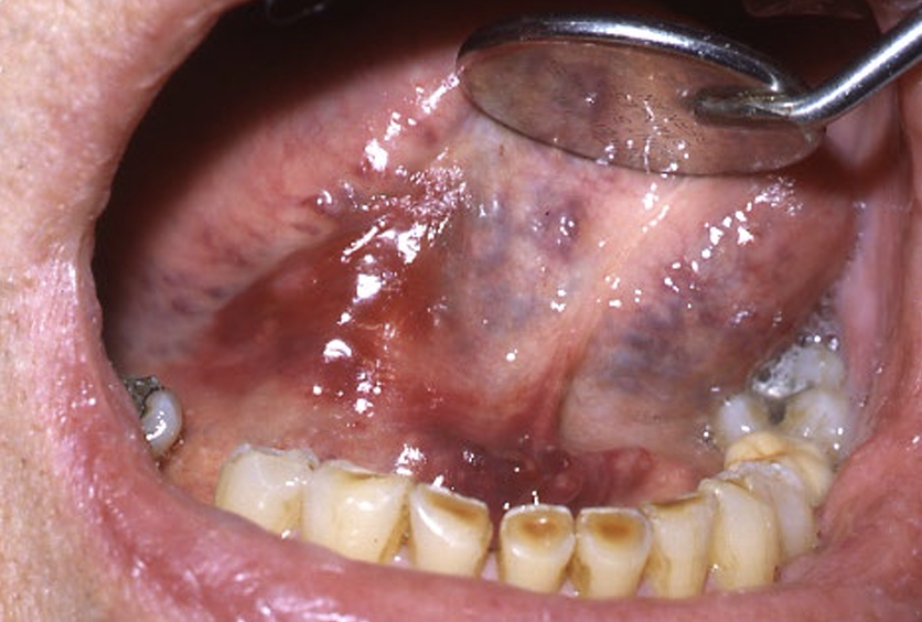
squamous cell carcinoma
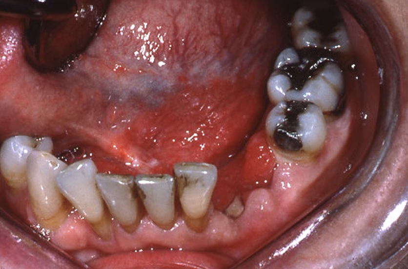
t/f once you treat a lesion like squamous cell carcinoma once, no need to follow up, it won’t come back
false- life long follow up at routine intervals is indicated
t/f there is a fancy machine that can help diagnose squamous cell carcinoma
true HOWEVER nothing is better than the unaided eye