9. Surgical diseases of the pinna and external ear canal. Methods of the treatment. Lateral resection of the external ear canal. Partial and total ablation of the EEC. Osteotomy of the bulla tympanica.
1/52
There's no tags or description
Looks like no tags are added yet.
Name | Mastery | Learn | Test | Matching | Spaced |
|---|
No study sessions yet.
53 Terms
What are examples of diseases of the pinna?
Aural haematoma (othaematoma auris)
Pinna trauma and lacerations
Neoplasia
Trauma
Violent head shaking or scratching due to otitis externa
Coagulopathies
Cushing’s disease
Vessel fragility
What types of dogs and cats are more predisposed to aural haematomas?
Long-eared breeds
Subcutaneous, subperichondral, or intrachondral locations
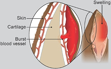
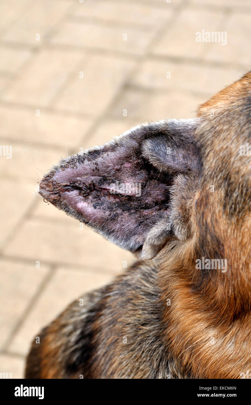
Needle Aspiration & local corticosteroids.
Passive Drain: Penrose drain placed through incisions, cavity lavaged, sutures secure the drain. Left for 2 weeks.
Active Drain: Butterfly catheter inserted, creating suction with a vacuum tube, secured with sutures.
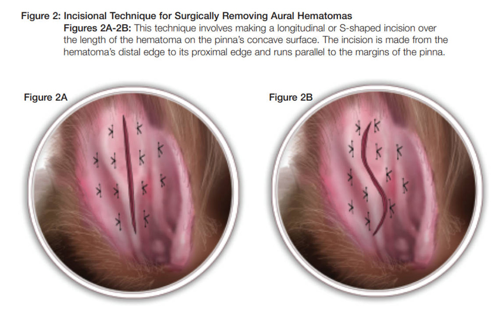
Incisional Drainage: S-shaped incision removes the clot. Mattress sutures with gauze stents prevent over tightening. Bandage ear, remove sutures after 2 weeks
What are causes of pinna trauma and lacerations?
Fighting, head trauma, frostbite, and other chronic injuries
How can pinna wounds be classified?
Superficial (skin only)
Deep (skin and cartilage)
Penetration (through the entire ear)
How are superficial pinna wounds treated?
Wound cleansing (chlorhexidine)
Debridement
Either suturing or healing by second intention (granulation, contraction, epithelialization)
How are deep pinna wounds treated?
Cleanse with chlorhexidine and suture if needed
Align cartilage and skin with simple interrupted sutures on both sides or a combination of simple interrupted and vertical mattress sutures.
Drainage at the outer edge of the wound
Cryotherapy to reduce oedema (first 48 hours of injury)
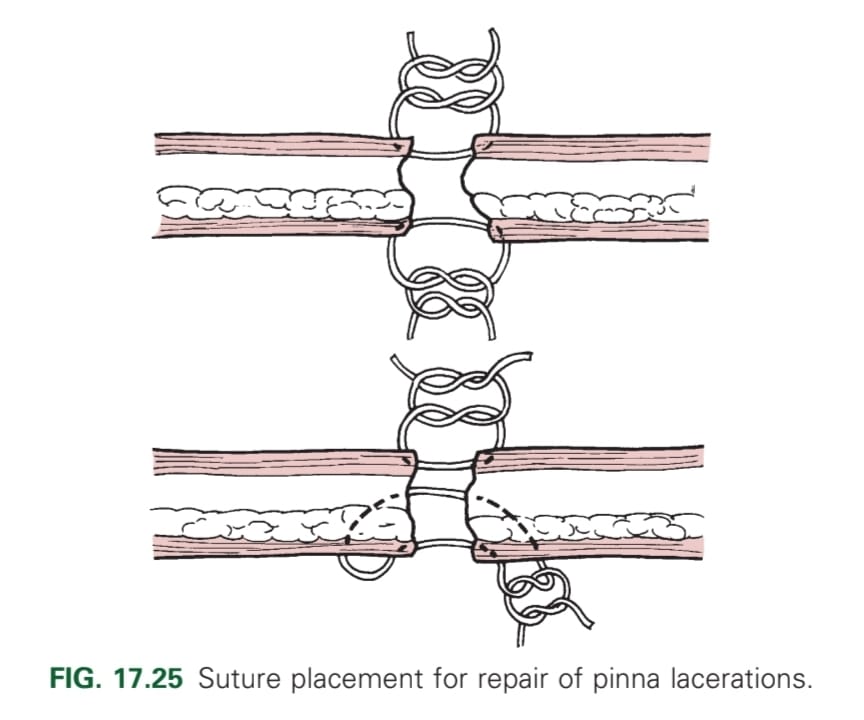
Which surgical method can be used to treat pinna wounds, and how should it be carried out?
Skin flaps. Multi-sided flaps should be sutured along the margins and center of the flap
Actinic keratosis
Squamous cell carcinoma
Haemangioma/haemangiosarcoma
Basal cell tumours
Mast cell tumours
Histiocytomas
Sebaceous adenomas
Soft tissue sarcoma
Fibrosarcoma
Rhabdomyoma
Melanoma
Subtotal or total pinnectomy
Cryotherapy
Ligation
Cauterisation
The pinna is being amputated with scissors at least 1 to 2 cm from the margin of neoplasm. Bleeding is cauterised. The skin on the convex surface is pulled over the cut surface of the auricular cartilage and sutured to the skin on the concave surface with a simple continuous pattern of fine gauze, nonabsorbable monofilament. Do not suture cartilage.
What are examples of diseases of the external ear canal?
Otitis externa
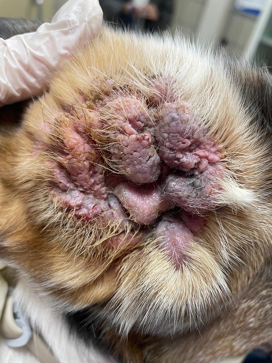
Otitis hyperplastica
Otitis ossificans
Neoplasia
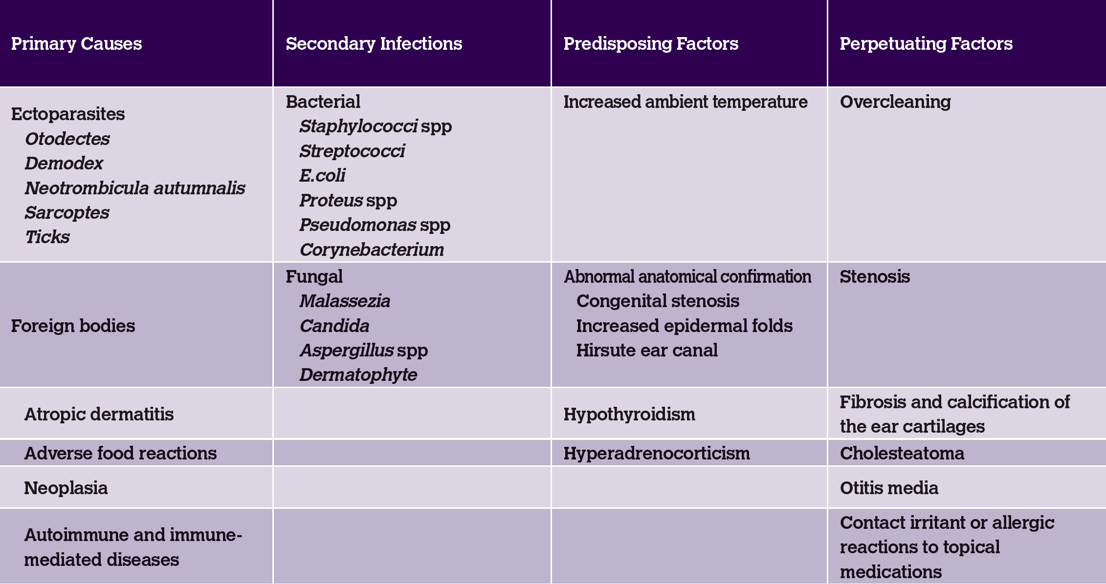
Parasites (Otodectes cynotis, Sarcoptes scabiei)
Allergies (food, atopy, contact)
Bacteria (Staphylococcus pseudointermedius, Proteus, Pseudomonas)
Yeast (Malassezia)
Foreign bodies (grass seeds)
Endocrinopathies (hypothyroidism)
Neoplasia
Autoimmune disorders (pemphigus)
Allergic dermatitis in dogs
Polyps and ectoparasites in cats
Medical history, otoscopy, cytology, biopsy, culture, and imaging (X-ray)
Ear cleaning (ceruminolytics)
Topical anti-inflammatories (corticosteroids)
Topical antibiotics (gentamycin, neomycin), antifungals (imidazoles), antiparasitics (pyrethrin, ivermectin)
Drying agents
Antiseptics (chlorhexidine)
Often epithelial and malignant (60%).
In dogs: ceruminous carcinoma, squamous cell carcinoma, melanoma.
In cats: ceruminous gland adenocarcinoma, squamous cell carcinoma.
Lateral wall resection (Zepp's method)
Partial ear canal ablation (vertical canal ablation)
Total ear canal ablation (TECA) with lateral bulla osteotomy
Osteotomy of the bulla tympanica
Chronic otitis externa, ear canal hyperplasia, congenital canal stenosis, and small tumours
NB. Not in animals with obstruction/stenosis of horizontal ear canal, concurrent otitis media or with severe epithelial hyperplasia
Improve ventilation and drainage, and reduce moisture, humidity, and temperature in the ear canal
What are the consequences of complete removal of the ear canal in TECA?
Facial nerve paralysis
Infection
Haemorrhage
Vestibular disease
Deafness
What is the procedure for lateral wall resection of the external ear (Zepp’s procedure)?
Animal is in lateral recumbency.
Skin incisions are made along the vertical ear canal to expose the underlying cartilage.
Vertical canal cartilage is exposed, cut, reflected ventrally and partially removed.
The remaining cartilage forms a drainage flap, and the skin is sutured to the ear canal epithelium
What is the procedure for a partial ear canal ablation?
Animal is in lateral recumbency.
A T-shaped skin incision is made over the vertical canal.
The vertical canal is separated from soft tissues and removed at the level of the horizontal canal.
Flaps are created to aid drainage, and the wound is closed in a T or inverted L shape.
What is the procedure for a total ear canal ablation?
The ear canal is fully removed, including the horizontal canal to the level of the external acoustic meatus. Use a curette to remove secretory tissue adherent to the rim of the external acoustic meatus.
A lateral bulla osteotomy is performed to remove inflamed tissue in the middle ear.
Bluntly dissect soft tissue from lateral wall
Use rongeurs to remove external osseous prominence ventrally to create a “keyhole”
Curette the bulla floor with intermittent warm saline lavage
The facial nerve is carefully preserved.
Complete removal of the ear canal, tympanic membrane, and sometimes auditory ossicles results in permanent hearing loss.
What is the indication for an osteotomy of the bulla tympanica?
Otitis media and neoplasia, especially in cats
What is the procedure for an osteotomy of the bulla tympanica?
Access to the tympanic bulla via a ventral or lateral approach.
Palpate bulla- incise 2 cm laterally towards the affected side from the midline on the axis of mandibular rami.
Cut platysma muscle, then dissect the digastricus muscle from the hyoglossal muscle (NB hypoglossal nerve)
Make a hole in the ventral bulla and enlarge the opening to examine the interior- remove anything that should be there and flush
If there is evidence of infection, place a drain
In cats, the procedure preserves hearing by retaining the tympanic membrane
What is the bulla tympanica attached to?
Horizontal external ear canal (external ear canal) → bulla tympanica (middle ear) → inner ear (auditory ossicles, semicircular canals, bony labyrinth)
How can problems in the bulla tympanica be assessed?
X-ray
Which positions for x-rays are important for assessing the bulla tympanica?
Rostrocaudal/frontal with open mouth
Lateral oblique (10-15 degrees)
Ventrodorsal/dorsoventral
What are some of the post operative complications that could occur with this osteotomy of the bulla tympanica that you NEED to discuss with the owner?
Deafness, Horner’s syndrome, facial nerve paralysis (sympathetic trunk)
Which nerve can be damaged by osteotomy of the bulla tympanica surgery?
Auriculopalpebral branch of the facial nerve (CNVII)
What type of Horner’s syndrome can be caused during ostectomy of the bulla tympanica?
Post ganglionic