respiratory tract
1/23
Earn XP
Description and Tags
lab practical
Name | Mastery | Learn | Test | Matching | Spaced |
|---|
No study sessions yet.
24 Terms

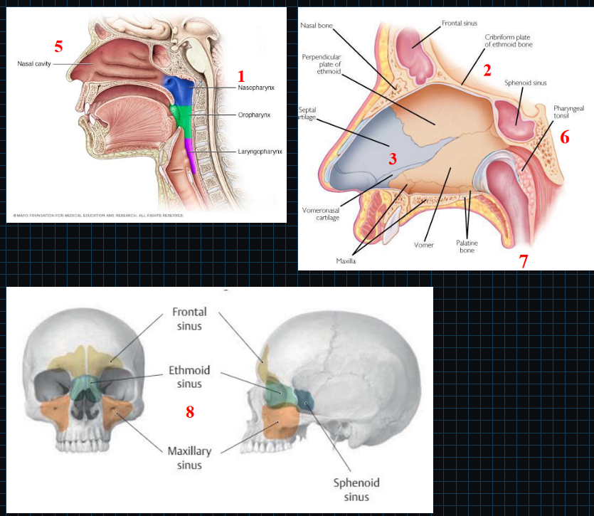
nasal cavity
Nasopharynx, oropharynx, laryngopharynx
Stratified squamous epithelium
Cribriform plate of ethmoid bone
Houses olfactory bulb
Nasal septum
Uvula
Produces mucus and saliva to keep throat moist
Nasal conchae → superior, middle, inferior
Warms and moisturizes air
Pharyngeal, palatine, and lingual tonsils
Lymphatic nodules and tissue of the immune system
Eustachian tube connects middle ear to nasopharynx
Paranasal sinuses
Four paired air-filled spaces that surround nasal cavity
Named after the bones in which they are located
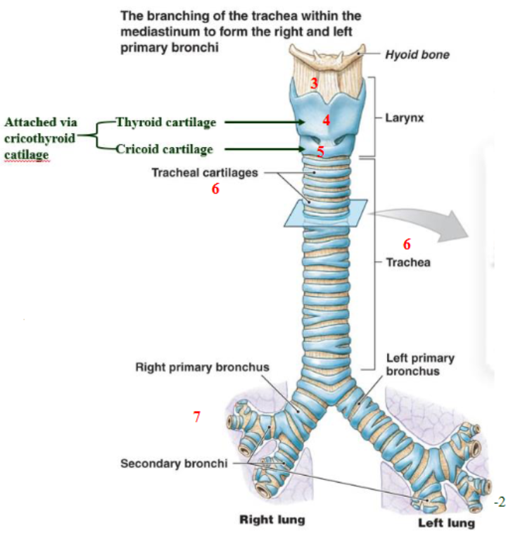
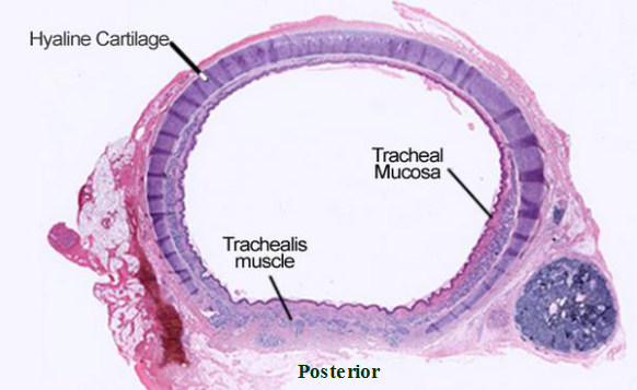
trachea structure
Larynx (voice box)
Epiglottis prevents aspiring food; made up of elastic cartilage
thyroid membrane and ligament connects hyoid bone to thyroid cartilage
thyroid cartilage is part of anterior part of larynx (hyaline cartilage)
cricoid cartilage (hyaline cartilage)
tracheal rings (hyaline cartilage)
primary, secondary, and tertiary bronchi
pseudostratified ciliated columnar epithelium secretes mucus → traps debris, preventing entry into lower respiratory tract
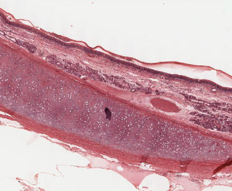
trachea histology
Facing lumen → ciliated pseudostratified columnar epithelium
Goblet cells secrete mucus
Lamina propria of the mucosa: contains areolar loose CT
Anterior trachea → rings of hyaline cartilage
Posterior trachea → smooth trachealis muscle
Inferior to trachea: esophagus
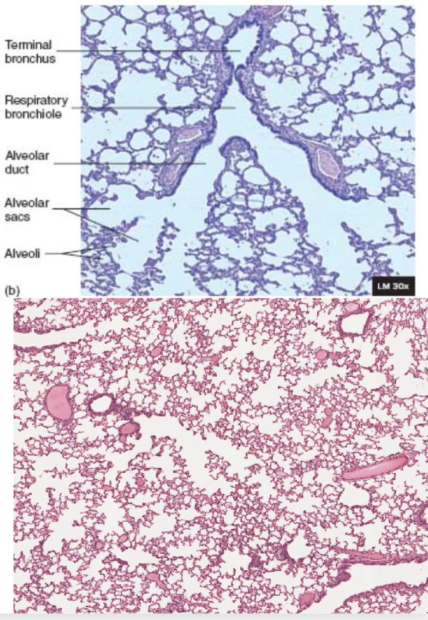
lung tissue histology
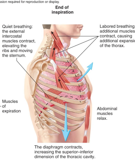
muscles of inspiration
Muscles of Inspiration:
External intercostal muscles contract → elevates ribs and moves sternum out
Diaphragm moves down → increase space in thoracic cavity
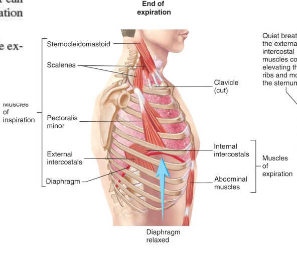
muscles of expiration
Muscles of Expiration:
External intercostals relax → everything moves back in
Diaphragm moves up → decrease space in thoracic cavity
tidal volume (TV)
amount of air inhaled or exhaled with each breath under resting conditions
inspiratory reserve volume (IRV)
amount of air that can be forcefully inhaled after a normal tidal volume inhalation
Purposefully taking a deep breath
expiratory reserve volume
amount of air that can be forcefully exhaled after a normal tidal volume exhalation
Purposefully exhaling
vital capacity (VC)
maximum amount of air that can be exhaled after a maximal inspiration
vital capacity (VC) equation
VC = TV + IRV + ERV
vital capacity = tidal volume + internal reserve volume + external reserve volume
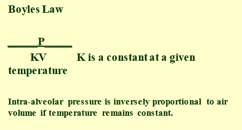
boyle’s law
Boyle's Law: P/KV
Intra-alveolar pressure is inversely proportional to volume if temperature remains constant (K)
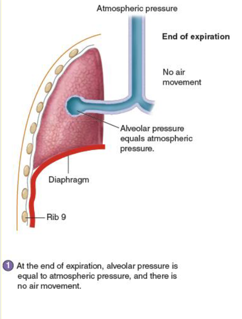
end of expiration
alveolar pressure is equal to atmospheric pressure → no air movement
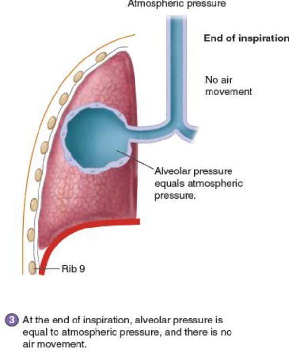
end of inspiration
alveolar pressure is equal to atmospheric pressure → no air movement
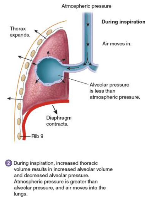
during inspiration
increased thoracic volume results in increased alveolar volume
Results in decreased alveolar pressure
Atmospheric pressure greater than alveolar pressure → air can move into the lungs
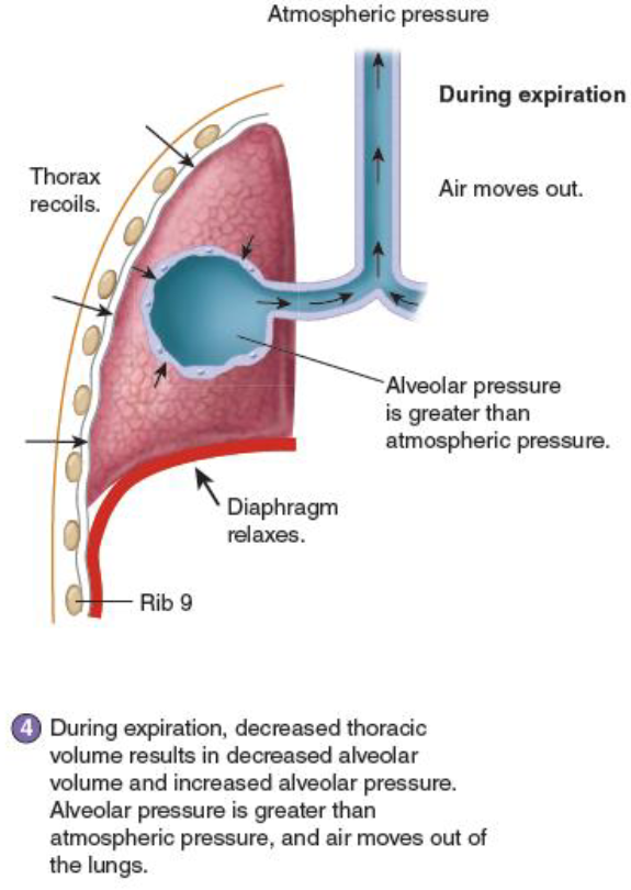
during expiration
decreased thoracic volume results in decreased alveolar volume
Results in increased alveolar pressure
Alveolar pressure greater than atmospheric pressure → air can move out of the lungs
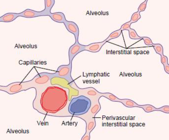
alveolar walls
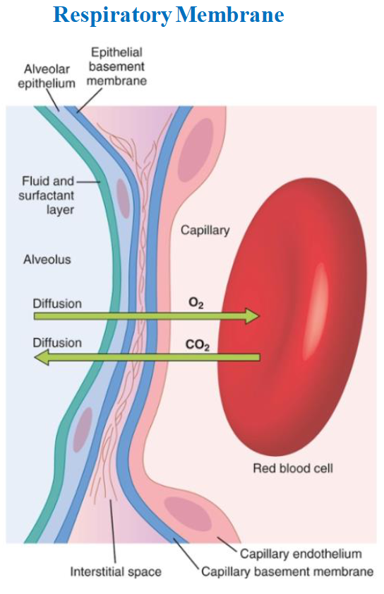
respiratory membrane layers
Respiratory membrane is the point at which the capillaries meet the alveolar sacs
Layers
Layer of liquid lining alveolus containing pulmonary surfactant → reduces surface tension
Alveolar epithelium composed of simple squamous
Basement membrane of epithelium
Thin interstitial space between basement membrane and capillary basement membrane
Basement membrane of the capillary endothelium
Capillary endothelium composed of simple squamous
what type of cells are suited for gas exchange
Simple squamous
what type of cells are suited for fluid exchange
simple cuboidal
type 1 pneumocytes
thin squamous epithelial cells
Form 90% of surface of alveoli
type 2 pneumocytes
secretory cells
Produces surfactant that minimizes surface tension at the alveolar air-liquid interface
Optimizes mechanics of breathing
Avoid alveolar collapse at the end of expiration
alveolar macrophage (dust cells)
protection against bacteria
Primary phagocytes of innate immune system
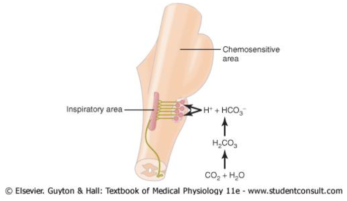
chemical control of respiration
In the brain stem, there is an area that controls respiration by responding to hydrogen ions
Problem: hydrogen ions cannot get through the blood brain barrier, but CO2 can
CO2 dissolves in cerebrospinal fluid (mainly water) → combines to form H2CO3
Immediately dissociates to form hydrogen and bicarbonate ions
Hydrogen ions sensed by the chemosensitive sensory area of the medulla → stimulate the inspiratory center to increase respiration
Bicarbonate ions will be broken down to release CO2 during expiration