Adult II Module 6: Infections and Inflammations
 Call Kai
 Learn
Learn Practice Test
 Spaced Repetition
 Match
Match Flashcards
 Knowt Play
1/37

There's no tags or description
Looks like no tags are added yet.
Name | Mastery | Learn | Test | Matching | Spaced | Call with Kai | Chat |
|---|
No analytics yet
Send a link to your students to track their progress
38 Terms
Emerging and Reemerging Infectious Diseases
Primary care providers are often on the front lines of the global battle to identify, manage, and control emerging and reemerging disease
Emerging infectious diseases result from newly discovered and previously unknown infections that threaten public health: COVID-19
Reemerging infectious diseases are those that had formerly caused so few infections that they were no longer considered a public health threat but have recently reactivated: Polio, Zika, Dengue Fever, Ebola
Primary care clinicians should always screen patients for any relevant travel history in order to institute appropriate infection control practices
If suspected, expert consultation should be sought to diagnose and manage these diseases
Coronaviruses
Coronaviruses are pathogens in animals and humans, well known as a cause of the common cold
SARS is a coronavirus that emerged in 2002 from Guangdong Province, China
Symptoms included severe and often fatal pneumonia
Fever, an influenza-like illness
Diarrhea, leukopenia, thrombocytopenia, and characteristically lymphopenia
Incubation period of 4 to 7 days
About 25% of patients developed severe pneumonia complicated by acute respiratory distress syndrome (ARDS)
Mortality was as high as 50% in older patients and hosts with underlying disease and survivors had significantly reduced exercise capacity and health status compared with the general population
Middle East Respiratory Syndrome (MERS)
Middle East respiratory syndrome (MERS) is caused by a previously unknown coronavirus now called MERS Co-V
All cases have been linked directly or indirectly to countries in the Arabian Peninsula, most notably Saudi Arabia, Qatar, Jordan, and the United Arab Emirates
Incubation period of 2 to 14 days
S/S fever, cough, and shortness of breath, gastrointestinal symptoms including diarrhea and/or nausea and vomiting
As with SARS, most patients progress to ARDS with multi-organ system failure. The mortality rate is approximately 55%.
Advise patients traveling to the affected region: remind strict adherence to routine measures to prevent respiratory illnesses, including washing hands, avoiding personal contact such as kissing and sharing eating utensils with ill individuals, and disinfecting frequently used surfaces such as doorknobs
Infectious Diarrhea
Diarrhea is very common, usually self-limiting
There are risks of morbidity and mortality related to diarrhea, especially in the very young, old, and in developing nations
Sources usually food-borne, water-borne, or fecal/oral
Acute Infectious Diarrhea
Non-inflammatory vs. inflammatory vs. penetrating
Acute Infectious Diarrhea: Non-Inflammatory
Noninflammatory diarrhea is caused by enterotoxin-producing organisms or by viruses that adhere to the mucosa and disrupt absorptive and/or secretory processes without causing acute inflammation or mucosal destruction
Proximal small bowel
Can be abrupt in onset, can be small volume (Ex: acute gastroenteritis) or large volume (Ex: cholera)
Acute Infectious Diarrhea: Inflammatory
Inflammatory diarrhea is characterized by frequent, larger-volume, bloody stools and may be accompanied by fever, severe abdominal pain, tenesmus
Colon
Inflammatory diarrhea is suspected with the demonstration of leukocytes or leukocyte proteins on stool examination
Ex: Dysentery
Acute Infectious Diarrhea: Penetrating
Penetrating diarrhea refers to organisms that cause an inflammatory process, with eventual invasion into the bloodstream associated with systemic manifestations referred to as enteric fever
Distal small bowel
Ex: Salmonella typhi or typhoid fever
Travelers Diarrhea
Likely to be non-inflammatory and of short duration
Fecal-oral contamination of food or water is usually implicated
E. coli (Enteroaggregative E. coli (EAEC) and Enterotoxigenic E. coli (ETEC) are the most likely pathogens, followed by Campylobacter jejuni, Shigella species, and Salmonella species
Prevention is the cornerstone of treatment. Traveler's diarrhea can be avoided by not drinking untreated water or ice cubes or unpasteurized milk and not eating raw fruits and vegetables and undercooked meat. Travelers should drink only sealed or carbonated beverages.
Can occur during or up to 10 days after travel.
Generally lasts approximately 3 to 7 days
Travelers Diarrhea Treatment
Treatment includes oral fluid replacement, Loperamide (Imodium) 4 mg orally at onset and 2 mg after each loose stool up to 16 mg/day
Bismuth subsalicylate (Pepto-Bismol) has both antimicrobial and anti-inflammatory effects and may be taken as 2 tablets every 30 to 60 minutes up to eight doses per day- avoid in Gout, ASA allergy, renal insufficiency
Antimicrobial therapy:
Ciprofloxacin 500 mg orally twice daily for 3-5 days
Azithromycin 500 mg orally daily for 3 days
Antibiotics are typically not prescribed as a preventive measure for travelers
Vibrio Cholera
Approximately 1.3 billion people at risk, resulting in 2.9 million cases and 95,000 deaths annually
V. cholerae is a bacterium naturally found in water, attached to algae, crustaceans, and plankton. Warmer-than-usual waters facilitate its growth. In this activated state, humans are more likely to become infected
Transmitted through fecally-contaminated food or water
S/s watery diarrhea which can lead to dehydration, electrolyte imbalance, renal failure, and metabolic acidosis
Cholera Assessment
Cholera is a potential cause of any case of severe watery diarrhea with or without vomiting, especially in patients who develop rapid and severe volume depletion
Specifically, according to the World Health Organization, cholera should always be suspected when a patient five years or older develops severe volume depletion from acute watery diarrhea, even in an area where cholera is not known to be endemic
Volume status should be assessed through examination of mental status, eyes, mouth, skin, and pulse
Cholera Treatment
Fluid resuscitation
Patients with severe volume depletion or hypovolemic shock need intravenous fluids should be urgently administered to restore circulation
Antibiotics are an adjunctive therapy for patients with cholera and moderate to severe volume depletion. Studies have demonstrated that in such patients, effective antibiotics for cholera can shorten the duration of diarrhea, reduce the volume of stool losses, and lessen the duration of shedding of V. cholerae
Antibiotics can be administered once the initial volume deficit is corrected and vomiting has ceased.

Clostridium Difficile
Broad-spectrum antibiotics alter anaerobic and enteric bowel flora, allowing antibiotic-resistant C. difficile to grow and to produce antibiotic associated diarrhea and more severe pseudomembranous colitis
Assess for recent antibiotic use, especially fluoroquinolones, clindamycin
Treatment of C. difficile colitis is multifactorial.
Broad-spectrum antibiotics should be stopped if possible or changed to more narrow-spectrum agents.
Hydration and electrolyte replacement need to be maintained
Oral fidaxomicin and vancomycin are preferred for initial episodes of nonsevere disease
Fidaxomicin (Dificid) 200 mg orally twice daily for 10 days, or
Vancomycin 125 mg orally 4 times a day for 10 days, or
Alternative: Metronidazole (Flagyl) 500 mg orally 3 times a day for 10-14 days
Severe, complicated C. difficile colitis with ileus will not receive adequate levels of oral vancomycin to the colon. In these cases, intravenous metronidazole may result in detectable colonic levels. Rectal administration of vancomycin may also be beneficial, but should be avoided if there is concern for colonic perforation
Fecal microbiota transplant is used quite successfully in severe cases and in immunocompromised patient. Cure rates up to 90%
In the most severe cases, colectomy is life-saving. It should be performed in the setting of toxic megacolon, colonic perforation, and acute abdomen, and may need to be considered in septic shock.
Fidaxomicin is an oral antibiotic that specifically targets C. difficile and has minimal impact on the intestinal flora. It has similar efficacy to vancomycin in clinical cure but lower rates of C. difficile recurrence
Bezlotoxumab is a monoclonal antibody directed against the B toxin of C. difficile and is FDA-approved for prevention of recurrent C. difficile infection when added to standard therapy
Antibiotic stewardship: Antibiotic use should be limited to the shortest effective course and the narrowest spectrum antibiotic for each pathogen
Remind patients that alcohol scrubs do not inactivate C. difficile spores, so soap and water are required to remove them from hands.
Role of the Primary Care Provider: Patients Presenting with Acute Diarrhea
Obtain a good medical history with OLDCARTS (symptom duration, frequency, and characteristics of the stool and any associated symptoms)
Identify any epidemiologic clues
Recent travel, drinking untreated water, community outbreak (are there a cluster of cases?) , animal or pet contact, occupational exposure such as daycare worker, food history- ingestion of shellfish, drinking untreated water, unpasteurized milk or dairy product, raw or undercooked food (eggs, meat, fish), recent antibiotic use, new medications
Most episodes of diarrhea in the United States are caused by noroviruses
self-limited, noninflammatory gastroenteritis
typically resolves in 2 to 3 days
diarrhea, nausea, vomiting, stomach pain, fever, and head and body aches
Give antibiotics only for presentations not consistent with norovirus infection
Consider bacterial causes if the clinical presentation is one of inflammatory diarrhea
fever, abdominal pain, tenesmus, large-volume diarrhea (more than six stools per day), and mucus or blood in stool
Order diagnostic testing while treating empirically with antibiotics
Assess for the presence of chronic illness, chemotherapy, tube feedings, immune deficiency, or human immunodeficiency virus (HIV) infection with low CD4 counts:
Consider severe manifestations of norovirus, rotavirus, adenovirus, or astrovirus infection and/or atypical presentation of bacterial and parasitic diarrhea
Chronic diarrhea lasting more than 14 days?
Consider protozoan parasites
Systemic manifestations of fever, chills, rigors, night sweats, and weight loss?
Consider penetrating bacteria
Physical Examination: Acute Diarrhea
Signs of dehydration (weight, temperature, orthostatic vital signs, poor poor skin turgor, dry mucous membranes, dark urine)
Mental status
Abdominal assessment: Carefully assess for bowel sounds, rigidity, guarding, tenderness, distention
Noninfectious Causes of Diarrhea
Hyperthyroidism
In chronic diarrhea: assess for the presence of thyromegaly, tachycardia, and proptosis -> may be suggestive of hyperthyroidism
Lymphoma or bowel cancer
Assess for lymphadenopathy, especially cervical node (and supraclavicular) -> may suggest lymphoma or bowel cancer
In the female patient with lower abdominal symptoms, a pelvic examination is imperative
In the geriatric patient, fecal impaction must be ruled out
Consider testing for HIV infection -> diarrhea is common in HIV
Diagnostics: Acute Diarrhea
Usually for self-limiting cases, diagnostics are not indicated
If concern for dehydration or electrolyte imbalance: CBC, CMP
Stool testing for bacterial pathogens: if febrile, bloody diarrhea, abdominal pain, signs of sepsis, more than six unformed stools in a 24-hour period, frail and elderly, or immunocompromised patients
If concern for obstruction, bowel perforation: CT Abdomen/Pelvis
Patient Education/Health Promotion: Acute Diarrhea
Good handwashing after each bowel movement reduces the possibility of spreading disease.
Immunocompromised patients are at greater risk of severe infection and should be diligent about proper safe food handling and preparation.
Drinking frequent, small sips of fluids (water, tea, bouillon, flat cola, flat ginger ale, or sports drink) helps to avoid dehydration.
Avoiding foods and letting your stomach rest is advised until bowel movements begin to return to normal or until the patient begins to feel better. Gradually adding small amounts of food (e.g., crackers, toast, rice, bananas) and avoiding those that may aggravate symptoms (e.g., dairy products, caffeine, high-fat or high-fiber foods, carbonated beverages, sugar-free products, and alcohol) are helpful steps to recovery.
It is better to avoid antidiarrheal products because most cases of diarrhea are self-limited.
Diverticulosis
Diverticular disease is the 8th most common outpatient diagnosis in the United States
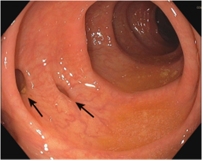
Outpouching of mucosa through the colon wall resulting from structural alterations, defined by the presence of numerous diverticula
Asymptomatic diverticulosis is typically an incidental finding on imaging, found on routine colonoscopy
Clinical manifestations vary from diverticulosis, diverticulitis, and diverticular bleeding or hemorrhage
Presentation depends greatly on the severity of the inflammatory process and the presence or absence of complications
80% to 85% of patients will remain asymptomatic, 10% to 25% will experience diverticulitis
Diverticulosis: Pathophysiology
Colonic diverticula are defects of the bowel wall
They are saclike herniations of the mucosa through the muscularis propria and are actually
pseudodiverticula because they do not contain the muscle layer)
Exact pathogenesis of the disease is unknown but is thought to include environmental, genetic, and dietary factors
Occurs more often with increasing age and as diets including more refined foods (increased refined sugar/white flour, decreased fiber)
Low-fiber diets decrease the amount of intraluminal bulk in the colon. Lack of fecal bulk is thought to produce uncoordinated and irregular colonic peristalsis, which creates sacculations in the colon wall. This results in diverticular outpouchings at weak points.
Diverticulosis: Clinical Presentation and Physical Examination
Asymptomatic patients
Asymptomatic diverticula are typically an incidental finding when the colon is studied for another reason with a barium enema, colonoscopy, CT scan, or ultrasound
Do not need treatment or further follow-up
Symptomatic patients
Signs and symptoms:
Intermittent abdominal pain, bloating, excessive flatulence, or irregular defecation (stool- flattened or ribbonlike to hard pellets)
Associated symptoms: urinary dysfunction, anorexia, nausea, vomiting, and heartburn
Older adults: left lower quadrant steady or crampy pain, in combination with constipation or alternating diarrhea/constipation
Physical exam:
Mild left-lower-quadrant tenderness with a thickened palpable sigmoid colon
Rectal bleeding that is bright red bleeding or maroon-colored -> may suggest a diverticular bleed
Diverticulosis: Diagnostics
Asymptomatic diverticulosis:
Require no diagnostic workup
Symptomatic diverticulosis:
CBC indicated if there is rectal bleeding
Microcytic anemia can be present in patients with chronic bleeding from diverticular disease
Stool for occult blood
CT scan of the abdomen/pelvis
Plain abdominal X-ray films will be normal and are unnecessary (can be ordered to exclude the presence of free air in the abdomen)
After resolution of symptoms: colonoscopy, flexible sigmoidoscopy, or barium enema (x-ray of lower GI tract and colon)
Diverticulitis
Most common complication of diverticulosis
Severity of diverticulitis can range from mild, self-limiting uncomplicated disease to a life-threatening condition complicated by perforation, abscess, fistula, or bleeding
Characterized by the inflammation of one or more colonic diverticula
Thought to result from the stagnation of fecal material in a diverticulum. This produces a fecalith, which leads to pressure necrosis of the mucosa and subsequent inflammation. As the inflammatory process progresses, perforation can occur.
Diverticulitis: Signs and Symptoms
Mild to moderate aching left lower quadrant abdominal pain
Localized guarding and rigidity
Mild abdominal distention
Fever
Leukocytosis
Nausea/vomiting (1/3 of patients)
Change in bowel habits (constipation or loose stools)
Rectal bleeding (uncommon)
Diffuse abdominal pain -> indicate possible macroperforation
Rigid board-like abdomen -> severe disease associated with peritonitis
Diverticulitis: Diagnostics
Acute diverticulitis can be made clinically based on history and physical examination alone
Diagnostic testing and clinical management are guided by symptom severity, signs of peritonitis, and the patient’s ability to tolerate oral intake
CT scan of the abdomen with IV contrast is the test of choice
Colonoscopy should be avoided in acute diverticulitis because of risk of perforation
Useful after the inflammatory process subsides to rule out a malignant etiology of symptoms
Diverticulitis: Pharmacologic Management (Outpatient)
Conservative treatment with bowel rest (e.g., clear liquids and follow-up in 48 to 72 hours), increased fluid intake, and oral antibiotics to cover colonic flora.
Oral antibiotic therapy for 7-14 days
amoxicillin-clavulanate (Augmentin, 875/125 mg three times daily) or
trimethoprim-sulfamethoxazole (Bactrim DS) 160/800 mg twice daily) plus metronidazole (500 mg three times daily) or
ciprofloxacin (500 mg twice daily) plus metronidazole (500 mg three times daily)
Diverticulitis: Nonpharmacologic Management
After an episode of acute diverticulitis: low-fiber diet consisting of 15 g or less of dietary fiber to reduce the volume of fecal material in the lower bowel and to prevent irritation of the colon
Whole-grain breads and cereals, raw fruits and vegetables, and legumes should be avoided
Once the patient is asymptomatic: gradual modification to a diet high in fiber
Do not need not avoid nuts, seeds, or popcorn
No relationship has been found between consumption of those foods and the development of diverticulitis
Colonoscopy is recommended 4 to 6 weeks after symptoms resolve to exclude malignancy
Diverticulitis: Indications for Referral and Hospitalization
Hospitalization in patients with:
temperature of 38.5°C (101.3°F) or higher
marked tenderness
signs of localized peritonitis, intestinal obstruction, or suspected intraabdominal or pelvic abscess
Hospitalization is recommended for patients with diabetes or who are immunosuppressed, older adults, and patients with chronic renal failure
Referral for surgical consultation
American Gastroenterological Association and American Society of Colon and Rectal Surgeons guidelines both suggest against elective colonic resection in patients with uncomplicated diverticulitis
In patients with complicated disease, the decision regarding surgical intervention is based on factors including the patient’s age, comorbidities, frequency and severity of attacks, and CT-graded severity of attack
Urinary Tract Infections (UTIs)
Pathogen within the urinary system and inflammatory response
Acute or chronic infection and/or inflammation
Bladder (cystitis), urethra, prostate, ureter, or kidney (pyelonephritis) with microbial colonization of the urine
UTI Types
Uncomplicated
Complicated (all UTIs in males are considered complicated)
Isolated
Unresolved-persistent infection, drug resistance
Reinfection
Relapse
UTIs: Types
Uncomplicated: UTI in healthy, nonpregnant women with no significant history of UTIs, no underlying urologic or gynecologic abnormalities.
Mild to moderate symptoms
Complicated: Associated with a condition, such as a structural or functional abnormality of the urinary tract
Infection may be more serious, treatment less effective
Isolated: First occurrence; or repeat occurrence at least 6 months from the previous episode.
Recurrent: Symptomatic UTIs that occur after resolution of a previously treated UTI
≥ 2 in 6 months
≥ 3 in 12 months
Relapse: Bacterial persistence of same pathogen; not completely eradicated
Reinfection: New bacterial strain or same bacteria after previous eradication
UTIs: Definition
UTIs in women are secondary to ascending infection from the periurethral or perianal area
Bacteria from the colon, vagina, or skin are the usual organisms causing the infection
Cystitis is more common in women than in men because of the short length of the urethra and the proximity of the urethral opening and vagina to the perianal area
Bacteria reaches the bladder through the urethra and can ascend to the kidneys through the ureters
Precipitating factors
Sexual intercourse, use of spermicidal agents, a new sexual partner, maternal history of UTI, and history of UTI during childhood
Older women: history of UTI before menopause, urinary incontinence, atrophic vaginitis due to estrogen deficiency, cystocele, increased postvoid urine residual
Men: lack of circumcision, anal intercourse, HIV infection, and prostatic hypertrophy, anatomical abnormalities of the urinary tract
UTIs are more common in young, sexually active women and older adults
Approximately 40% of women develop a UTI in their lifetime
Uncommon in men < 50 years of age with normal anatomy
Most common pathogen- Escherichia coli
Other pathogens- Staphylococcus saprophyticus and the Enterococcus, Klebsiella, Enterobacter, and Proteus genus
Asymptomatic bacteriuria (ABU)
Significant levels of bacteriuria (colony count >100K) without symptoms
Risk factors- pregnancy, history of indwelling catheterization, instrumentation, urinary incontinence, diabetes, medical illnesses, obstructive uropathy, postmenopausal status, impaired functional and mental status
Risk factors- decreased fluid intake, irregular bladder emptying, comorbidities- diabetes, etc
UTIs: Clinical Presentation
Symptoms vary based on the type of UTI
Screen and assess for
Urinary frequency, nocturia, dysuria, pruritus, fever or chills, hematuria, vaginal discomfort or discharge, pelvic discomfort, back or flank pain, date of last menstrual period, any prior history of UTIs, cervicitis, or pelvic inflammatory disease (PID), dyspareunia
Male- urethral discharge, penile lesions, history of UTIs, STIs, and prior treatment
Acute UTIs include symptoms of
Increased frequency, urgency, dysuria, suprapubic pain, odorous urine, and occasionally hematuria
Complicated UTI symptoms include
High fever, chills, flank pain, costovertebral angle tenderness, nausea, and vomiting, pyuria (pyelonephritis)
UTIs: Physical Exam
Vital signs
Thorough abdominal exam, pelvic, periurethral and perianal exam, penile, prostate
Females
Assess: vulva, vagina, cervix, periurethral and perianal areas for discharge, excoriations, tenderness, and ulcerations.
Males:
Assess penis for discharge, lesions, ulcerations, and swelling
Assess prostate for tenderness, swelling, masses, or nodules.
DRE: tender prostate may be indicative of acute prostatitis
normal or enlarged prostate can indicate chronic bacterial prostatitis
UTIs: Diagnostics
Urinalysis
Clean-voided midstream specimen
Nitrates, leukocyte esterase, bacteria, WBCs, RBCs, and blood in the urine are (+) signs of infection
Urine culture is the definitive test
Urine cultures should be obtained from patients with the following:
History of frequent UTIs
Empirical treatment failed
Febrile or seriously ill
Live in a community with high rates of antibiotic resistance,
Recently been hospitalized
Pregnant women
Young men (UTIs are unusual and may suggest underlying problems)
Sterile pyuria- negative urine culture despite a positive urinalysis, e.g. positive leukocyte esterase, WBCs)
Multiple bacterial species identified by culture- suggest contamination of the specimen unless catheterized specimen
Large numbers of skin flora, such as Staphylococcus epidermidis, diphtheroids, and β-hemolytic streptococci, can usually be ignored.
Candida organisms usually suggest vaginal contamination.
Beta strep should not be ignored in pregnancy
Test of cure urine culture should be obtained in men and whenever there is suspicion that an infection may not have been eradicated
Routine testing of cure cultures is not indicated unless a persistent UTI is suspected
Renal ultrasounds- structural abnormalities, calculi, masses, and hydronephrosis

UTIs: Management in Nonpregnant Adults
Empiric antimicrobial management in the outpatient setting
Acute simple cystitis in nonpregnant adults
First-line therapy
Nitrofurantoin (Macrobid) 100mg orally twice daily (5 days for females, 7 days for males)
Trimethoprim-sulfamethoxazole (Bactrim) 160/800mg orally twice daily (3 days for females, 7 days for males)
Fosfomycin 3g orally single dose for 1 day
Beta-Lactam Alternatives:
Amoxicillin-clavulanate 500 mg orally twice daily (5-7 days for females; 7 days for males)
Cephalexin 500 mg orally twice daily (5-7 days for females; 7 days for males)
Fluoroquinolone Alternatives: useful for males concerned with prostatitis
Ciprofloxacin 250mg orally twice daily (3 days for females, 5 days for males)
Levofloxacin 250mg orally once daily (3 days for females, 5 days for males)
Phenazopyridine (Pyridium)- urinary analgesic
Prophylaxis: initiated when the existing UTI has been eradicated, confirmed with negative culture 1 to 2 weeks after treatment
Topical estrogen cream- symptomatic relief in postmenopausal women
Chronic/recurrent UTIs
Refer to urology consult

UTIs: Complications
Pyelonephritis
Most common
Bacterial infection of the kidney resulting from ascending, untreated, or inadequately treated lower UTI
Uncomplicated pyelonephritis- can be treated outpatient
Complicated- inpatient management
Urosepsis
Life-threatening systemic complication
requires hospitalization with high-dose parenteral antimicrobial therapy, IV volume repletion, etc
Rare complications
Emphysematous cystitis
Abscess formation
Renal papillary necrosis
UTIs: Referral
Presence of macroscopic hematuria, suspected malignancy, recurrent UTIs or infections that do not respond to standard antimicrobial therapy
Urinary tract anomalies or obstructions, acute scrotum, and all forms of prostatitis. Hospitalization is recommended for pregnant women with pyelonephritis.
Older adults and those with acute, severe symptoms are candidates for hospitalization and often require parenteral therapy
Comorbidities -diabetes mellitus, sickle cell anemia, nephrolithiasis,
Excessive analgesic use increase risk of renal papillary necrosis and obstruction
UTIs: Patient Education
Void 10 to 15 minutes after sexual intercourse
Adequate fluid intake (64 to 80 ounces daily)
Regular urination(at least every 4 hours)
Wiping from front to back, tampons use during menstruation
Avoiding wiping more than once with the same tissue, extended soaking in a bathtub, wearing tight-fitting underwear made of nonbreathable fabric
Avoid spermicidal products
Seek treatment as soon as symptoms are recognized if previous hx UTI
Educate about the possible benefits of antimicrobial suppression or postcoital prophylaxis
Intravaginal estrogen cream for postmenopausal women with recurrent UTIs